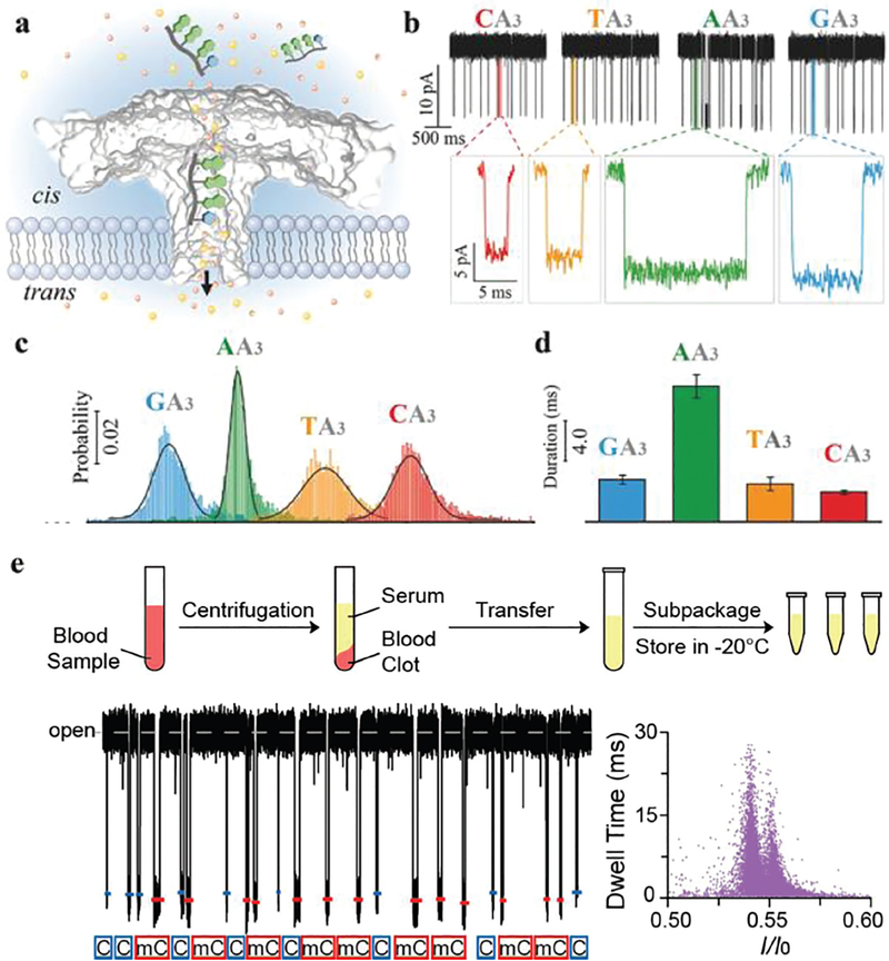
Fig. 3.
(a) Aerolysin inserted into the lipid bilayer from the cis chamber. The ssDNA oligomers were added into the cis solution after applying a positive potential from the trans side. With the cis solution grounded, the negatively charged ssDNA was able to traverse the aerolysin from the cis to the trans chamber and induce a blockade current signal. (b) Single-channel recording of CA3 (red), TA3 (orange), AA3 (green), and GA3 (blue), and the corresponding typical current traces. (c) The residual current histograms (top) and scatter plots (bottom) of the duration versus residual current of CA3, TA3, AA3, and GA3, respectively. All residual current histograms were fitted to Gaussian distributions. (d) Durations recorded for the additions of CA3, TA3, AA3, and GA3 into the aerolysin nanopore, respectively. Reprinted with permission from DOI: 10.1002/smll.201702011, Small, 2017. (e) Detection of methylated and unmethylated DNA in serum. Upper panel: Preparation of serum samples. Human serum was centrifuged, apportioned, and stored at −20 °C before usage. Lower panel: Assignments of C-DNA (blue) and mC-DNA (red) in raw current traces. Two colored short lines and marks were assigned to C-DNA and mC-DNA, respectively. Reprinted with permission from DOI: 10.1021/acs.analchem.7b03133, Anal. Chem., 2017.

