Abstract
We aimed to test if early, perfusion phase tau-PET imaging with [18F]THK5351 might substitute for [18F]FDG PET information on neurodegeneration, as has been previously shown for amyloid-tracers. A patient with cognitive impairment and positive amyloid-PET was examined by [18F]THK5351 tau-PET and [18F]FDG PET. The pattern of early phase of [18F]THK5351 uptake was compared to [18F]FDG visually and by the dice similarity coefficient. Visual inspection of axial slices and stereotactic-surface projection indicated a striking agreement between combined 0–2 min p.i. perfusion images of [18F]THK5351 and standard [18F]FDG 30–60 min p.i. A two-phase protocol of [18F]THK5351 PET might give information on neurodegeneration and tau pathology in a single session.
Keywords: Alzheimer’s disease, [18F]FDG, [18F]THK5351, neurodegeneration, perfusion, PET
INTRODUCTION
Assessments of biomarkers in vivo by means of positron emission tomography (PET) for amyloidosis, tau aggregates, and neurodegeneration are becoming indispensable to characterize individual underlying neuropathology in Alzheimer’s disease (AD) and other disorders. We and others have previously shown that perfusion-phase amyloid-PET imaging is a potential surrogate biomarker of neurodegeneration, possibly with a capability to substitute for [18F]fluorodeoxyglucose (FDG) PET [1–4]. With respect to cost and radiation exposure, such “one-stop shops” giving two channels of pathophysiological information in one session are highly desirable. Recently-developed radioligands for imaging of hyperphosphorylated tau [5, 6] promise to assume a role in future classification schemes for AD, which would comprise a three-fold characterization of neurodegeneration, amyloid and tau pathology for the individual patient [7]. A recent study showed that the initial uptake phase for the arylquinoline tau ligand [18F]THK5317 provides information closely related to the pattern of [18F]FDG uptake [8], presumably due to the general relationship between perfusion and energy metabolism in AD [9]. Thus, we aimed to investigate whether perfusion-phase tau-PET acquisition with the successor arylquinoline ligand [18F]THK5351 also affords this opportunity in clinical routine.
MATERIAL AND METHODS
Patient characteristics and study protocol
A 59-year-old female patient with progressive cognitive impairment over three years (Mini-Mental State Examination: 24) and a diagnosis of probable AD was examined with [18F]THK5351 PET [10] and four months later with [18F]FDG PET. An amyloid PET scan conducted earlier had given a positive visual read.
PET acquisition and preparation
[18F]THK5351 PET was performed in a full dynamic 0-90-min setting initiated upon intravenous injection (∼10 s) of 180 MBq of the ligand. The static 40-60-min frame was reconstructed for the clinical read-out of cerebral tau pathology [11]. The perfusion-phase of [18F]THK5351 was reconstructed in a series of 15 frames of 1 min duration each; to account for their lower count statistics, a 6 mm Gaussian filter was applied to each 1-min frames. [18F]FDG PET was performed in a static 30-60-min setting after injection of 125 MBq of the ligand, with reconstruction of a single static frame.
A T1 weighted MRI sequence was used for co-registration of both PET datasets by use of the PNEURO processing pipeline (PMOD technologies, V 3.5, Basel, Switzerland). Data were analyzed in the MNI space after normalization to the global mean uptake.
PET data evaluation
Uptake data of both tracers were compared in three steps:
-
1)
Each globally normalized one minute [18F]THK5351 frame of the initial 15-min sequence was compared with the static 30-60-min [18F]FDG frame by dice similarity coefficients (DSC). A public available Matlab script (https://github.com/rordenlab/spmScripts/blob/master/nii_dice.m) was used for binarization of both PET images at a 50% maximum intensity threshold. DSCs are given as values between 0% and 100% , with 100% representing perfect anatomical overlap of binary images. Extracerebral voxels were masked in this analysis to avoid artificial maxima.
-
2)
Single 1-min [18F]THK5351 frames and successive summed frames over 2 min were compared visually with [18F]FDG uptake projected in axial slices.
-
3)
[18F]THK5351 and [18F]FDG data were contrasted against an in-house database of age-matched cognitively healthy subjects by 3D stereotactic surface projection (3DSSP).
RESULTS
Late-phase [18F]THK5351-PET revealed elevated binding in the bilateral hippocampi, and in the bilateral temporal and parietal cortices. According to recent publications, this tau PET result corresponds to an AD-typical Braak Stage of III-IV.
[18F]FDG PET revealed widespread hypometabolism indicative of synaptic dysfunction in the lateral parietal and temporal cortices, with a minor accentuation of the right hemisphere. Further synaptic dysfunction was observed in the bilateral posterior cingulate gyrus, all of which was indicated an AD-typical glucose metabolism pattern.
DSC were highest in the first minute after tracer injection (DSC: 95% ), but decline linearly (R2 = 0.99) towards the 15-min frame (DSC: 89% ) (Fig. 1).
Fig.1.
Axial slices show the decrease of visual similarity between the single [18F]FDG static frame and [18F]THK5351 as a function of frame time in minutes. The middle part of the figure shows dice similarity coefficients for each of the 15 initial 1-min [18F]THK5351 frame relative to the 30-60-min [18F]FDG frame. SUVR TACs of [18F]THK5351 for affected parietal and preserved frontal brain regions in this AD case, which are illustrated in the lower part of the figure, indicate the widest gap at the very earliest perfusion phase.
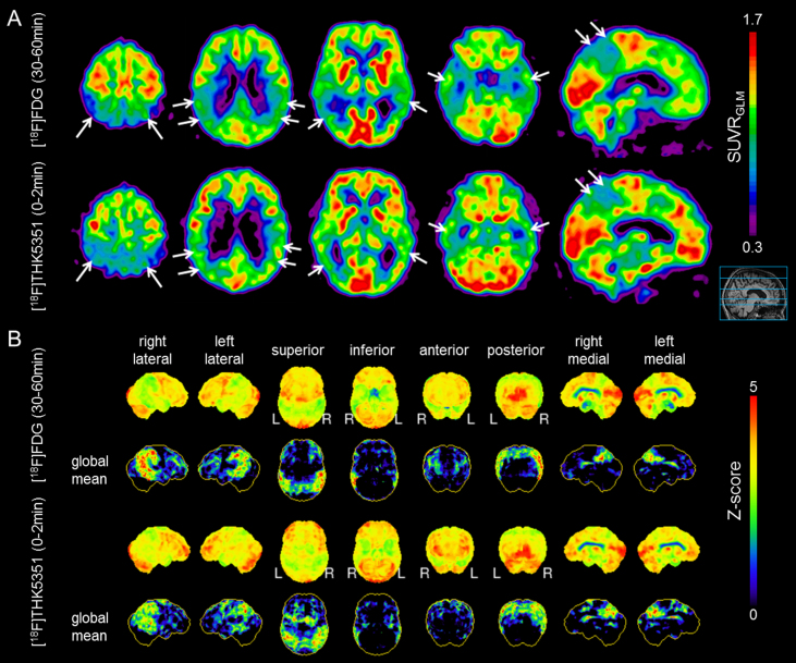
Visual inspection of axial and sagittal slices indicated a striking agreement between the 0–2 min p.i. acquisition of [18F]THK5351 and standard 30-60-min p.i. [18F]FDG uptake (Fig. 2A). Visual comparison of [18F]THK5351 frames ≤5 min p.i. were judged similar, whereas frames >5 min p.i. did not suggest a compelling congruency (Fig. 1) to visual read, although DSC values remained high at later frames. 3D-SSP likewise resulted in a highly congruent regional hypometabolism/-perfusion pattern, which was most pronounced in parieto-temporal and posterior cingulate cortices (Fig. 2B).
Fig.2.
A) Visual comparison of axial and sagittal slices of 30-60-min [18F]FDG PET (upper row) and 0-2-min [18F]THK5351 PET (lower row) frames. White arrows indicate congruent regions of hypometabolism/-perfusion. B) 3DSSP comparison with a normative FDG database (age matched) of 30-60-min [18F]FDG PET (upper two rows) and 0-2-min [18F]THK5351 PET (lower two rows) data. Positive contrast and Z-scores of hypometabolism/-perfusion are projected on a standardized brain surface.
DISCUSSION
We provide first preliminary data suggesting that very early, perfusion-weighted [18F]THK5351 frames can be a surrogate for conventional [18F]FDG PET information indicative of neurodegeneration in a probable AD case. Our analysis revealed a linear decrease of DSC ([18F]THK5351 versus [18F]FDG) with circulation time after [18F]THK5351 injection, which implies that the initial distribution of the tau ligand in the first few minutes provides FDG-like information. Similarly, uptake in a very early interval between 0-3-min was proposed for the predecessor arylquinoline ligand [18F]THK5317 as an [18F]FDG PET surrogate [8]. The more transient relationship in the present study may be related to the very fast washout of [18F]THK5351 from human brain [5]; we found that the 0-2-min p.i. [18F]THK5351 frame was largely congruent with [18F]FDG to visual inspection of axial slices and 3DSSP. Importantly, this read out is widely used in clinical routine [18F]FDG imaging of neurodegenerative disorders, and should therefore serve as a standard procedure. Inspection of the 0-2-min p.i. [18F]THK5351 PET data suggested a pattern of reduced perfusion matching the parieto-temporal hypometabolism to [18F]FDG. Recent [18F]THK5351 PET data from cognitively healthy subjects indicated a perfusion-like pattern in the 0-10-min phase after injection [12]. This physiological finding fits to our present observation, although earlier summation images are seemingly better-suited to identify relevant hypoperfusion/-metabolism in neurodegenerative disorders like AD. Thus, the clinically relevant perfusion-weighted images of [18F]THK5351 uptake should be acquired at a briefer phase of earlier uptake compared to amyloid tracers like [18F]-florbetaben [1]. Compartmental analysis relative to arterial input function might reveal that the differing time-frames are related to the similarly high extraction fractions of the two tracers, but faster tissue clearance of [18F]THK5351 [10], which causes a more transient stability of the perfusion-weighted image.
While derived from a single AD patient, the present findings emphasize the need for further research focusing on the very early perfusion phase of [18F]THK5351, given that the congruency with [18F]FDG is potentially missed in times >5 min p.i. Findings from the predecessor arylquinoline tau ligand [18F]THK5317 further strengthen the validity of the present pilot result [8].
Conclusion
A two-phase protocol of dynamic [18F]THK5351 PET may give combined neurodegeneration and tau pathology biomarker information, with savings in cost, time and radiation exposure in clinical routine PET imaging of AD. These pilot results derived from a single patient encourage us to analyze the very early perfusion phase after tracer administration in a large study population.
CONFLICT OF INTEREST
N. Okamura is a consultant for Clino Corp., receives royalties from GE Healthcare Corp., and is funded by Grant-in-Aid for Scientific Research (B) (15H04900) and Scientific Research on Innovative Areas (26117003) of the Ministry of Education, Culture, Sports, Science and Technology (MEXT), Japan. J. Levin received research support from DZNE, speaker’s fees from Bayer and serves as a consultant for Aesku and Hexal. A. Rominger received speaking honoraria from GE Healthcare and Piramal Imaging. All other authors report no financial disclosure.
REFERENCES
- [1]. Daerr S, Brendel M, Zach C, Mille E, Schilling D, Zacherl MJ, Burger K, Danek A, Pogarell O, Schildan A, Patt M, Barthel H, Sabri O, Bartenstein P, Rominger A (2017) Evaluation of early-phase [18F]-florbetaben PET acquisition in clinical routine cases. Neuroimage Clin 14, 77–86. [DOI] [PMC free article] [PubMed] [Google Scholar]
- [2]. Hsiao IT, Huang CC, Hsieh CJ, Hsu WC, Wey SP, Yen TC, Kung MP, Lin KJ (2012) Correlation of early-phase 18F-florbetapir (AV-45/Amyvid) PET images to FDG images: Preliminary studies. Eur J Nucl Med Mol Imaging 39, 613–620. [DOI] [PubMed] [Google Scholar]
- [3]. Meyer PT, Hellwig S, Amtage F, Rottenburger C, Sahm U, Reuland P, Weber WA, Hull M (2011) Dual-biomarker imaging of regional cerebral amyloid load and neuronal activity in dementia with PET and 11C-labeled Pittsburgh compound B. J Nucl Med 52, 393–400. [DOI] [PubMed] [Google Scholar]
- [4]. Tiepolt S, Hesse S, Patt M, Luthardt J, Schroeter ML, Hoffmann KT, Weise D, Gertz HJ, Sabri O, Barthel H (2016) Early [(18)F]florbetaben and [(11)C]PiB PET images are a surrogate biomarker of neuronal injury in Alzheimer’s disease. Eur J Nucl Med Mol Imaging 43, 1700–1709. [DOI] [PubMed] [Google Scholar]
- [5]. Okamura N, Furumoto S, Harada R, Tago T, Yoshikawa T, Fodero-Tavoletti M, Mulligan RS, Villemagne VL, Akatsu H, Yamamoto T, Arai H, Iwata R, Yanai K, Kudo Y (2013) Novel 18F-labeled arylquinoline derivatives for noninvasive imaging of tau pathology in Alzheimer disease. J Nucl Med 54, 1420–1427. [DOI] [PubMed] [Google Scholar]
- [6]. Schwarz AJ, Yu P, Miller BB, Shcherbinin S, Dickson J, Navitsky M, Joshi AD, Devous MD Sr, Mintun MS (2016) Regional profiles of the candidate tau PET ligand 18F-AV-1451 recapitulate key features of Braak histopathological stages. Brain 139, 1539–1550. [DOI] [PubMed] [Google Scholar]
- [7]. Jack CR Jr., Bennett DA, Blennow K, Carrillo MC, Feldman HH, Frisoni GB, Hampel H, Jagust WJ, Johnson KA, Knopman DS, Petersen RC, Scheltens P, Sperling RA, Dubois B (2016) A/T/N: An unbiased descriptive classification scheme for Alzheimer disease biomarkers. Neurology 87, 539–547. [DOI] [PMC free article] [PubMed] [Google Scholar]
- [8]. Rodriguez-Vieitez E, Leuzy A, Chiotis K, Saint-Aubert L, Wall A, Nordberg A (2017) Comparability of [18F]THK5317 and [11C]PIB blood flow proxy images with [18F]FDG positron emission tomography in Alzheimer’s disease. J Cereb Blood Flow Metab 37, 740–749. [DOI] [PMC free article] [PubMed] [Google Scholar]
- [9]. Mielke R, Pietrzyk U, Jacobs A, Fink GR, Ichimiya A, Kessler J, Herholz K, Heiss WD (1994) HMPAO SPET and FDG PET in Alzheimer’s disease and vascular dementia: Comparison of perfusion and metabolic pattern. Eur J Nucl Med 21, 1052–1060. [DOI] [PubMed] [Google Scholar]
- [10]. Harada R, Okamura N, Furumoto S, Furukawa K, Ishiki A, Tomita N, Tago T, Hiraoka K, Watanuki S, Shidahara M, Miyake M, Ishikawa Y, Matsuda R, Inami A, Yoshikawa T, Funaki Y, Iwata R, Tashiro M, Yanai K, Arai H, Kudo Y (2016) 18F-THK5351: A novel PET radiotracer for imaging neurofibrillary pathology in Alzheimer disease. J Nucl Med 57, 208–214. [DOI] [PubMed] [Google Scholar]
- [11]. Lockhart SN, Baker SL, Okamura N, Furukawa K, Ishiki A, Furumoto S, Tashiro M, Yanai K, Arai H, Kudo Y, Harada R, Tomita N, Hiraoka K, Watanuki S, Jagust WJ (2016) Dynamic PET measures of tau accumulation in cognitively normal older adults and Alzheimer’s disease patients measured using [18F] THK-5351. PLoS One 11, e0158460. [DOI] [PMC free article] [PubMed] [Google Scholar]
- [12]. Hsiao IT, Lin KJ, Huang KL, Huang CC, Chen HS, Wey SP, Yen TC, Okamura N, Hsu JL (2017) Biodistribution and radiation dosimetry for the tau tracer 18F-THK-5351 in healthy human subjects. J Nucl Med. 10.2967/jnumed.116.189126 [DOI] [PubMed] [Google Scholar]


