Abstract
Background
Analysis of the initial experience on learning curve, technical differences and perioperative or early postoperative complications using lumbar hyperlordotic anterior and lateral interbody cages for the correction of lumbar lordosis as compared with the usage of regular lordotic cages.
Methods
Initial 21 consecutive patients were treated with 13 hyperlordotic anterior lumbar interbody fusion (ALIF) cages and 8 hyperlordotic extreme lateral interbody fusion (XLIF) cages. The mean patient age was 64 years, and the mean lumbar hypolordosis was 23°.
Results
No significant procedure-related technical differences were found between the hyperlordotic and nonhyperlordotic ALIF cages. Slightly significant procedure-related technical differences were found between hyperlordotic and nonhyperlordotic XLIF cages. The complication type and occurrence were comparable.
Conclusions
Sagittal balance correction of lumbar lordosis using hyperlordotic ALIF and XLIF cages is a relatively safe surgical procedure with a short learning curve for those surgeons already familiar with anterior and lateral retroperitoneal procedures.
Keywords: sagittal imbalance, lumbar lordosis, hyperlordotic cages, deformity correction
INTRODUCTION
Adult spinal deformity can be defined as a 3-dimensional deviation of the physiological alignment of the spinal column and can have a major impact on a patient's quality of life. Symptoms can range from slight low back pain up to significant back and leg pain with significant reduction of mobility and function. Adult spinal deformity, specifically the sagittal imbalance, is a major source of low back pain in the elderly population. Not all patients affected by this pathological condition need surgery; however, if surgery is required, shortening procedures with posterior 2- or 3-column osteotomy are usually used for that purpose.1,2 Posterior vertebral osteotomies are highly invasive surgical procedures with a relevant percent of perioperative and postoperative complications correlated to large volumes of blood loss and prolonged surgical times. Neurological deficits, non-union cases, and hardware failure are the most frequent postoperative complications that often require one or more repeated surgeries.3,4 Infection is another frightening complication frequently seen in long posterior surgeries.5 Recently developed, hyperlordotic anterior and lateral lumbar cages have been shown to be powerful correction tools that are frequently able to replace posterior osteotomies having, at the same time, with reduced perioperative and postoperative complication incidence.
MATERIALS AND METHODS
We have analyzed our first 21 cases of patients for whom hyperlordotic cages were used for the correction of lumbar lordosis in a sagittal imbalance setting. The study was centered on the evaluation of the learning curve and technical dissimilarities when compared with nonhyperlordotic cages for anterior lumbar interbody fusion (ALIF) and extreme lateral interbody fusion (XLIF). Length of surgery, blood loss, peristalsis restoration, walk out of bed, and perioperative and immediate postoperative complications were also noted. Thirteen hyperlordotic ALIF (HLALIF) procedures were performed at the L5-S1 level, and 8 hyperlordotic XLIF (HLXLIF) procedures were performed at the L3-L4 level. The mean patient's age was 64 years (minimum 57, maximum 78). There were 15 female and 6 male patients. The mean preoperative lumbar sagittal imbalance was 23° (minimum 10°, maximum 36°). The materials used were Brigade hyperlordotic cages for ALIF anterior column realignment and CoRoent XL hyperlordotic cages for XLIF anterior column realignment (Nuvasive Inc, San Diego, California).
RESULTS
Hyperlordotic ALIF
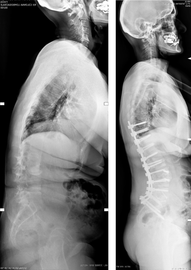
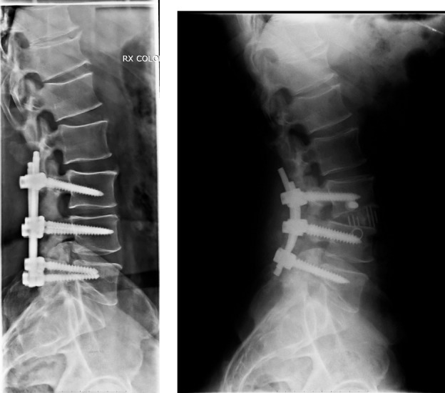
Figures 1 and 2 show examples of preoperative and postoperative patients undergoing the HLALIF procedure. Our regular ALIF procedure comprises a miniopen access with 5 cm- to 7 cm-midline, horizontal skin opening, horizontal fascia incision, blunt muscle dissection, and left retroperitoneal approach to the promontorium. No significant differences were found comparing the approach for the HLALIF cage and the approach for regular ALIF cages. The access itself did not require modification of skin or fascia incision; neither required a more extensive retroperitoneal dissection. The disc removal and the endplate preparation were performed in the same way. The annulus required a more-extensive, circumferential resection so as to enable full mobilization of the vertebral bodies. Initially, a certain degree of difficulty was encountered while choosing the appropriate height and degree of lordosis of the cage to be useful for an adequate correction and distraction. For that reason, initially, frequent C-arm controls increased the patient's and surgeon's X-ray exposure. However, after the initial three cases, the cage selection procedure proceeded much faster, with consequently less C-arm usage.
Figure 1.
Preoperative and postoperative antero-posterior and latero-lateral whole spine standing X-ray: hyperlordotic anterior lumbar interbody fusion.
Figure 2.

Preoperative and postoperative latero-lateral whole spine standing X-ray: hyperlordotic anterior lumbar interbody fusionand hyperlordotic extreme lateral interbody fusion.
No vascular lesions were observed, and no peritoneal or urethral lesions were encountered. No significant differences were found between the positioning of HLALIF and nonhyperlordotic ALIF cages regarding postoperative restoration of normal bowel function and walk-out-of-bed timing, although this last parameter was more influenced by the extension of the concomitant posterior approach done in each of the patients. No sexual dysfunction was reported, either. The mean length of surgery was higher for HLALIF cage insertion (mean difference 20 minutes) when compared with regular-cage insertion. The blood loss was slightly more elevated in the HLALIF procedures, mainly due to frequently observed bleeding from the epidural plexus during detachment of the posterior annulus for distraction purposes (Table 1).
Table 1.
Regular and hyperlordotic ALIF cages: mean surgical differences.
|
Length of Surgery |
Blood Loss |
Walk-Out-of-Bed Timing |
Peristalsis Function Restoration |
|
| Regular ALIF cages | 58 min | < 50 ml | 12 h | 18 h |
| Hyperlordotic ALIF cages | 70 min | 80 ml | 28 h | 18 h |
Abbreviation: ALIF, anterior lumbar interbody fusion.
Overall, the surgical procedure for HLALIF didn't differ significantly when compared with the regular ALIF approach.
Hyperlordotic XLIF
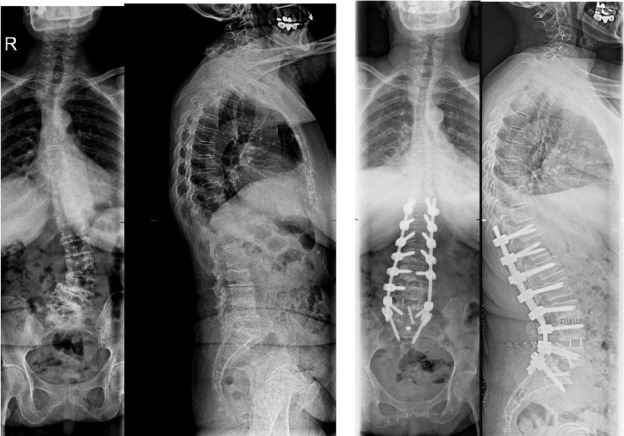
Figures 2 and 3 show examples of preoperative and postoperative patients undergoing the HLXLIF procedure. Some slightly significant differences were found when comparing the insertion of HLXLIF cages and positioning of regular XLIF cages (Table 2). The access itself did not require any modification of skin or fascia incision or blunt muscle dissection. The exposure of the vertebral column segment, however, differed slightly because a better view of the anterior part of the lateral aspect of the disc was judged mandatory due to the necessity of positioning the anterior blade between the anterior longitudinal ligament and the vascular elements (aorta and the cava vein). A right-sided approach was usually used (unless the scoliosis curve dictated a left-side approach) so as to have a visual of an eventual vein lesion. Once the anterior blade was positioned appropriately, covering the whole of the anterior aspect of the disc (as seen on the C-arm image), the anterior longitudinal ligament and three quarters of the annulus were released by a cutting blade and a Cobb dissector. A further difference consisted of the retractor positioning that, unlike regular XLIF, should be placed further posteriorly over the disc space so as to wholly cover, once opened, the lateral aspect of the disc. More attention, too, has to be paid during the trial and cage insertion with respect to the endplate violation. A maneuver we found to be useful was to exert fist pressure over the spine at the treated level so as to increase lordotization during the trial or cage positioning. All of the HLXLIF cages were placed on levels different than L4-5 (all done at L3-4) where the risk of vascular lesion, due to the presence of aorta and cava bifurcation and a poorly defined adventitial layer, is extremely high. However, this is not an absolute contraindication, as some of the cases might involve a high or low bifurcation and a different vascular pattern at this specific level; thus this should be evaluated case by case.
Figure 3.

Preoperative and postoperative latero-lateral whole spine standing X-ray: hyperlordotic extreme lateral interbody fusion.
Table 2.
Regular and hyperlordotic XLIF cages: mean surgical differences.
|
Length of Surgery |
Blood Loss |
Walk-Out-of-Bed Timing |
|
| Regular XLIF cages (single level) | 27 min | < 50 ml | 12 h |
| Hyperlordotic XLIF cages | 55 min | < 50 ml | 24 h |
Abbreviation: XLIF, extreme lateral interbody fusion.
No vascular nor peritoneal or bowel lesions were observed. Bowel peristalsis was never a problem, while the walk-out-of-bed timing was similar to the regular XLIF procedure, though it was more dependent on the concomitant posterior approach procedure that was done in all of the cases. Postoperative neurological dysfunction was never observed, and in just 1 case we had a prolonged (2 months) crural neuropathy with hypersensitivity and pain that resolved completely. The mean length of surgery for positioning of the HLXLIF cages was almost double when compared with nonhyperlordotic cage insertion (55 minutes versus 27 minutes).
DISCUSSION
The recent development of lumbar hyperlordotic cages implantable both through anterior and through lateral retroperitoneal approaches provide for the possibility of powerful sagittal balance correction through lengthening of the spine instead of its shortening.6,7 This type of sagittal correction resembles a more physiological spine morphology than the osteotomies do while at the same time providing for a better distribution of vertical loading forces and a higher level of useful bone fusion. Moreover, the anterior approach to the lumbar spine holds a lower incidence of perioperative and postoperative complications such as infection, non-union, and hardware failure. Other authors found correction capability of the hyperlordotic cages through the anterior and lateral approach highly efficient.8–10 Although not much could be found in the literature, the complication rate is seemingly low and not significantly dissimilar to ours.11,12 In our experience, we had fewer postoperative neurological dysfunctions after using the hyperlordotic XLIF cages as compared with regular XLIF cages. A possible explanation could have been the fewer cases we had and that the procedure was performed at a level different than L4 through L5, where most of the neurological dysfunctions occur after a regular XLIF surgery.
CONCLUSION
Anterior correction of sagittal lumbar imbalance with HLALIF and HLXLIF cages is a reasonably safe surgical procedure with complications comparable to regular anterior and lateral retroperitoneal procedures, except for the higher possibility of vascular lesion during the HLXLIF. No procedure-specific complications were encountered during the initial learning curve. The learning curve was short, and the procedure can be regarded as safe for surgeons already familiar with anterior and lateral retroperitoneal approaches to the lumbar spine.
REFERENCES
- 1.Dorward IG, Lenke LG. Osteotomies in the posterior-only treatment of complex adult spinal deformity: a comparative review. Neurosurg Focus. 2010;28(3):E4. doi: 10.3171/2009.12.FOCUS09259. [DOI] [PubMed] [Google Scholar]
- 2.Enercan M, Ozturk C, Kahraman S, Sarıer M, Hamzaoglu A, Alanay A. Osteotomies/spinal column resections in adult deformity. Eur Spine J. 2013;22(suppl 2):254–264. doi: 10.1007/s00586-012-2313-0. [DOI] [PMC free article] [PubMed] [Google Scholar]
- 3.Buchowski JM, Bridwell KH, Lenke LG, et al. Neurologic complications of lumbar pedicle subtraction osteotomy: a 10-year assessment. Spine (Phila Pa 1976) 2007;32(20):2245–2252. doi: 10.1097/BRS.0b013e31814b2d52. [DOI] [PubMed] [Google Scholar]
- 4.Kim YJ, Bridwell KH, Lenke LG, Cheh G, Baldus C. Results of lumbar pedicle subtraction osteotomies for fixed sagittal imbalance: a minimum 5-year follow-up study. Spine (Phila Pa 1976) 2007;32(20):2189–2197. doi: 10.1097/BRS.0b013e31814b8371. [DOI] [PubMed] [Google Scholar]
- 5.Pull ter Gunne AF, van Laarhoven CJHM, Cohen DB. Incidence of surgical site infection following adult spinal deformity surgery: an analysis of patient risk. Eur Spine J. 2010;19(6):982–988. doi: 10.1007/s00586-009-1269-1. [DOI] [PMC free article] [PubMed] [Google Scholar]
- 6.Saville P, Kadam A, Smith H, Arlet V. Anterior hyperlordotic cages: early experience and radiographic results. J Neurosurg Spine. 2016;25(6):713–719. doi: 10.3171/2016.4.SPINE151206. [DOI] [PubMed] [Google Scholar]
- 7.Berjano P, Cecchinato R, Sinigaglia A, et al. Anterior column realignment from a lateral approach for the treatment of severe sagittal imbalance: a retrospective radiographic study. Eur Spine J. 2015;24(suppl 3):433. doi: 10.1007/s00586-015-3930-1. [DOI] [PubMed] [Google Scholar]
- 8.Marchi L, Oliveira L, Amaral R, et al. Anterior elongation as a minimally invasive alternative for sagittal imbalance—a case series. HSS Journal. 2012;8(2):122–127. doi: 10.1007/s11420-011-9226-z. [DOI] [PMC free article] [PubMed] [Google Scholar]
- 9.Manwaring JC, Bach K, Ahmadian AA, Deukmedjian AR, Smith DA, Uribe JS. Management of sagittal balance in adult spinal deformity with minimally invasive anterolateral lumbar interbody fusion: a preliminary radiographic study. J Neurosurg: Spine. 2014;20(5):515–522. doi: 10.3171/2014.2.SPINE1347. [DOI] [PubMed] [Google Scholar]
- 10.Anand N, Cohen RB, Cohen J, Kahndehroo B, Kahwaty S, Baron E. The influence of lordotic cages on creating sagittal balance in the CMIS treatment of adult spinal deformity. Int J Spin Surg. 2017;11(3):183–192. doi: 10.14444/4023. [DOI] [PMC free article] [PubMed] [Google Scholar]
- 11.Saville PA, Kadam AB, Smith HE, Arlet V. Anterior hyperlordotic cages: early experience and radiographic results. J Neurosurg Spine. 2016;25(6):713–719. doi: 10.3171/2016.4.SPINE151206. [DOI] [PubMed] [Google Scholar]
- 12.Deukmedjian AR, Dakwar E, Ahmadian A, Smith DA, Uribe JS. Early outcomes of minimally invasive anterior longitudinal ligament release for correction of sagittal imbalance in patients with adult spinal deformity. Sci World J. 2012 doi: 10.1100/2012/789698. 789698. [DOI] [PMC free article] [PubMed]


