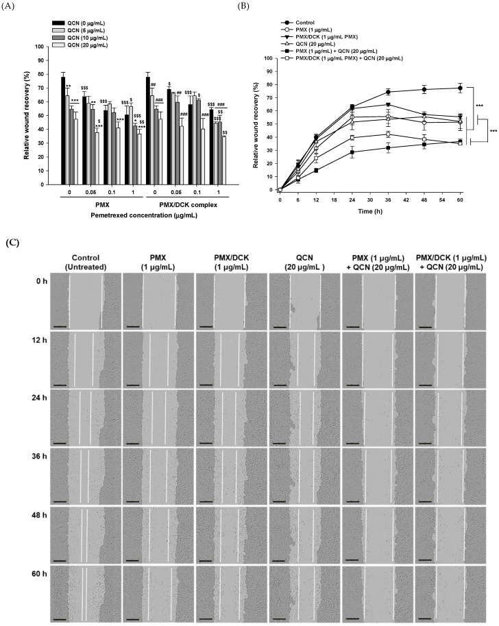
Figure 3.
In vitro effects of PMX, PMX/DCK, QCN, and combinations of PMX or PMX/DCK with QCN on the proliferation/migration of A549 cells. (A) Wound closure after 48 h treatment (n = 5; * p < 0.05, ** p < 0.01, *** p < 0.001 compared with PMX alone at the same concentration; ## p < 0.01, ### p < 0.001 compared with PMX/DCK alone at the same concentration equivalent of PMX; $ p < 0.05, $$ p < 0.01, $$$ p < 0.001 compared with QCN alone at the same concentration); (B) Time course of closure of the wounded areas after treatment of the cells with PMX, PMX/DCK, QCN, and combinations of PMX or PMX/DCK with QCN (n = 5; *** p < 0.001); (C) Representative images taken at different time points after treatment of the cells with PMX, PMX/DCK, QCN, and combinations of PMX or PMX/DCK with QCN. White lines represent the cell-proliferation/migration progress. Scale bar represents 300 μm.

