Abstract
Background:
The pathogenesis of ventilator-associated pneumonia (VAP) is not clearly known. Recently, the role of gastric bacterial colonization has been proposed. The role of gastric colonization with Helicobacter pylori in pathogenesis of VAP was determined by comparing the prevalence of H. pylori in patients with VAP and control participants.
Materials and Methods:
One hundred and eighteen mechanically ventilated patients were divided into two groups; 59 participants with VAP and 59 without VAP. Serologic tests for H. pylori were registered.
Results:
Mean age in seropositive patients was significantly higher. About 71.2% in VAP group and 61.01% in controls were IgG seropositive (P = 0.24). IgM seropositivity was 23.73% versus 8.47% in VAPs and controls, respectively (P = 0.024). By increasing the time of intubation, more patients became seropositive for IgM (Pearson's correlation coefficient = 0.4, P = 0.002).
Conclusion:
IgM seropositivity and serum levels were significantly higher in VAP patients. Furthermore, by increasing the duration of intubation, serum levels for IgM increased significantly.
Keywords: Helicobacter pylori, pathogenesis, serology, ventilator-associated pneumonia
INTRODUCTION
Recently, the role of gastric colonization in pathogenesis of the VAP has been proposed.[1] Few studies have demonstrated that Helicobacter pylori can gain access to the bronchial tree and may cause pneumonia due to Gram-negative bacilli.[2]
We aimed to evaluate the prevalence of H. pylori by serology in patients with VAP and compare it with control participants to determine the role of H. pylori and gastric colonization in pathogenesis of VAP.
MATERIALS AND METHODS
Study design
This retrospective case–control study was conducted in Imam Reza Hospital (Tehran, Iran), from August 2014 to November 2015. Study population was patients with or without VAP who were at least 48 h mechanically ventilated in Intensive Care Unit.
Patient selection
Diagnosis of VAP was made based on the presence of at least three from four major clinical criteria including leukocytosis, fever, purulent tracheal secretions, and development of new or progressive infiltrate on chest radiograph. Fifty-nine patients with VAP were selected. There were also 59 controls who were matched with cases in gender and age. Totally, 118 patients were enrolled in the study. All patients aged between 18 and 75 years old.
Sampling
Anti-H. pylori IgM and IgG serologic tests were done with enzyme-linked immunosorbent assay. Equivocal results were excluded from further analysis. Results of the serologic tests, demographic characteristics of the patients, and time of blood sampling (days after intubation) were registered in data entry form.
Ethical consideration
Informed consent was obtained from all participants’ trustees before the study. Patients’ information remained confidential. All aspects of this study were approved by the Institutional Review Board of the AJA University of Medical Sciences.
Statistical analysis
Analysis was performed using the SPSS ver. 18 software (SPSS Inc., Chicago, IL, USA). P < 0.05 was considered statistically significant.
RESULTS
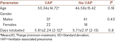
Demographic characteristics of the participants are shown in Table 1. There was no significant difference in age or gender distribution and intubated days between the groups (P > 0.05).
Table 1.
Demographic characteristics of the participants

From all 118 participants, 66.1% (78 patients) and 16.1% (19 patients) were seropositive for IgG and IgM, respectively. Mean age in seropositive patients was significantly higher than others (53.59 vs. 38.45 for IgG+ and IgG− participants, respectively, P < 0.001 and 60.74 vs. 46.1 for IgM+ and IgM− participants, respectively, P < 0.001).
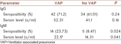
About 71.2% in VAP group and 61.01% in controls were IgG seropositive which the difference was not significant (P = 0.24). Furthermore, the serum levels of IgG were not significantly different between the groups (P = 0.16). More information is shown in Table 2.
Table 2.
Detailed information about serology results in cases and controls

About 23.73% of VAPs and 8.47% of controls were IgM seropositive, respectively (P = 0.024). Furthermore, there was significant difference in serum level of IgM between groups (P = 0.041, 95% confidence interval [CI] = −15.41, −0.31).
Among all patients, intubation days were higher in seropositive patients for both IgG and IgM, but the difference was only significant for IgM positive patients (6 ± 2.42 for IgG+ vs. 5.3 ± 1.6 for IgG−, P = 0.1 and 6.89 ± 2.26 for IgM+ vs. 5.55 ± 2.13 for IgM−, P = 0.014, 95% CI = −2.96, −0.34).
Analysis of the results in VAP group showed that there is direct and significant relationship between IgM results and intubation days (Pearson's correlation coefficient = 0.4, P = 0.002), in other words by increasing the time of intubation, more patients became seropositive for IgM.
DISCUSSION
In the recent study, the prevalence of seropositive patients for anti-H. pylori IgM and its serum levels were significantly higher in VAP patients.
Mean age of IgM and IgG - seropositive patients were significantly higher. These findings are consistent with several studies which found older patients have higher IgG levels in developed countries.[3,4,5,6]
Although IgG and IgM seropositivity and serum levels were higher in VAP patients, only IgM results were significant. Furthermore, by increasing the duration of intubation, serum levels and seropositivity for IgM increased significantly. These findings demonstrate that there are higher rates of acute H. pylori infection in VAP patients. Higher IgG levels could be explained by older ages and probably more comorbidity in VAP patients, but there is no explanation for higher IgM levels because it increases in acute infection. Furthermore, it is unlikely to consider IgM rise due to acute primary H. pylori infection because of patient's age and condition. To explain these findings, there are three possible hypotheses as follows:
In VAP patients, H. pylori colonizes the stomach and leads to GNB colonization by gastric PH alteration.[7,8] Hence, gastroesophageal reflux and oropharyngeal colonization of GNB occurs and aspiration of colonized bacteria might cause VAP in these patients. This stomach-pharynx-lower respiratory tract infection route has been shown in many studies[2,7,9]
Gastroesophageal reflux and aspiration of H. pylori could help it to reach the tracheobronchial tree. It made the environment susceptible for GNB colonization and the development of VAP
Activation of systemic or respiratory membrane inflammatory mediators by H. pylori proteins or inhalation of H. pylori or its exotoxins into the respiratory tract and development of VAP is other hypotheses.
CONCLUSIONS
IgM seropositivity and serum levels were significantly higher in VAP patients. Furthermore, by increasing the duration of intubation, serum levels and seropositivity for IgM increased significantly. H. pylori infection, subsequent GNB colonization and aspiration to tracheobronchial tree, and systemic or local inflammation are possible causes. Further studies with larger sample size, detailed and precise laboratory, and microbiologic assessment are needed to evaluate the possible role of H. pylori in pathogenesis of VAP.
Financial support and sponsorship
Nil.
Conflicts of interest
There are no conflicts of interest.
Acknowledgments
Research project number from AJA University of Medical Sciences: NB/141/7168/3.
REFERENCES
- 1.Joseph NM, Sistla S, Dutta TK, Badhe AS, Parija SC. Ventilator-associated pneumonia: A review. Eur J Intern Med. 2010;21:360–8. doi: 10.1016/j.ejim.2010.07.006. [DOI] [PubMed] [Google Scholar]
- 2.Mitz HS, Farber SS. Demonstration of Helicobacter pylori in tracheal secretions. J Am Osteopath Assoc. 1993;93:87–91. [PubMed] [Google Scholar]
- 3.Souto FJ, Fontes CJ, Rocha GA, de Oliveira AM, Mendes EN, Queiroz DM, et al. Prevalence of Helicobacter pylori infection in a rural area of the state of Mato Grosso, Brazil. Mem Inst Oswaldo Cruz. 1998;93:171–4. doi: 10.1590/s0074-02761998000200006. [DOI] [PubMed] [Google Scholar]
- 4.Kim N. Epidemiology and transmission route of Helicobacter pylori infection. Korean J Gastroenterol. 2005;46:153–8. [PubMed] [Google Scholar]
- 5.Lim SH, Kwon JW, Kim N, Kim GH, Kang JM, Park MJ, et al. Prevalence and risk factors of Helicobacter pylori infection in Korea: Nationwide multicenter study over 13 years. BMC Gastroenterol. 2013;13:104. doi: 10.1186/1471-230X-13-104. [DOI] [PMC free article] [PubMed] [Google Scholar]
- 6.Miranda AC, Machado RS, Silva EM, Kawakami E. Seroprevalence of Helicobacter pylori infection among children of low socioeconomic level in São Paulo. Sao Paulo Med J. 2010;128:187–91. doi: 10.1590/S1516-31802010000400002. [DOI] [PMC free article] [PubMed] [Google Scholar]
- 7.Li HY, He LX, Hu BJ, Wang BQ, Zhang XY, Chen XH, et al. The impact of gastric colonization on the pathogenesis of ventilator-associated pneumonia. Zhonghua Nei Ke Za Zhi. 2004;43:112–6. [PubMed] [Google Scholar]
- 8.Torres A, El-Ebiary M, Soler N, Montón C, Fàbregas N, Hernández C, et al. Stomach as a source of colonization of the respiratory tract during mechanical ventilation: Association with ventilator-associated pneumonia. Eur Respir J. 1996;9:1729–35. doi: 10.1183/09031936.96.09081729. [DOI] [PubMed] [Google Scholar]
- 9.Estes RJ, Meduri GU. The pathogenesis of ventilator-associated pneumonia: I. Mechanisms of bacterial transcolonization and airway inoculation. Intensive Care Med. 1995;21:365–83. doi: 10.1007/BF01705418. [DOI] [PubMed] [Google Scholar]


