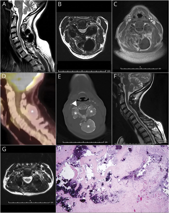
Figure. Calcified cervical mass lesion (asterisks).
(A–C) Sagittal, axial T2- weighted, and T1-weighted gadolinium-enhanced cervical spine MRI show an intrinsic spinal cord abnormality (arrow in A) related to compression by a large mass involving the C4 vertebral body. (D) The bulky lesion is demonstrated as photopenic by means of 18FDG-PET. (E) Axial neck CT angiogram identifies a bony matrix within the lesion resulting in segmental narrowing of the right vertebral artery at C4 (arrowhead). (F, G) Sagittal and axial T2-weighted postoperative MRI shows the effects of resection of the mass and nearly complete resolution of intrinsic cord signal abnormality. (H) Hematoxylin & eosin stain of the biopsy specimen shows dense regular fibroconnective tissue with reactive fibrovascular proliferation and multiple deposits of calcium hydroxyapatite (original magnification × 40).

