Abstract
In this study, Ag@SiO2 nanoparticles were synthesized by a modified Stöber method for preparing the TiO2 mesoporous layer of carbon counter electrode-based perovskite solar cells (PSCs) without a hole transporting layer. Compared with normal PSCs (without Ag@SiO2 incorporated in the TiO2 mesoporous layer), PSCs with an optimal content of Ag@SiO2 (0.3 wt. % Ag@SiO2-TiO2) show a 19.46% increase in their power conversion efficiency, from 12.23% to 14.61%, which is mainly attributed to the 13.89% enhancement of the short-circuit current density, from 20.23 mA/cm2 to 23.04 mA/cm2. These enhancements mainly contributed to the localized surface Plasmon resonance effect and the strong scattering effect of Ag@SiO2 nanoparticles. However, increasing the Ag@SiO2 concentration in the mesoporous layer past the optimum level cannot further increase the short-circuit current density and incident photon-to-electron conversion efficiency of the devices, which is primarily ascribed to the electron transport pathways being impeded by the insulating silica shells inside the TiO2 network.
Keywords: Ag@SiO2 nanoparticles, perovskite solar cell, localized surface Plasmon resonance effect, scattering effect
1. Introduction
Over the past few years, much significant progress has been made in the development of perovskite solar cells (PSCs), such as the low-cost but high-efficiency CuSCN replacing spiro-OMeTAD [1], breakthroughs in large-area perovskite films fabrication [2] and constant improvements in power conversion efficiencies (PCEs), from an initial 3.9% to 23.2% [3,4]. Due to the excellent photovoltaic properties of perovskite, such as large light-harvesting coefficient, long carrier diffusion distance and high carrier mobility [5,6,7,8,9], PSCs have a bright prospect of partly replacing conventional energy. Recently, Bella et al. [10] made a 360-degree overview focusing on Cs-doping for PSCs to illustrate the excellent properties of Cs in perovskite-based devices. Bella et al. [11] explained the different electrochemical behavior of the amorphous and anatase phases of TiO2 and clarified the controversial mechanism of reversible Na storage in TiO2 nanotube arrays. Zhang et al. [12] prepared TiO2@C aerogel as superior anode for lithium-ion batteries which exhibited a superior high rate and stable performance. Bella et al. [13] introduced the photoanodes and cathodes exclusively based on polymeric materials into dye-sensitized solar cells (DSSCs) and obtained a 5.33% power conversion efficiency. Galliano et al. [14] made a multivariate study on aqueous DSSCs to provide a new approach of finding the optimal operating conditions of any new energy device with solid mathematical and statistical bases. Abate et al. [15] analyzed the critical points about currently limiting the industrial production of PSCs.
However, being high efficient light harvesters, the thickness of organo-metal halide perovskites should be limited, reducing the lead content as much as possible, due to environmental and health concerns [16,17]. Therefore, the light capture capability of perovskite absorbers (PA) must be appropriately regulated to reach optimum efficiency. One strategy to improve the utilization rate of sunlight of PA could be incorporating metal nanoparticles (NPs) into PSCs.
When the photon frequency matches the natural oscillation frequency of the metal NP surface electrons, the conduction band (CB) electrons of noble metal NPs are induced to the coherent surface Plasmon oscillations [18], which is termed localized surface Plasmon resonance (LSPR). Consequently, this causes a huge increase in light absorption and scattering [19,20]. LSPR is macroscopically expressed as a strong characteristic absorption peak in the ultraviolet-visible (UV-vis) absorption spectrum. Mixing metal NPs into PSCs could not only enhance the optical properties related to size, shape, structure and environment [21] but also make use of electrical characteristics which are reported to help dissociate excitons and transport carriers more efficiently [22].
Certainly, there are numerous academic papers on the incorporation of metal nanomaterials into photovoltaic devices for better performance via their optical and electrical properties [23,24,25]. For example, Wang et al. [26] significantly improved the PCEs by adding 70 nm-Au NPs into polymer bulk heterojunction solar cells. Kulkarni et al. [27] utilized photoinduced absorption spectroscopy to improve the generation of charge carrier by using silver nanoprisms in organic photovoltaic devices. Batmunkh et al. [28] introduced the plasmonic Au nanostars into PSCs for enhancing photon absorption and thus improving device performance. Furthermore, an insulating SiO2 shell was added onto the surface of metal NPs to inhibit corrosion [29] and exciton quenching [30], however, this did not influence the LSPR effect. Metal-oxide core-shell NPs have widespread application in PSCs [31,32,33,34,35,36], as well as in DSSCs [37] and organic solar cells [38,39]. However, there has been little research on incorporating Ag@SiO2 NPs into low-cost free-hole-conductor PSCs with carbon counter electrodes. Ag NPs show superior scattering efficiency compared to any other metal NPs [40].
In this work, we adopted a modified Stöber [41] method to prepare Ag@SiO2 NPs for manufacturing the TiO2 mesoporous layer of PSCs. Through ultrasound treatment and continuous stirring, Ag@SiO2 NPs and mesoporous TiO2 pulp suspension were thoroughly homogenized. PSCs with Ag@SiO2 NPs exhibited better performance, evidenced by their UV-vis absorption spectra, J-V curves and incident photon-to-electron conversion efficiency (IPCE) spectra, because of the strong LSPR and scattering effects. With the hole-conductor-free and carbon-based counter electrode, the PSC devices gained a 19.46% enhancement of PCE, from 12.23% to 14.61% and a 13.89% increase in short-circuit current density (JSC), from 20.23 mA/cm2 to 23.04 mA/cm2.
2. Results and Discussion
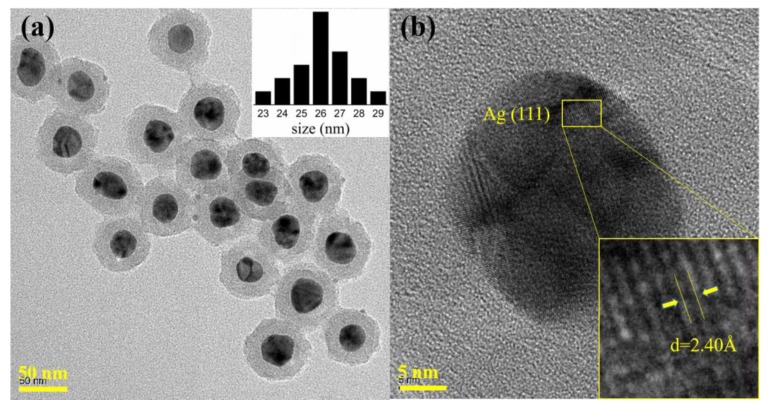
We successfully synthesized Ag NPs with a polyol method, whereas the silica shells coated on the Ag NPs were deposited uniformly with a modified Stöber method. The obtained NPs were then characterized by field emission transmission electron microscopy (FE-TEM, JEOL Ldt., Tokyo, Japan), high-resolution TEM (HRTEM, JEOL Ldt., Tokyo, Japan) and UV-vis absorption spectra (UV3600, Shimadzu Corporation, Tokyo, Japan). As depicted in Figure 1a, Ag NPs were evenly dispersed in ethanol, with the size distribution results in the upper right corner of the image. The nucleation sizes of the Ag NPs were almost equal and basically concentrated at 26 nm, while the thickness of the silica shells averaged 14 nm. Figure 1b shows the HRTEM image of a Ag@SiO2 NP. The lattice fringes of the Ag NP can be observed and were measured at about 2.40 Å matched with the (111) crystallographic planes of Ag NPs, which is consistent with the available literature [42].
Figure 1.
(a) TEM image and size distribution results of Ag@SiO2 NPs; (b) HRTEM image of a Ag@SiO2 NP.
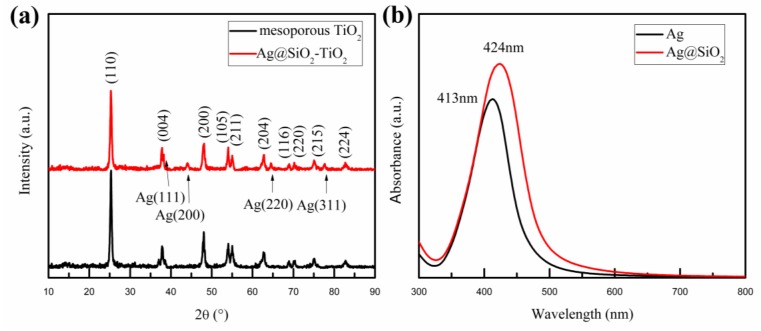
Figure 2a shows the X-ray diffraction (XRD) patterns of mesoporous TiO2 and the mesoporous TiO2 mixed with Ag@SiO2 NPs. A series of matching diffraction peaks characteristic for TiO2 were observed at 25°, 38°, 48°, 54°, 55°, 63°, 69°, 70°, 75°, 83°, which correspond to the anatase phase according to the standard PDF card (JCPDS Card No. 89-4921). These characteristic diffraction peaks are respectively attributed to the (101), (004), (200), (105), (211), (204), (116), (220), (215), (224) crystal planes of anatase phase TiO2. Simultaneously, there are several additional diffraction peaks in Figure 2a at the angles of 38°, 44°, 65° and 78°; these correspond to the reflections of the (111), (200), (220) and (311) crystal planes of the Ag NPs [43]. Furthermore, no other characteristic diffraction peaks of Ag oxide and SiO2 were observed, which implies that Ag was present in crystal form and SiO2 in an amorphous state in the prepared samples. Figure 2b exhibits the optical absorption spectra of 26 nm-Ag NPs and Ag@SiO2 NPs dispersed in alcohol solution. Compared to Ag NPs, the Plasmon resonance peak of Ag@SiO2 NPs had an obvious red-shift of around 10 nm, which was caused by the large refractive index of the silica shell [44,45]. The red-shifted distances of Ag@SiO2 NPs are related to the thickness of the silica shells, which are relative to the added amount of ethylsilicate [46]. The dielectric SiO2 shells coated around Ag NPs could prevent the recombination of carriers on the surface of metal NPs [30], even further enhancing the LSPR effect [47], as already reported.
Figure 2.
(a) XRD patterns of mesoporous TiO2 and the Ag@SiO2-TiO2; (b) Optical absorption spectra of 26 nm-Ag NPs and Ag@SiO2 NPs dispersed in alcohol.
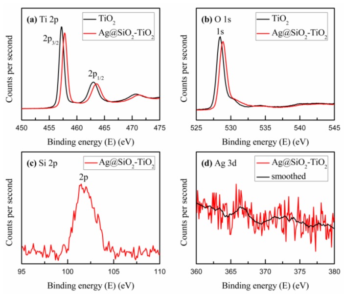
To confirm the physical state of chemical elements in the mesoporous TiO2 mixed with Ag@SiO2 NPs, X-ray photoelectron spectroscopy (XPS) was performed to assess the binding energy of chemical elements in the sample. Figure 3 shows the Ti, O, Si, Ag binding states and the surface composition of the NPs. As pictured in Figure 3a,b, the binding energies of 457.2 eV, 463.0 eV and 528.9 eV, correspond to the peaks of Ti 2p3/2, Ti 2p1/2 and O 1s respectively. However, compared with the pristine TiO2, the binding energies of the Ti 2p3/2, Ti 2p1/2 and O 1s electrons increased to 0.6 eV, 0.6 eV and 0.4 eV, respectively, after the addition of Ag@SiO2 NPs, which implies that a few electrons of the Ti and O atoms were trapped by Ag@SiO2 NPs due to the local electromagnetic field [48]. The decrease in electrons around Ti and O atoms implies a reduction in electron cloud densities around the Ti and O atoms and thus an increase in the binding energies. Figure 3c shows the peak of Si 2p at the binding energy of 103 eV, which is in accord with the standard database. In Figure 3d, no distinct peak can be found, which suggests that the 10 nm silica shells around the Ag NPs are sufficiently thick to attenuate the Ag photoelectrons generated by the X-ray [49]. Nonetheless, after a simple smoothing treatment, a weak peak was observed at the binding energy of 367 eV, corresponding to the peak of Ag 3d5/2. Compared with the standard binding energy of Ag0 at 368.25 eV, there is a small decrease that may be attributed to the few Ag NPs not covered by the silica shells. While calcinating the samples, the increased temperature led to the growth of the Ag NPs, which decreased the Ag 3d5/2 electronic binding energy [48]. Both the XRD patterns and XPS survey spectra indicate that the Ag@SiO2 NPs were successfully mixed into the TiO2 mesoporous layer.
Figure 3.
XPS survey spectra acquired from mesoporous TiO2 and the Ag@SiO2-TiO2: (a) Ti 2p; (b) O 1s; (c) Si 2p; (d) Ag 3d.
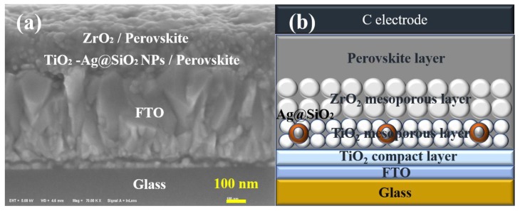
As depicted in Figure 4, a whole PSC device includes glass, FTO, a TiO2 compact layer, TiO2 mesoporous layer, ZrO2 mesoporous layer, perovskite layer and carbon counter electrode from the bottom up. According to the scale plate in Figure 4a, the thickness of each layer of the solar cell could be diagrammatically calculated. The thickness of the FTO, TiO2 compact layer, TiO2 mesoporous layer/perovskite and ZrO2 mesoporous layer/perovskite was roughly 500 nm, 30 nm 100 nm and 100 nm, respectively. In addition, the carbon counter electrode film was almost 30 μm, which is upon the perovskite film by screen printing. In the FTO/c-TiO2/m-TiO2/m-ZrO2/perovskite/carbon architecture-based PCSs, the ZrO2 layer acting as a scaffold to host the perovskite absorber with TiO2 layer, can block the flow of photogenerated electrons to the back contact and prevent recombination with the holes from the perovskite at the back contact [50].
Figure 4.
(a) Scanning Electron Microscope (SEM) image of a cross section of a complete PSC mixed with Ag@SiO2 NPs; (b) Structural representation of a whole PSC device mixed with Ag@SiO2 NPs.
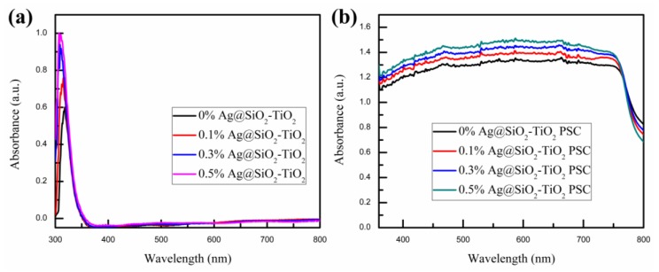
The UV-vis absorption spectra of pristine TiO2 and TiO2 mixed with Ag@SiO2 NPs mesoporous film samples are presented in Figure 5a. The black absorption curve represents the absorption intensity of pristine mesoporous TiO2 in the wavelength of 300–800 nm, which has an absorption peak at approximately 325 nm and is nearly zero in 360–800 nm, as previously reported by Yue et al. [51]. Compared with the other absorption curves, it is evident that the intensity of the absorption peak at around 325 nm became stronger as the content of Ag@SiO2 NPs in the mesoporous TiO2 layer gradually increased. As pictured in Figure 5b, the UV-vis absorption spectra of PSCs with pristine TiO2 and TiO2 mixed with Ag@SiO2 NPs followed the same trend in Figure 5a. The enhancement could be attributed to the LSPR effect and scattering effect of Ag@SiO2 NPs. By absorbing photons in the wavelength of the plasmonic resonance band, Ag@SiO2 NPs produced an intense LSPR effect which stimulated a strong electromagnetic field [52]. The intense local electromagnetic field could cause bandgap excitation for the nearby TiO2 NPs, enhancing their photocatalytic abilities, which could generate more electron-hole pairs in the TiO2 NPs [53]. The scattering effect of Ag@SiO2 NPs is mainly due to the scattering of incident light by the Ag NPs, which increases the length of the light propagation path and then further improves light trapping and the light utilization rate [34]. Macroscopically, the LSPR and scattering effects of Ag@SiO2 NPs are shown in the absorption spectrum, as depicted in Figure 5. As the content of Ag@SiO2 NPs increases, the LSPR effect and scattering effect increase gradually, which correlates with the absorption curves. According to previous research, the LSPR effect and scattering effect are dependent on the size of the metal NPs, with the LSPR effect decreasing as the metal NPs grow and the scattering effect increasing as the metal NPs grow [21,40]. Based on Zhang’s calculations, for Ag NPs, the LSPR effect reaches the maximum absorption efficiency at 20 nm, while the scattering effect achieves maximum scattering efficiency at 40 nm [54]. For the relatively small 26 nm-Ag NPs, the LSPR effect plays a more significant role than the scattering effect in the PSC devices [40,54].
Figure 5.
UV-vis absorption spectra of (a) pristine TiO2 and TiO2 mixed with Ag@SiO2 NPs mesoporous film samples and (b) PSCs with pristine TiO2 and TiO2 mixed with Ag@SiO2 NPs mesoporous film.
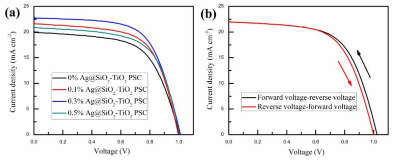
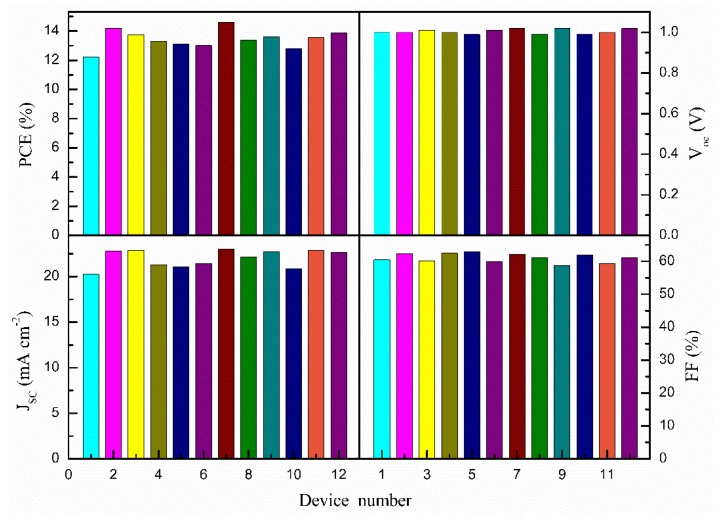
The PSCs based on mesoporous TiO2 film mixed with different amounts of Ag@SiO2 NPs were measured via scanning from −1.1 V to short circuit at a scan rate of 150 mV/s under AM 1.5G irradiation (100 mW/cm2) in ambient air to characterize their photovoltaic performance. Except for the process of spin-coating the perovskite solution, which was accomplished inside a nitrogen glovebox, all other processes, including spin-coating the precursor solution of the compact TiO2 layer, spin-coating the pulp suspension of mesoporous TiO2 mixed with different amounts of Ag@SiO2 NPs and mesoporous ZrO2 and screen-printing the carbon counter electrode, were performed in ambient air, at room temperature. As shown in Figure 6a, the PSC devices containing 0.3 wt. % Ag@SiO2 NPs had the most outstanding photovoltaic performance. Summarized photovoltaic parameters of PSCs based on mesoporous TiO2 film mixed with different amounts of Ag@SiO2 NPs are described in Table 1, which corresponds to Figure 6a. Figure 7 shows the histograms of photovoltaic parameters for 12 devices (the same batch of samples) based on mesoporous TiO2 film mixed with 0.3 wt. % Ag@SiO2 NPs. Compared with normal PSCs (with no plasmonic NPs), the photovoltaic performance of cells containing 0.3 wt. % Ag@SiO2 NPs was enhanced 19.46% regarding their PCE (from 12.23% to 14.61%) and 13.89% at JSC, from 20.23 mA/cm2 to 23.04 mA/cm2, while the open-circuit voltages (VOC) and fill factor (FF) of the PSCs rose and fell slightly at 1.00 V and 61.00%, respectively. The enhancements could be attributed to the incorporation of Ag@SiO2 NPs, which generated strong LSPR and scattering effects, improving the light trapping and utilization rate. The VOC of the PSC devices are related to the CBs of TiO2 and perovskite [55] and were not influenced by the Ag@SiO2 NPs. As described in Figure 6a and Table 1, as the number of Ag@SiO2 NPs increased gradually, the JSC and PCE initially increased, reaching the optimal value with the addition of 0.3 wt. % Ag@SiO2 NPs and then decreased. The enhancement was due to the strong increase in LSPR effect and scattering effect, while the diminution may be attributed to the offset between plasmonic light trapping effects and the decrease in electron transmission routes with the increased Ag@SiO2 loading [56]. Certainly, PSCs usually suffer from a hysteresis effect in J-V measurements, which is related with measurement settings and device properties [57,58,59]. In the FTO/c-TiO2/m-TiO2/m-ZrO2/perovskite/carbon architecture-based PCSs, the properties of the c-TiO2 layer and corresponding interfaces significantly affect the J-V hysteresis [60]. As depicted in Figure 6b and Table 2, the photocurrent, voltage, efficiency and fill factor for the forward-reverse scan were a little higher than those for the reverse-forward scan, which indicated that the hysteresis effect was a little influence in this architecture.
Figure 6.
(a) J-V curves of PSC devices based on mesoporous TiO2 film mixed with different amounts of Ag@SiO2 NPs; (b) J-V curves scanned form forward voltage to reverse voltage and scanned form reverse voltage to forward voltage.
Table 1.
Summarized photovoltaic parameters of PSC devices based on mesoporous TiO2 film mixed with different amounts of Ag@SiO2 NPs.
| Samples | VOC (V) | JSC (mA/cm2) | PCE (%) | FF (%) |
|---|---|---|---|---|
| 0% Ag@SiO2-TiO2 | 1.00 | 20.23 | 12.23 | 60.45 |
| 0.1% Ag@SiO2-TiO2 | 0.99 | 21.90 | 13.65 | 62.96 |
| 0.3% Ag@SiO2-TiO2 | 1.02 | 23.04 | 14.61 | 62.17 |
| 0.5% Ag@SiO2-TiO2 | 0.99 | 21.16 | 13.20 | 63.01 |
Figure 7.
Histograms of photovoltaic parameters for 12 devices (the same batch of samples) based on mesoporous TiO2 film mixed with 0.3 wt. % Ag@SiO2 NPs.
Table 2.
Summarized photovoltaic parameters of PSC devices scanned form forward voltage to reverse voltage and scanned form reverse voltage to forward voltage.
| Samples | VOC (V) | JSC (mA/cm2) | PCE (%) | FF (%) |
|---|---|---|---|---|
| Forward-reverse | 1.02 | 22.23 | 13.88 | 61.21 |
| Reverse- forward | 1.00 | 22.22 | 13.50 | 60.76 |
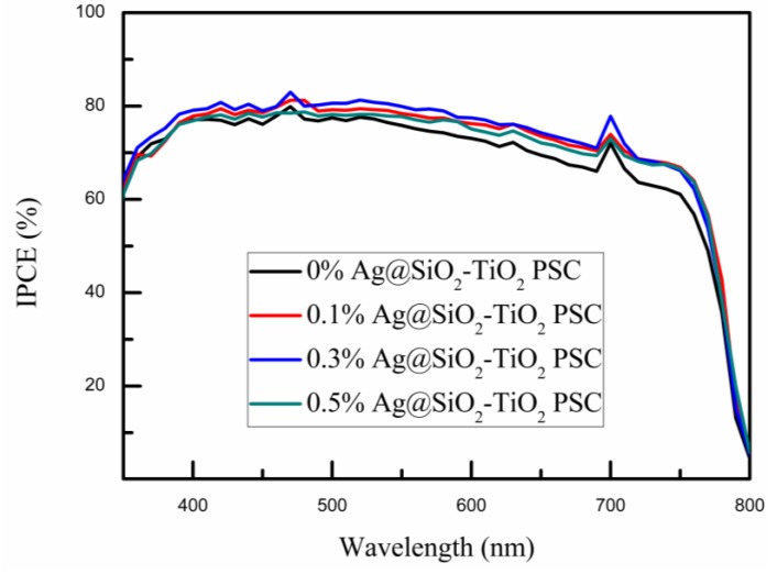
The IPCE spectra of PSC devices were measured to further demonstrate the impact of Ag@SiO2 NPs on the photoelectric properties of PSCs. It is evident that the trends shown in Figure 8 were consistent with the J-V curves depicted in Figure 6. With increased loading of Ag@SiO2 NPs, the IPCE reached its maximum at the concentration of 0.3 wt. %, followed by a decrease as the Ag@SiO2 NPs content increased past the optimal value. As pictured in Figure 8, in the wavelength of 500–750 nm, substantial increases were observed compared to the reference device (the black curve). This may be attributed to the perovskite material, which can efficiently use light in the short wavelength but behaves relatively poor in the long wavelength region of 600–800 nm [61,62]. Therefore, the LSPR effect and scattering effect of Ag@SiO2 NPs performed relatively better in the long wavelength. According to previous research, it is crucial to obtain the electron transport paths simple, straight and with few crystal boundaries to collect more electrons [63,64]. However, with increased concentration of Ag@SiO2 NPs, electrons became impeded by the insulating silica shells on the way to the electrode inside the TiO2 network [56]. Hence, as the Ag@SiO2 concentration increased past the optimal value, the JSC and IPCE curves presented a downward trend.
Figure 8.
IPCE spectra of PSC devices based on mesoporous TiO2 film mixed with different amounts of Ag@SiO2 NPs.
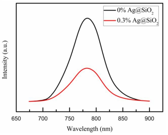
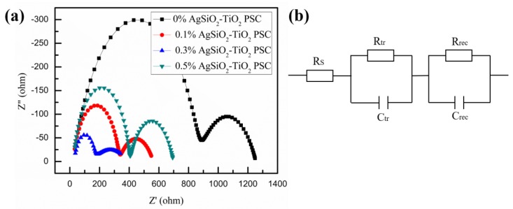
To understand the charge transport properties of the devices in more details, we studied the photoluminescence (PL) at room temperature of 296 K. In Figure 9, it was obvious that there was a clear quenching of the steady-state PL for the compact TiO2/Ag@SiO2-TiO2/ZrO2/perovskite films compared with the films without Ag@SiO2 NPs, which was contributed from ionization of the excitons and enhanced charge separation [65]. The electrochemical impedance spectroscopy (EIS) analyses were performed for frequencies of 10 mHz to 10 MHz at a bias of 0.8 V under simulated AM 1.5G radiation (irradiance of 100 mW/cm2) to further understand charge transport and charge recombination in the devices. As described in Figure 10a, there were two semicircles in the Nyquist plots, which corresponded to the high-frequency region and low-frequency region, respectively. The left semicircle represented charge transfer and charge recombination at the perovskite/C electrode interface associated with the charge transfer resistance (Rtr), while the right one represented charge transfer and charge recombination at the perovskite/TiO2 interface related to the recombination resistance (Rrec) [66,67]. The series resistance (RS) value is inversely related to FF value [68]. In Table 3, the RS of 0% Ag@SiO2-TiO2 PSC was slightly larger than the others, which was in keeping with the FF values in Table 1 basically. For the recombination resistance Rrec at the perovskite/TiO2 interface, it was clear that with the Ag@SiO2 NPs mixed into the films Rrec values decreased immediately, which indicated that there were more electrons generated duo to the LSPR effect and scattering effect of Ag@SiO2 NPs and sequentially more electrons were recombined at the perovskite/TiO2 interface. To analyze the charge transfer at the perovskite/C electrode interface, the Rtr values were shown in Table 3, which decreased as the content of Ag@SiO2 NPs decreased gradually and increased when the concentration was higher than the optimal value. The results of Rtr indicated that there were more holes transported to the C electrode and with increased concentration of Ag@SiO2 NPs higher than the optimal value, holes were impeded by the insulating silica shells on the way to the C electrode inside the TiO2/ZrO2 network. Therefore, 0.3% Ag@SiO2-TiO2 PSC got the best photovoltaic properties in this work, in keeping with J-V curves and IPCE spectra.
Figure 9.
PL spectra of compact TiO2/Ag@SiO2-TiO2/ZrO2/perovskite films at room temperature of 296 K.
Figure 10.
(a) Nyquist curves of the PSC devices based on different amounts of Ag@SiO2 NPs films and (b) the equivalent circuit employed to fit the plots.
Table 3.
Summary of EIS parameters of PSCs based on different amounts of Ag@SiO2 NPs films.
| Samples | RS (Ω) | Rtr (Ω) | Rrec (Ω) |
|---|---|---|---|
| 0% Ag@SiO2-TiO2 PSC | 29.75 | 842.4 | 381.6 |
| 0.1% Ag@SiO2-TiO2 PSC | 22.77 | 308.2 | 231.1 |
| 0.3% Ag@SiO2-TiO2 PSC | 25.02 | 125.8 | 243.0 |
| 0.5% Ag@SiO2-TiO2 PSC | 24.84 | 379.0 | 294.1 |
3. Materials and Methods
3.1. Synthesis of Ag NPs
The Ag NPs were prepared via a polyol method, according to previously reported methods with slight modifications [40,53,69,70]. First, 3 g polyvinylpyrrolidone (PVP) was dissolved in 40 mL ethylene glycol and heated up to 120 °C slowly, in an oil bath, with continuous stirring, until it dissolved completely. Subsequently, 0.5 g silver nitrate (AgNO3) were dissolved in 20 mL ethylene glycol with vigorous stirring. After the AgNO3 was fully dissolved, the mixture was added to the ethylene glycol solution of PVP dropwise and the resulting solution was kept at 120 °C, for 1 h, under stirring. After the colloidal solution cooled down to room temperature, moderate deionized water was added to the solution, followed by centrifugation. Finally, the precipitate was washed with deionized water and acetone for several times, then dried at 40 °C to obtain the Ag NPs.
3.2. Synthesis of Ag@SiO2 NPs
The Ag@SiO2 NPs were synthesized by a modified Stöber method [40,53,71]. Firstly, 120 mg Ag NPs were uniformly dispersed in 120 mL alcohol with vigorous stirring. Secondly, after 20 min of ultrasonication, 2.5 mL ammonium hydroxide, 2.5 mL deionized water and 0.3 mL ethylsilicate were added to the solution dropwise. Finally, the solution was stirred for 12 h at room temperature and Ag@SiO2 NPs were obtained by centrifugation, wash (ethyl alcohol for several times) and desiccation.
3.3. Device Fabrication
First, to prepare the Ag@SiO2-TiO2 with different ratios (0.1–0.5 wt. % ), 4 g alcohol were added to 1 g TiO2 sizing agent (solid content: 20%) followed by stirring for 6 h. Then, 0.2–1 mg Ag@SiO2 NPs powder were added to the mixture followed by stirring for at least 48 h. Normally, Ag@SiO2 NPs disperse in a mixture more uniformly after 30 min of ultrasonication. Second, fluorine-doped tin oxide-patterned glass substrates (7 Ω/sq) were cleaned sequentially with detergent, acetone, isopropanol and ethanol, each followed by 30 min of ultrasonication. Third, the TiO2 compact layer was deposited by spin coating 35 μL TiO2 precursor solution (1 mL titanium disopropoxide bis added into 19 mL ethanol) at 4000 r/min for 25 s and annealed at 150 °C for 10 min. Then, 35 μL TiO2 precursor solution was spin-coated on the substrate again at 4000 r/min for 25 s and annealed at 150 °C for 10 min, followed by annealing at 500 °C for 30 min. Fourth, after cooling down to room temperature, the TiO2 mesoporous layer was deposited on the top of the TiO2 compact layer by spin coating appropriate TiO2 sizing agent mixed with Ag@SiO2 NPs at 3500 r/min for 25 s, after which the glass substrates were heated at 150 °C for 10 min and annealed at 500 °C for 30 min. Fifth, the ZrO2 paste was spun onto the substrates at 5000 r/min for 25 s, then the glass substrates were heated at 150 °C for 10 min and sintered at 500 °C for 30 min. Lastly, the (CH3NH3)PbI3 perovskite film was fabricated by spin coating 35 μL perovskite precursor solution at 1000 r/min for 10 s and 4000 r/min for 20 s, followed by annealing at 100 °C for 5–10 min. The perovskite precursor solution was prepared by dissolving 462 mg PbI2 and 178 mg (CH3NH3)I in 600 mg dimethylformamide (DMF) and 78 mg dimethyl sulfoxide (DMSO). During 5 s at 4000 r/min, about 200 μL methylbenzene were dropped on the spinning substrates to ensure the fast crystallization of perovskite by extracting the solvent of the perovskite precursor solution.
3.4. Characterization Techniques
FE-TEM images were photographed by a JEM-2100F (JEOL Ldt., Tokyo, Japan). XRD patterns were acquired from an X-ray diffractometer (Advance D8, AXS, Rigaku Corporation, Tokyo, Japan). XPS (ESCALAB 250Xi, Thermo Fisher Scientific, Waltham, Massachusetts, USA) was employed to analyze the chemical elements of the samples. SEM (JSM-IT300, JEOL Ldt., Tokyo, Japan) was used to scan the surface and section of the as-prepared samples. The light-absorption spectra were obtained via UV-vis spectrophotometry (UV3600, Shimadzu Corporation, Tokyo, Japan). The photocurrent-voltage (J-V) characteristics were acquired from an electrochemical workstation (Zahner Company, Kronach, Germany) with a solar light simulator (Oriel Sol3A, Newport Corporation, Irvine, CA, USA), under simulated AM 1.5G illumination, at 100 mW/cm2 intensity. Finally, IPCE (Newport Corporation, Irvine, CA, USA) was employed to study the quantum efficiency of the PSCs. The PL spectra were measured with a fluorescence spectrometer (RF-6000, Shimadzu Corporation, Tokyo, Japan). The EIS analyses were conducted on an electrochemical workstation (Zahner Company, Kronach, Germany) for frequencies of 10 mHz to 10 MHz at a bias of 0.8 V under simulated AM 1.5G radiation (irradiance of 100 mW/cm2) with an alternating current (AC) signal amplitude of 10 mV at room temperature.
4. Conclusions
In this study, we successfully synthesized Ag@SiO2 NPs with a modified Stöber method and developed PSCs with different contents of Ag@SiO2 NPs. TEM images, XRD patterns and XPS survey spectra showed that Ag was present in nanocrystal form and that Ag@SiO2 NPs were thoroughly mixed into the TiO2 mesoporous layer. UV-vis absorption spectra demonstrated that the optical absorption capacity of mesoporous TiO2 films and PSC devices can be improved with increased loading of Ag@SiO2 NPs (0–0.5 wt. %), which could be attributed to the strong LSPR and scattering effects. Finally, the variation of J-V, IPCE and EIS between devices with different Ag@SiO2 concentrations was assessed and the results indicated that the electron transporting paths were blocked by the Ag@SiO2 NPs when their concentration was higher than the optimal value (0.3 wt. %). Compared to the normal PSCs (0 wt. % Ag@SiO2-TiO2), the PSCs incorporating the optimal content of NPs exhibited a 19.46% improvement of the PCE (from 12.23% to 14.61%) and 13.89% improvement of JSC (from 20.23 mA/cm2 to 23.04 mA/cm2). Our method of using Ag@SiO2 NPs provides a new approach to boosting highly efficient hole-conductor-free PSCs based on carbon counter electrodes.
Author Contributions
Conceptualization, B.W., X.Z., S.L. and M.C.; Data curation, B.W., X.Z., S.L. and M.C.; Investigation, B.W., X.Z., S.L. and M.C.; Methodology, B.W., X.Z., S.L. and M.C.; Software, B.W., X.Z., S.L. and M.C.; Writing-original draft, B.W., X.Z., S.L. and M.C.; Writing–review and editing, H.L. and Y.Y.
Funding
This work was funded by the National Natural Science Foundation of China (NSFC) (11704293), the Fundamental Research Funds for the Central Universities under Grant WUT (2018IB017) and The Excellent Dissertation Cultivation Funds of Wuhan University of Technology (2017-YS-77).
Conflicts of Interest
The authors declare no conflict of interest.
References
- 1.Arora N., Dar M.I., Hinderhofer A., Pellet N., Schreiber F., Zakeeruddin S.M., Grätzel M. Perovskite solar cells with CuSCN hole extraction layers yield stabilized efficiencies greater than 20% Science. 2017;358:768–771. doi: 10.1126/science.aam5655. [DOI] [PubMed] [Google Scholar]
- 2.Chen H., Ye F., Tang W., He J., Yin M., Wang Y., Xie F., Bi E., Yang X., Grätzel M., et al. A solvent- and vacuum-free route to large-area perovskite films for efficient solar modules. Nature. 2017;550:92. doi: 10.1038/nature23877. [DOI] [PubMed] [Google Scholar]
- 3.Kojima A., Teshima K., Shirai Y., Miyasaka T. Organometal Halide Perovskites as Visible-Light Sensitizers for Photovoltaic Cells. J. Am. Chem. Soc. 2009;131:6050–6051. doi: 10.1021/ja809598r. [DOI] [PubMed] [Google Scholar]
- 4.Jeon N.J., Na H., Jung E.H., Yang T.Y., Lee Y.G., Kim G., Shin H.W., Seok S.I., Lee J., Seo J. A fluorene-terminated hole-transporting material for highly efficient and stable perovskite solar cells. Nat. Energy. 2018;3:682–689. doi: 10.1038/s41560-018-0200-6. [DOI] [Google Scholar]
- 5.Green M.A., Hobaillie A., Snaith H.J. The emergence of perovskite solar cells. Nat. Photonics. 2014;8:506–514. doi: 10.1038/nphoton.2014.134. [DOI] [Google Scholar]
- 6.Chen C.W., Hsiao S.Y., Chen C.Y., Kang H.W., Huang Z.Y., Lin H.W. Optical Properties of Organometal Halide Perovskite Thin Films and General Device Structure Design Rules for Perovskite Single and Tandem Solar Cells. J. Mater. Chem. A. 2014;3:9152–9159. doi: 10.1039/C4TA05237D. [DOI] [Google Scholar]
- 7.Stoumpos C.C., Malliakas C.D., Kanatzidis M.G. Semiconducting tin and lead iodide perovskites with organic cations: Phase transitions, high mobilities, and near-infrared photoluminescent properties. Inorg. Chem. 2013;52:9019–9038. doi: 10.1021/ic401215x. [DOI] [PubMed] [Google Scholar]
- 8.Hu J., Qiao Y., Yang Y., Zhao L., Liu W., Li S., Liu P., Chen M. Enhanced Performance of Hole-Conductor-Free Perovskite Solar Cells by Utilization of Core/Shell-Structured β-NaYF4:Yb3+, Er3+@SiO2 Nanoparticles in Ambient Air. IEEE J. Photovolt. 2017;8:132–136. doi: 10.1109/JPHOTOV.2017.2762585. [DOI] [Google Scholar]
- 9.Qiao Y., Li S., Liu W., Ran M., Lu H., Yang Y. Recent Advances of Rare-Earth Ion Doped Luminescent Nanomaterials in Perovskite Solar Cells. Nanomaterials. 2018;8:43. doi: 10.3390/nano8010043. [DOI] [PMC free article] [PubMed] [Google Scholar]
- 10.Bella F., Renzi P., Cavallo C., Gerbaldi C. Caesium for perovskite solar cells: an overview. Chem. Eur. J. 2018;24:12183–12205. doi: 10.1002/chem.201801096. [DOI] [PubMed] [Google Scholar]
- 11.Bella F., Garcia A.B.M., Meligrana G., Lamberti A., Destro M., Pavone M., Gerbaldi C. Unveiling the controversial mechanism of reversible Na storage in TiO2 nanotube arrays: Amorphous versus anatase TiO2. Nano Res. 2017;10:2891–2903. doi: 10.1007/s12274-017-1656-6. [DOI] [Google Scholar]
- 12.Zhang C., Liu S., Qi Y., Cui F., Yang X. Conformal carbon coated TiO2 aerogel as superior anode for lithium-ion batteries. Chem. Eng. J. 2018;351:825–831. doi: 10.1016/j.cej.2018.06.125. [DOI] [Google Scholar]
- 13.Bella F., Verna A., Gerbaldi C. Patterning dye-sensitized solar cell photoanodes through a polymeric approach: A perspective. Mater. Sci. Semicond. Process. 2018;73:92–98. doi: 10.1016/j.mssp.2017.07.030. [DOI] [Google Scholar]
- 14.Galliano S., Bella F., Piana G., Giacona G., Viscardi G., Gerbaldi C., Grätzel M., Barolo C. Finely tuning electrolytes and photoanodes in aqueous solar cells by experimental design. Sol. Energy. 2018;163:251–255. doi: 10.1016/j.solener.2018.02.009. [DOI] [Google Scholar]
- 15.Abate A., Baena J.P.C., Saliba M., Suait M.S., Bella F. Perovskite solar cells from the lab to the assembly line. Chem. Eur. J. 2018;24:3083–3100. doi: 10.1002/chem.201704507. [DOI] [PubMed] [Google Scholar]
- 16.Babayigit A., Ethirajan A., Muller M., Conings B. Toxicity of organometal halide perovskite solar cells. Nat. Mater. 2016;15:247–251. doi: 10.1038/nmat4572. [DOI] [PubMed] [Google Scholar]
- 17.Babayigit A., Thanh D.D., Ethirajan A., Manca J., Muller M., Boyen H.G., Conings B. Assessing the toxicity of Pb- and Sn-based perovskite solar cells in model organism Danio rerio. Sci. Rep. 2016;6:18721. doi: 10.1038/srep18721. [DOI] [PMC free article] [PubMed] [Google Scholar]
- 18.Huang X., Neretina S., Elsayed M.A. Gold nanorods: from synthesis and properties to biological and biomedical applications. Adv. Mater. 2010;21:4880–4910. doi: 10.1002/adma.200802789. [DOI] [PubMed] [Google Scholar]
- 19.Stratakis E., Kymakis E. Nanoparticle-based plasmonic organic photovoltaic devices. Mater. Today. 2013;16:133–146. doi: 10.1016/j.mattod.2013.04.006. [DOI] [Google Scholar]
- 20.Petridis C., Savva K., Kymakis E., Stratakis E. Laser generated nanoparticles based photovoltaics. J. Colloid Interface Sci. 2017;489:28–37. doi: 10.1016/j.jcis.2016.09.065. [DOI] [PubMed] [Google Scholar]
- 21.Kelly K.L., Coronado E., Zhao L.L., Schatz G.C. The Optical Properties of Metal Nanoparticles: The Influence of Size, Shape, and Dielectric Environment. Cheminform. 2003;34:668–677. doi: 10.1002/chin.200316243. [DOI] [Google Scholar]
- 22.Chan K., Wright M., Elumalai N., Uddin A., Pillai S. Plasmonics in Organic and Perovskite Solar Cells: Optical and Electrical Effects. Adv. Opt. Mater. 2017;5:1600698. doi: 10.1002/adom.201600698. [DOI] [Google Scholar]
- 23.Choy W.C.H. Using plasmonic-electrical effects for high-performance organic solar cells. Spienewsroom. 2013;23:4255–4261. doi: 10.1117/2.1201311.005125. [DOI] [Google Scholar]
- 24.Ren X., Cheng J., Zhang S., Li X., Rao T., Huo L., Hou J., Choy W.C. High Efficiency Organic Solar Cells Achieved by the Simultaneous Plasmon-Optical and Plasmon-Electrical Effects from Plasmonic Asymmetric Modes of Gold Nanostars. Small. 2016;12:5102. doi: 10.1002/smll.201670187. [DOI] [PubMed] [Google Scholar]
- 25.Paci B., Generosi A., Albertini V.R., Spyropoulos G.D., Stratakis E., Kymakis E. Enhancement of photo/thermal stability of organic bulk heterojunction photovoltaic devices via gold nanoparticles doping of the active layer. Nanoscale. 2012;4:7452–7459. doi: 10.1039/c2nr32799f. [DOI] [PubMed] [Google Scholar]
- 26.Wang D.H., Kim D.Y., Choi K.W., Seo J.H., Im S.H., Park J.H., Park O.O., Heeger A.J. Enhancement of Donor–Acceptor Polymer Bulk Heterojunction Solar Cell Power Conversion Efficiencies by Addition of Au Nanoparticles. Angew. Chem. Int. Ed. Engl. 2011;123:5519–5523. doi: 10.1002/anie.201101021. [DOI] [PubMed] [Google Scholar]
- 27.Kulkarni A.P., Noone K.M., Munechika K., Guyer S.R., Ginger D.S. Plasmon-enhanced charge carrier generation in organic photovoltaic films using silver nanoprisms. Nano Lett. 2010;10:1501–1505. doi: 10.1021/nl100615e. [DOI] [PubMed] [Google Scholar]
- 28.Batmunkh M., Macdonald T.J., Peveler W.J., Bati A.S.R., Carmalt C.J., Parkin I.P., Shapter J.G. Plasmonic Gold Nanostars Incorporated into High-Efficiency Perovskite Solar Cells. ChemSuSChem. 2017;10:3750–3753. doi: 10.1002/cssc.201701056. [DOI] [PubMed] [Google Scholar]
- 29.Wang Y., Zhai J., Song Y., He L. The Ag shell thickness effect of Au@Ag@SiO2 core-shell nanoparticles on the optoelectronic performance of dye sensitized solar cells. Chem. Commun. 2016;52:2390–2393. doi: 10.1039/C5CC08827E. [DOI] [PubMed] [Google Scholar]
- 30.Jankovic V., Yang Y.M., You J., Dou L., Liu Y., Cheung P., Chang J.P., Yang Y. Active layer-incorporated, spectrally tuned Au/SiO2 core/shell nanorod-based light trapping for organic photovoltaics. ACS Nano. 2013;7:3815–3822. doi: 10.1021/nn400246q. [DOI] [PubMed] [Google Scholar]
- 31.Wu R., Yang B., Zhang C., Huang Y., Cui Y., Liu P., Zhou C., Hao Y., Gao Y., Yang J. Prominent Efficiency Enhancement in Perovskite Solar Cells Employing Silica-Coated Gold Nanorods. J. Phys. Chem. C. 2016;120:6996–7004. doi: 10.1021/acs.jpcc.6b00309. [DOI] [Google Scholar]
- 32.Cui J., Chen C., Han J., Cao K., Zhang W., Shen Y., Wang M. Surface Plasmon Resonance Effect in Inverted Perovskite Solar Cells. Adv. Sci. 2016;3:1500312–1500320. doi: 10.1002/advs.201500312. [DOI] [PMC free article] [PubMed] [Google Scholar]
- 33.Fan R., Wang L., Chen Y., Zheng G., Li L., Li Z., Zhou H. Tailored Au@TiO2 Nanostructures for Plasmonic Effect in Planar Perovskite Solar Cells. J. Mater. Chem. A. 2017;5:12034–12042. doi: 10.1039/C7TA02937C. [DOI] [Google Scholar]
- 34.Zhang W., Saliba M., Stranks S.D., Sun Y., Shi X., Wiesner U., Snaith H.J. Enhancement of perovskite-based solar cells employing core-shell metal nanoparticles. Nano Lett. 2013;13:4505–4510. doi: 10.1021/nl4024287. [DOI] [PubMed] [Google Scholar]
- 35.Hao H., Wang L., Ma X., Cao K., Yu H., Wang M., Gu W., Zhu R., Anwar M.S., Chen S., et al. Improved Efficiency of Inverted Perovskite Solar Cells Via Surface Plasmon Resonance Effect of Au@PSS Core–Shell Tetrahedra Nanoparticles. Sol. RRL. 2018;2:1800061. doi: 10.1002/solr.201800061. [DOI] [Google Scholar]
- 36.Luo Q., Zhang C., Deng S., Zhu H., Li Z., Wang Z., Chen X., Huang S. Plasmonic Effects of Metallic Nanoparticles on Enhancing Performance of Perovskite Solar Cells. ACS Appl. Mater. Interfaces. 2017;9:34821–34832. doi: 10.1021/acsami.7b08489. [DOI] [PubMed] [Google Scholar]
- 37.Brown M.D., Suteewong T., Kumar R.S.S., Innocenzo V.D., Petrozza A., Lee M., Wiesner U., Snaith H.J. Plasmonic Dye-Sensitized Solar Cells Using Core−Shell Metal−Insulator Nanoparticles. Nano Lett. 2011;11:438. doi: 10.1021/nl1031106. [DOI] [PubMed] [Google Scholar]
- 38.Wang Z., Hao Y., Wang W., Cui Y., Sun Q., Ji T., Li Z., Wang H., Zhu F. Incorporating silver-SiO2 core-shell nanocubes for simultaneous broadband absorption and charge collection enhancements in organic solar cells. Synth. Met. 2016;220:612–620. doi: 10.1016/j.synthmet.2016.08.004. [DOI] [Google Scholar]
- 39.Chan K.H., Elumalai N.K., Tayebjee M.J.Y., Uddin A., Pillai S. Dark carrier dynamics and electrical characteristics of organic solar cells integrated with Ag-SiO2 core-shell nanoparticles. Synth. Met. 2017;223:34–42. doi: 10.1016/j.synthmet.2016.11.033. [DOI] [Google Scholar]
- 40.Baek S.W., Noh J., Lee C.H., Kim B.S., Seo M.K., Lee J.Y. Plasmonic Forward Scattering Effect in Organic Solar Cells: A Powerful Optical Engineering Method. Sci. Rep. 2013;3:542. doi: 10.1038/srep01726. [DOI] [Google Scholar]
- 41.Stöber W., Fink A., Bohn E. Controlled growth of monodisperse silica spheres in the micron size range. J. Colloid Interface Sci. 1968;26:62–69. doi: 10.1016/0021-9797(68)90272-5. [DOI] [Google Scholar]
- 42.Wang X., Yu J.C., Ho C., Mak A.C. A robust three-dimensional mesoporous Ag/TiO2 nanohybrid film. Chem. Commun. 2005;17:2262–2264. doi: 10.1039/b500605h. [DOI] [PubMed] [Google Scholar]
- 43.Tom R.T., Nair A.S., Singh N., Aslam M., Nagendra C.L., Philip R., Vijayamohanan K., Pradeep T. Freely Dispersible Au@TiO2, Au@ZrO2, Ag@TiO2, and Ag@ZrO2 Core−Shell Nanoparticles: One-Step Synthesis, Characterization, Spectroscopy, and Optical Limiting Properties. Langmuir. 2003;19:3439–3445. doi: 10.1021/la0266435. [DOI] [Google Scholar]
- 44.Choi H., Lee J.P., Ko S.J., Jung J.W., Park H., Yoo S., Park O., Jeong J.R., Park S., Kim J.Y. Multipositional silica-coated silver nanoparticles for high-performance polymer solar cells. Nano Lett. 2013;13:2204. doi: 10.1021/nl400730z. [DOI] [PubMed] [Google Scholar]
- 45.Jain P.K., El-Sayed M.A. Noble metal nanoparticle pairs: effect of medium for enhanced nanosensing. Nano Lett. 2008;8:4347–4352. doi: 10.1021/nl8021835. [DOI] [PubMed] [Google Scholar]
- 46.Bardhan R., Grady N.K., Halas N.J. Nanoscale control of near-infrared fluorescence enhancement using Au nanoshells. Small. 2008;4:1716–1722. doi: 10.1002/smll.200800405. [DOI] [PubMed] [Google Scholar]
- 47.Mori K., Kawashima M., Che M., Yamashita H. Enhancement of the Photoinduced Oxidation Activity of a Ruthenium (II) Complex Anchored on Silica-Coated Silver Nanoparticles by Localized Surface Plasmon Resonance. Angew. Chem. Int. Ed. 2010;122:8780–8783. doi: 10.1002/ange.201004942. [DOI] [PubMed] [Google Scholar]
- 48.Luo K., Clair T.P.S., Lai X., Goodman D.W. Silver Growth on TiO2(110) (1 × 1) and (1 × 2) J. Phys. Chem. B. 2000;104:1374–1377. doi: 10.1021/jp993062o. [DOI] [Google Scholar]
- 49.Zarick H.F., Erwin W.R., Aufrecht J., Coppola A., Rogers B.R., Pint C.L., Bardhan R. Morphological modulation of bimetallic nanostructures for accelerated catalysis. J. Mater. Chem. A. 2014;2:7088–7098. doi: 10.1039/C3TA15228F. [DOI] [Google Scholar]
- 50.Mei A., Li X., Liu L., Ku Z., Liu T., Rong Y., Xu M., Hu M., Chen J., Yang Y., et al. A hole-conductor–free, fully printable mesoscopic perovskite solar cell with high stability. Science. 2014;345:295–298. doi: 10.1126/science.1254763. [DOI] [PubMed] [Google Scholar]
- 51.Yue L., Yan B., Attridge M., Wang Z. Light absorption in perovskite solar cell: Fundamentals and plasmonic enhancement of infrared band absorption. Sol. Energy. 2016;124:143–152. doi: 10.1016/j.solener.2015.11.028. [DOI] [Google Scholar]
- 52.Shi X., Ueno K., Takabayashi N., Misawa H. Plasmon-Enhanced Photocurrent Generation and Water Oxidation with a Gold Nanoisland-Loaded Titanium Dioxide Photoelectrode. J. Phys. Chem. C. 2013;117:2494–2499. doi: 10.1021/jp3064036. [DOI] [Google Scholar]
- 53.Lee J.E., Bera S., Choi Y.S., Lee W.I. Size-dependent plasmonic effects of M and M@SiO2, (M = Au or Ag) deposited on TiO2 in photocatalytic oxidation reactions. Appl. Catal. B Environ. 2017;214:15–22. doi: 10.1016/j.apcatb.2017.05.025. [DOI] [Google Scholar]
- 54.Zhang Y.J. Investigation of Gold and Silver Nanoparticles on Absorption Heating and Scattering Imaging. Plasmonics. 2011;6:393–397. doi: 10.1007/s11468-011-9216-5. [DOI] [Google Scholar]
- 55.Li S., Hu J., Yang Y., Zhao L., Qiao Y., Liu W., Liu P., Chen M. Ag/nano-TiO2 composite compact film for enhanced performance of perovskite solar cells based on carbon counter electrodes. Appl. Phys. A. 2017;123:628. doi: 10.1007/s00339-017-1240-7. [DOI] [Google Scholar]
- 56.Gangishetty M.K., Lee K.E., Scott R.W., Kelly T.L. Plasmonic Enhancement of Dye Sensitized Solar Cells in the Red-to-near-Infrared Region using Triangular Core-Shell Ag@SiO2 Nanoparticles. Appl. Mater. Interfaces. 2013;5:11044. doi: 10.1021/am403280r. [DOI] [PubMed] [Google Scholar]
- 57.Nemnes G.A., Besleaga C., Tomulescu A.G., Pintilie I., Pintilie L., Torfason K., Manolescu A. Dynamic electrical behavior of halide perovskite based solar cells. Sol. Energy Mater. Sol. Cells. 2017;159:197–203. doi: 10.1016/j.solmat.2016.09.012. [DOI] [Google Scholar]
- 58.Nemnes G.A., Besleaga C., Stancu V., Dogaru D.E., Leonat L.N., Pintilie L., Torfason K., Ilkov M., Manolescu A. Normal and Inverted Hysteresis in Perovskite Solar Cells. J. Phys. Chem. C. 2017;121:11207–11214. doi: 10.1021/acs.jpcc.7b04248. [DOI] [Google Scholar]
- 59.Snaith H.J., Abate A., Ball J.M., Eperon G.E., Leijtens T., Noel N.K., Stranks S.D., Wang J.T.W., Wojciechowski K., Zhang W. Anomalous Hysteresis in Perovskite Solar Cells. J. Phys. Chem. Lett. 2014;5:1511–1515. doi: 10.1021/jz500113x. [DOI] [PubMed] [Google Scholar]
- 60.Rong Y., Hu Y., Ravishankar S., Liu H., Hou X., Sheng Y., Mei A., Wang Q., Li D., Xu M., et al. Tunable hysteresis effect for perovskite solar cells. Energy Environ. Sci. 2017;10:2383–2391. doi: 10.1039/C7EE02048A. [DOI] [Google Scholar]
- 61.Kakavelakis G., Petridis K., Kymskis E. Recent advances in plasmonic metal and rare-earth-element upconversion nanoparticle doped perovskite solar cells. J. Mater. Chem. A. 2017;5:21604–21624. doi: 10.1039/C7TA05428A. [DOI] [Google Scholar]
- 62.Li S., Zhu X., Wang B., Qiao Y., Liu W., Yang H., Liu N., Chen M., Lu H., Yang Y. Influence of Ag Nanoparticles with Different Sizes and Concentrations Embedded in a TiO2 Compact Layer on the Conversion Efficiency of Perovskite Solar Cells. Nanoscale Res. Lett. 2018;13:210. doi: 10.1186/s11671-018-2626-y. [DOI] [PMC free article] [PubMed] [Google Scholar]
- 63.Kang S.H., Choi S.H., Kang M.S., Kim J.Y., Kim S.H., Hyeon T., Sung Y.E. Nanorod-Based Dye-Sensitized Solar Cells with Improved Charge Collection Efficiency. Adv. Mater. 2008;20:54–58. doi: 10.1002/adma.200701819. [DOI] [Google Scholar]
- 64.Ghadiri E., Taghavinia N., Zakeeruddin S.M., Gratzel M., Moser J.E. Enhanced electron collection efficiency in dye-sensitized solar cells based on nanostructured TiO2 hollow fibers. Nano Lett. 2010;10:1632–1638. doi: 10.1021/nl904125q. [DOI] [PubMed] [Google Scholar]
- 65.Wang Y., Zhou X., Liang C., Li P., Hu X., Cai Q., Zhang Y., Li F., Li M., Song Y. Enhanced Effciency of Perovskite Solar Cells by using Core–Ultrathin Shell Structure Ag@SiO2 Nanowires as Plasmonic Antennas. Adv. Electron. Mater. 2017;3:1700169. doi: 10.1002/aelm.201700169. [DOI] [Google Scholar]
- 66.Perez E.J.J., Wuβler M., Santiago F.F., Wollny K.L., Mankel E., Mayer T., Jaegermann W., Sero I.M. Role of the selective contacts in the performance of lead halide perovskite solar cells. J. Phys. Chem. Lett. 2014;5:680–685. doi: 10.1021/jz500059v. [DOI] [PubMed] [Google Scholar]
- 67.Rong Y., Ku Z., Mei A., Liu T., Xu M., Ko S., Li X., Han H. Hole-conductor-free mesoscopic TiO2/CH3NH3PbI3 heterojunction solar cells based on anatase nanosheets and carbon counter electrodes. J. Phys. Chem. Lett. 2014;5:2160–2164. doi: 10.1021/jz500833z. [DOI] [PubMed] [Google Scholar]
- 68.Gao X., Li J., Baker J., Hou Y., Guan D., Chen J., Yuan C. Enhanced photovoltaic performance of perovskite CH3NH3PbI3 solar cells with freestanding TiO2 nanotube array films. Chem. Commun. (Camb.) 2014;50:6368–6371. doi: 10.1039/C4CC01864H. [DOI] [PubMed] [Google Scholar]
- 69.Zhao T., Sun R., Yu S., Zhang Z., Zhou L., Huang H., Du R. Size-controlled preparation of silver nanoparticles by a modified polyol method. Colloid Surf. A. 2010;366:197–202. doi: 10.1016/j.colsurfa.2010.06.005. [DOI] [Google Scholar]
- 70.Saliba M., Zhang W., Burlakov V.M., Stranks S.D., Sun Y., Ball J.M., Johnston M.B., Goriely A., Wiesner U., Snaith H.J. Plasmonic-Induced Photon Recycling in Metal Halide Perovskite Solar Cells. Adv. Funct. Mater. 2015;25:5038–5046. doi: 10.1002/adfm.201500669. [DOI] [Google Scholar]
- 71.Bai Z., Chen R., Si P., Huang Y., Sun H., Kim D.H. Fluorescent pH sensor based on Ag@SiO2 core-shell nanoparticle. ACS Appl. Mater. Interfaces. 2013;5:5856–5860. doi: 10.1021/am401528w. [DOI] [PubMed] [Google Scholar]


