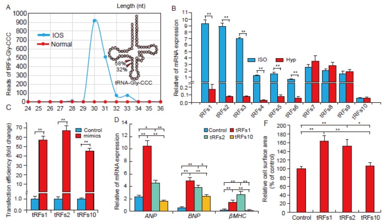
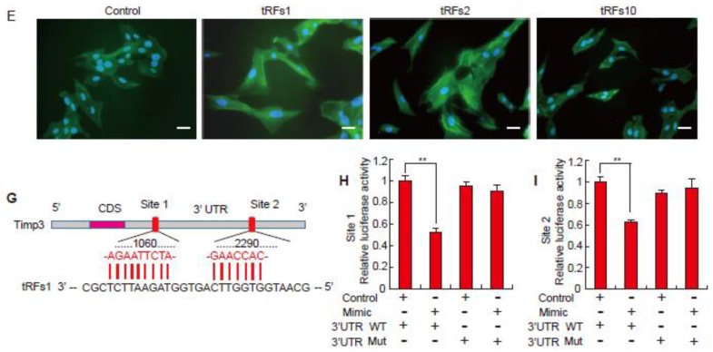
Figure 2.
tRFs promote cardiac hypertrophy in cultured cardiomyocytes. (A) The distribution of different lengths of tRFs derived from tRNA-Gly-CCC; (B) Verification of the expression of tRFs by qRT-PCR, n = 3; (C) Transfection efficiency of tRFs mimics in the H9c2 cells, n = 3; (D) Relative mRNA expression of ANP, BNP, and β-MHC in treated H9c2 cells, n = 3; (E) Immunofluorescence evaluation of cardiac hypertrophy in cells treated with tRFs mimics. Bar indicates 50 μm, n = 3; (F) The relative cell surface area in treated H9c2 cells, n = 3; (G) Sequence alignment of tRFs1 with 3′-UTR of Timp3; (H) The repressive effect of tRFs1 on the activity of site1 of Timp3 3′UTR measured by luciferase assay, n = 3. (I) The repressive effect of tRFs1 on the activity of site2 of Timp3 3′UTR measured by luciferase assay, n = 3. Data are means ± SD. * p < 0.05, ** p < 0.01.


