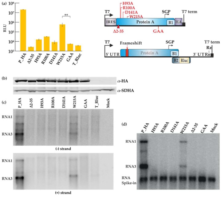
Figure 4.
Mutational analysis of the FHV RNA capping enzyme. (a) BSR T7/5 cells were co-transfected with each FHV protein A mutant construct and template construct T_Rluc. Rluc activity was measured 40 h post-transfection. Mock values have been subtracted, error bars represent the standard deviation and ** designates p < 0.01 (Student’s t test). The location of the mutations is shown schematically on the right. RLU: relative light units. (b) Expression of protein A variants as detected by Western blotting with anti-HA antibodies; SDHA was used as a control. (c) Viral RNA synthesis detected by Northern blotting for minus and plus strands as indicated. (d) CMP fractions were isolated from transfected cells and used in the IVRA. The spike-in of short radioactive RNA was used to ensure equal isolation and loading of RNA. The relative density of the bands was measured with ImageJ software.

