Abstract
Silver nanoparticles (Ag NPs) were synthesized using Juglans regia (J. regia) leaf extract, as both reducing and stabilizing agents through microwave irradiation method. The effects of a 1% (w/v) amount of leaf extract (0.1–0.9 mL) and an amount of 1 mM AgNO3 solution (15–25 mL) on the broad emission peak (λmax) and concentration of the synthesized Ag NPs solution were investigated using response surface methodology (RSM). Fourier transform infrared analysis indicated the main functional groups existing in the J. regia leaf extract. Dynamic light scattering, UV-Vis spectroscopy and transmission electron microscopy were used to characterize the synthesized Ag NPs. Fabricated Ag NPs with the mean particle size and polydispersity index and maximum concentration and zeta potential of 168 nm, 0.419, 135.16 ppm and −15.6 mV, respectively, were obtained using 0.1 mL of J. regia leaf extract and 15 mL of AgNO3. The antibacterial activity of the fabricated Ag NPs was assessed against both Gram negative (Escherichia coli) and positive (Staphylococcus aureus) bacteria and was found to possess high bactericidal effects.
Keywords: green synthesis, silver nanoparticles, microwave irradiation, Juglans regia, antibacterial activity
1. Introduction
Silver is known as strong antimicrobial agent due to its toxicity against most microorganisms including bacteria, fungi, and viruses [1]. As compared to the silver element in bulk state, silver nanoparticles (Ag NPs) exhibit unusual physico-chemical and antimicrobial properties due to their lower particle size (less than 100 nm) and higher surface area to volume ratio, which increase the proportion of high-energy surface atoms [1,2,3]. Because of the marvelous antimicrobial properties of Ag NPs, they have been widely utilized in numerous fields such as medicine, electronics, biotechnology, water disinfection, air treatment, and packaging [3].
Green chemistry is the use of chemistry principles to reduce or eliminate the use of toxic reagents, leading to lower amounts of undesirable residues, which in turn are harmful to human health or the environment [4,5]. Incorporation of green chemistry and nanotechnology is of great interest and has gained much attention over the past decade. Green synthesis of Ag NPs, using plants and their derivative extracts as an alternative method for the conventional physical and chemical NP fabrication methods, has attracted considerable attention in recent years [1,6]. In fact, various metabolites existing in the plants including sugars, alkaloids, phenolic acids, terpenoids, polyphenols, and proteins play an important role in the bioreduction of silver ions to Ag NPs and their stabilization [7]. Several studies have been completed on green synthesis of Ag NPs using Aloe vera and Pelargonium zonali leaf extract and chitosan [8,9,10].
Walnut belongs to Juglandaceae family and has the scientific name Juglans regia (J. regia). Numerous health benefits of walnut for human have been reported, some of which are related to the existence of its main bioactive compounds including polyphenolic compounds, flavonoids, proteins, carotenoids, lipids, and alkaloids. Leaf of walnut contains malic acid, sucrose, α-tocopherol, 3-O-caffeoylquinic acids, and quercetin O-pentoside as the most abundant organic acids, disaccharide, tocopherol isomer and phenolic compounds, respectively [11,12,13].
The present work focuses on (i) the potential of walnut leaf extract in the fabrication of Ag NPs; (ii) Ag NPs synthesized parameter optimization using microwave irradiation to achieve Ag NPs with more desirable physico-chemical properties and (iii) an antibacterial activity assessment of the produced Ag NPs.
2. Results
2.1. Formation of Ag NPs
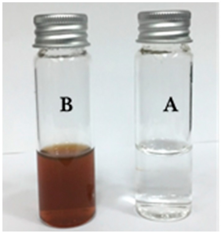
During the synthesis of Ag NPs, the color of the mixture solution containing silver salt and walnut leaf extract changed from colorless into brown and dark. In fact, the synthesized Ag NPs, due to their surface plasmon resonance (SPR), changed the color of the mixture solution and this color change verified the formation of Ag NPs using walnut leaf extract and microwave irradiation (Figure 1). As clearly observed in Table 1, λmax of the synthesized Ag NPs was varied 424–429 nm, which was in a favorable range for Ag NPs [8,10]. The particle size of the synthesized Ag NPs could be correlated with their broad emission peaks (λmax), where longer wavelengths in λmax of Ag NPs were associated with bigger size [9]. This indicated that J. regia leaf extract successfully reduced silver ions and formed Ag NPs.
Figure 1.
Color and appearance changes during synthesis of silver nanoparticles (Ag NPs) using J. regia leaf extract. J. regia leaf extract containing silver nitrate before (A) and after (B) exposure to microwave irradiation.
Table 1.
Experimental runs according to the central composite design (CCD) and response variables for synthesis of Ag NPs.
| Sample No. | Amount of Leaf Extract (mL) | Amount of Silver Salt (mL) | λmax (nm) | Concentration (ppm) | ||
|---|---|---|---|---|---|---|
| Exp | Pre | Exp | Pre | |||
| 1 | 0.10 | 20.00 | 425 | 424.896 | 104.813 | 101.702 |
| 2 | 0.78 | 16.46 | 429 | 429.104 | 100.003 | 97.802 |
| 3 | 0.78 | 23.53 | 424 | 424.00 | 93.503 | 96.614 |
| 4 | 0.50 | 15.00 | 427 | 426.854 | 72.443 | 74.643 |
| 5 | 0.50 | 25.00 | * | * | 48.393 | 43.081 |
| 6 | 0.50 | 20.00 | 426 | 427.250 | 56.453 | 61.835 |
| 7 | 0.50 | 20.00 | 426 | 427.250 | 56.453 | 61.835 |
| 8 | 0.217 | 23.53 | 426 | 426.043 | 50.343 | 55.654 |
| 9 | 0.50 | 20.00 | 428 | 427.250 | 66.073 | 61.835 |
| 10 | 0.90 | 20.00 | 427 | 426.957 | * | * |
| 11 | 0.217 | 16.46 | 424 | 424.146 | * | * |
| 12 | 0.50 | 20.00 | 429 | 427.250 | 66.723 | 61.835 |
| 13 | 0.50 | 20.00 | * | * | 63.473 | 61.835 |
Exp, Experimental values of studied responses; Pre, Predicted values of studied responses. *, Out of range.
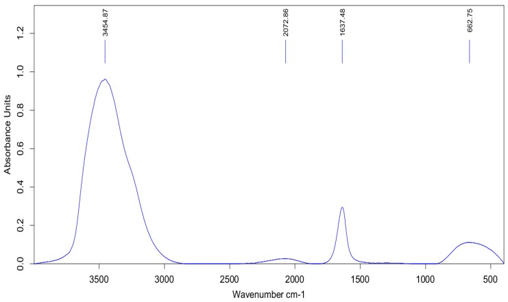
Figure 2 shows the Fourier transform-infrared (FT-IR) spectrum of J. regia leaf extract which was in the region range of 400–4000 cm−1. The IR spectrum of leaf extract absorption bands at 3454.87 and 2072.86 cm−1 represent the phenolic–OH and N=C groups. Absorption bands at 1637.48 cm−1 are related to the amino group, bending vibrations, and the –OH group. The obtained results indicated that the phenolic compounds and proteins were the two main components of the J. regia leaf extract which had key roles in the formation of the stabilized Ag NPs [11,12].
Figure 2.
Fourier transform-infrared (FT-IR) spectrum of J. regia leaf extract.
2.2. Models Generation and Synthesis Conditions Optimization
Based on experimental runs, the values of broad emission peaks (λmax, nm) and concentration (ppm) for the fabricated Ag NPs were achieved (Table 1) and according to these obtained data, models were created. Table 2 shows coefficients of the model terms and models accuracy based on R square and its adjusted (R2, R2-adj) and lack of fit. The high values of R2 (>77.80) and R2-adj (>85.61) and lack of fit (p > 0.05) verified higher suitability of the generated models to predict the synthesis response parameters. p-Values of the generated model terms are also described in Table 3. As can be seen, the interaction of the amounts of leaf extract and silver salt solution had significant (p < 0.05) effects on λmax and concentration of the formed Ag NPs.
Table 2.
Regression coefficients, R2, R2-adj, and probability values for the fitted models.
| Regression Coefficient | λmax (nm) | Concentration (ppm) |
|---|---|---|
| β0 (constant) | 386.44 | 249.7 |
| β1 (main effect) | −45,084 | −513.04 |
| β2 (main effect) | 3.09 | −3.68 |
| β11 (quadratic effect) | −8.27 | 336.8 |
| β22 (quadratic effect) | −0.061 | −0.119 |
| β12 (interaction effect) | −1.75 | 10.56 |
| R2 | 77.81% | 95.25% |
| R2-adj | 85.62% | 90.49% |
| Lack of fit (p-value) | 0.985 | 0.142 |
β0 is a constant, β1, β11 and β12 are the linear, quadratic, and interaction coefficients of the quadratic polynomial equation, respectively.
Table 3.
p-Values of the regression coefficients in the obtained models.
| Main Effects | Main Effects | Quadratic Effects | Interacted Effect | ||
|---|---|---|---|---|---|
| X1 | X2 | X11 | X22 | X1 X2 | |
| λmax (nm) (p-value) | 0.003 | 0.693 | 0.00 | 0.580 | 0.045 |
| Concentration (ppm) (p-value) | 0.018 | 0.153 | 0.224 | 0.247 | 0.030 |
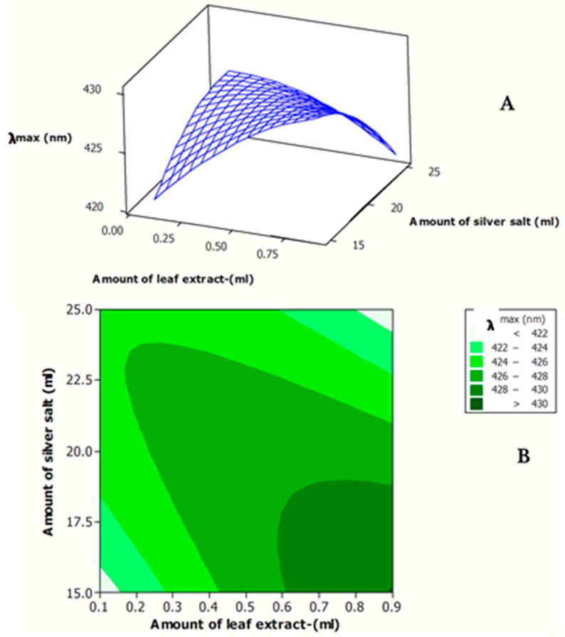
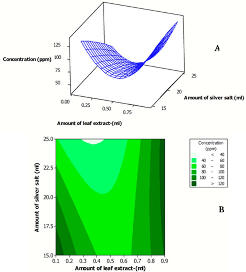
Figure 3A,B indicates the effects of the amount of J. regia leaf extract and amount of AgNO3 on the λmax of the synthesized Ag NPs. As clearly observed in Figure 3, the minimum λmax (particle size) was obtained at both minimum amount of J. regia leaf extract and of AgNO3 solution and the maximum amount of J. regia leaf extract and of AgNO3 solution. The experimental value of the concentration for the synthesized Ag NPs ranged 19–80 ppm (Table 1). The effects of the amount of J. regia leaf extract andof AgNO3 on the concentration of the fabricated Ag NPs are shown in Figure 4A,B.
Figure 3.
Response surface (A) and contour plots (B) for λmax of the synthesized Ag NP solution as function of the amount of J. regia leaf extract and amount of AgNO3 solution.
Figure 4.
Response surface (A) and contour plots (B) for the concentration of the synthesized Ag NP solution as function of amount of J. regia leaf extract and amount of AgNO3 solution.
As clearly observed in Figure 4, the maximum concentration of the synthesized Ag NPs was obtained using the highest amount of the J. regia leaf extract. The obtained results were in line with the findings of other research [8,9,10]. They found that by increasing the amount of plant extract, the concentration of the bioreductants increased in the extract, which in turn increased the concentration of the formed Ag NPs.
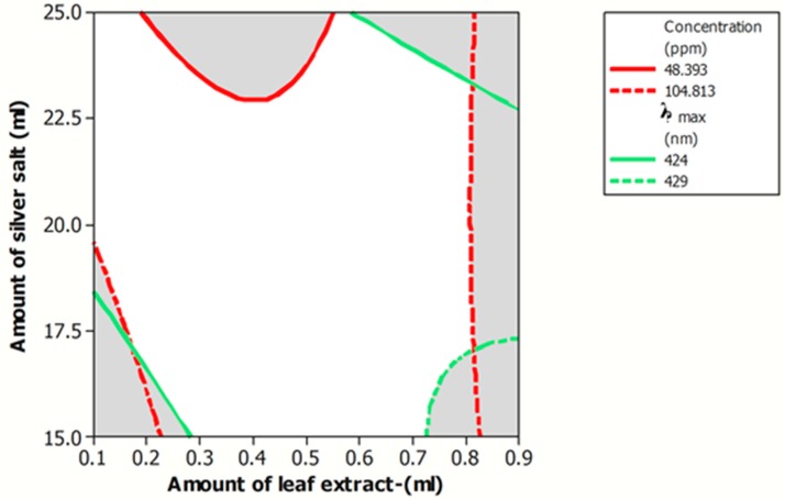
In the synthesis of Ag NPs, the main objective is formation of NPs with desirable physico-chemical properties, including minimum particle size (λmax) and maximum concentration. This synthesis process is known as optimized procedure. According to the generated models for the synthesis of Ag NPs, an overlaid contour plot, as graphical optimization (Figure 5), was plotted to better visualize of optimum area (white colored zone).
Figure 5.
Overlaid contour plot of Ag NPs λmax and concentration with acceptable levels as a function of amount of J. regia leaf extract and amount of AgNO3 solution.
The result of a numerical optimization also demonstrated that by using 0.1 mL of J. regia leaf extract and 15 mL of AgNO3, Ag NPs with minimum λmax of 421 nm and maximum concentration of 135.64 ppm were produced. A verification test using optimum synthesis parameters also indicated insignificant (p > 0.05) differences between the values of predicted and experimental of λmax and concentration of the fabricated Ag NPs and verified the suitability of the models.
2.3. Physico-Chemical Characteristics of the Synthesized Ag NPs at Obtained Optimum Conditions
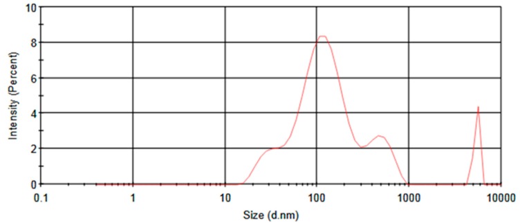
Formation of Ag NPs using J. regia leaf extract at obtained optimum conditions was confirmed by changes in the color of the mixture solution. Dynamic light scattering (DLS) analysis also indicated that the synthesized Ag NPs had particle size, polydispersity index (PDI), and zeta potential values of 168 nm, 0.419 and −15.6 mV, respectively. The particle size distributions (PSD) of the sample are also shown in Figure 6.
Figure 6.
Particle size distribution of synthesized Ag NPs at obtained optimum synthesis conditions using J. regia leaf extract.
2.4. Antibacterial Activity
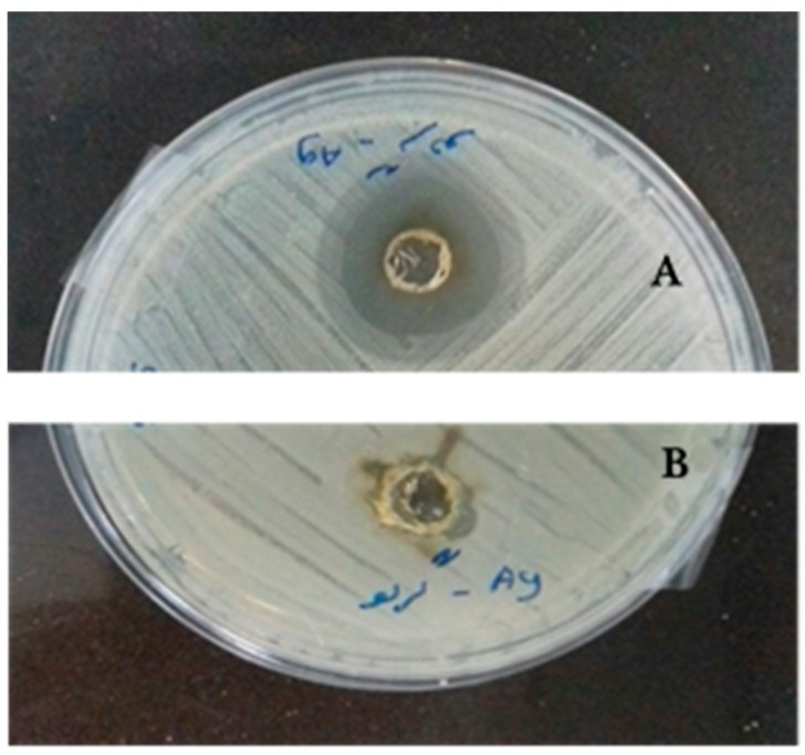
The antibacterial activity of synthesized Ag NPs on the growth of Gram-positive (S. aureus) and Gram-negative (E. coli) bacteria during incubation indicated that the diameter of the clear zone for synthesized Ag NPs in the plate containing S. aureus and E. coli were 16 and 10 mm, respectively (Figure 7). The results also indicated that the mean diameter of formed clear zone around the Ampicillin disc in the plates containing S. aureus and E. coli were 35 and 33 mm, respectively.
Figure 7.
Created zones of inhibition with S. aureus (A) and E. coli (B) incubated at 37 °C for 24 h for synthesized Ag NPs using J. regia leaf extract.
The obtained results indicated that the fabricated Ag NPs had higher antibacterial activity against Gram-positive bacteria compared to the Gram-negative bacteria. The obtained results were in agreement with findings of Ahmadi et al., Mohammadlu et al., and Torabfam and Jafarizadeh-Malmiri [8,9,10]. The main reason behind the bactericidal activity of the Ag NPs against the bacteria strains is related to their effects on the permeability of the cell wall and membrane. In fact, the released silver ions (Ag+) from the Ag NPs were attached into the anionic groups of the cell wall, such as polycyclic aromatic hydrocarbon and teichoic acids. They also formed polyelectrolyte complexes, which limited the transference of nutrients and provided metabolites into and outside the cell [9]. Furthermore, caffeoylquinic acid is the main phenylpropanoids in J. regia leaf extract, which has various bioactivities such as antioxidant, antibacterial, anticancer, antihistamic, and other biological effects. The direct antimicrobial activity of caffeoylquinic acid implies an array of possibilities, including effects in the cell envelope [13].
3. Materials and Methods
3.1. Materials
J. regia leaves were picked from walnut trees in Tabriz, Iran. AgNO3, as silver salt, was bought from Dr. Mojallali (Dr. Mojallali Chemical Complex Co., Tehran, Iran). Standard solution of Ag NPs (with particle size of 10 nm and concentration of 1000 ppm) was purchased from Tecnan-Nanomat (Spain). Escherichia coli (PTCC 1270) and Staphylococcus aureus (PTCC 1112) were attained from microbial Persian-type culture collection (PTCC, Tehran, Iran). There is no ATCC (American type culture collection) number for E. coli and this bacteria is clinical isolate. However, the ATCC number of S. aureus is 6538. Nutrient agar was bought from Biolife (Biolife Co., Milan, Italy).
3.2. Preparation of J. regia Leaf Extract
The J. regia leaf was washed, dried (in dark room), powdered, and 1 g of the prepared powder was added into 100 mL of boiling distilled water for 5 min. After cooling the solution, it was filtered (Whatman No. 1 filter paper) using a Buchner funnel under vacuum pressure and the clear walnut leaf extract was stored at a cold temperature (4 °C).
3.3. Ag NPs Synthesis Using J. regia Leaf Extract
The Ag NPs solution was obtained by a domestic microwave-assisted synthetic approach. AgNO3 solution (1 mM) was made by dissolving 0.017 g of its powder in 100 mL of deionized double-distilled water. In a typical synthesis, different amounts of AgNO3 solution (15–25 mL) were mixed with different amounts of J. regia leaf extract (0.1–0.9 mL) and the mixture solutions were put into a microwave oven (MG-2312W, LG Co., Seoul, Korea) at a constant power of 800 W and microwave exposure time (180 s).
3.4. Physico-Chemical Assay
3.4.1. Fourier Transform-Infrared (FT-IR) Spectra Analysis
In order to identify the possible reducing and stabilizing biomolecules of J. regia leaf extract, FT-IR measurements were carried out. The FT-IR spectrum of the extract was recorded on a Bruker Tensor27 spectrometer (Bruker Co., Karlsruhe, Germany) using KBr pellets in the 4000–400 cm−1 region.
3.4.2. Surface Plasmon Resonance
Ag NPs, due to their SPR, have a strong absorption of light, which is shown as broad emission peaks (λmax) in the wavelength ranging from 380 to 450 nm [8,9,10]. Therefore, formation of Ag NPs using J. regia leaf extract can be confirmed by the absorption spectrum of the mixture solutions containing fabricated Ag NPs by using a Jenway UV-Vis spectrophotometer 6705 (Cole-Parmer Co., Staffordshire, UK). Furthermore, by preparing several serial dilute Ag NPs solutions (10–1000 ppm) and establishing standard curves based on the defined concentrations of the Ag NPs solutions and their absorbance unit values, it is possible to determine the concentration of the formed Ag NPs using J. regia leaf extract [10].
3.4.3. Particle Size, Particle Size Distribution, Polydispersity Index and Zeta Potential of the Synthesized Ag NPs
In order to measure values of the mean particle size (nm), PDI (ranging 0–1) and zeta potential (mV) of the fabricated Ag NPs and their PSD, a DLS particle size analyzer (Nanotrac Wave, Microtrac, Montgomeryville, PA, USA) was utilized. The DLS technique scatters a laser light beam at the surface of dispersed NPs, which results in the detection of the backscattered light. PDI is a dimensionless value which shows that the uniformity of the synthesized NPs and their surface electric charge are related to the PDI and zeta potential values of the synthesized NPs [14,15].
3.5. Antibacterial Assay
Bactericidal activity of the formed Ag NPs was evaluated using the well diffusion method. For this reason, bacterial suspensions containing 1.5 × 10⁸ colony-forming units of bacteria was prepared based on a 0.5 McFarland standard and 0.1 mL of that amount was spread on the surface of solid nutrient agar in the plates and a hole, 5 mm in diameter, was made in the solid agar. 10 µL of the produced Ag NPs solution was then poured into the created well and put in incubator at 37 °C for 24 h. The antibacterial activity of the synthesized Ag NPs correlated to the diameter of created clear zones around the holes. An Ampicillin disc 5mm in diameter (Oxoid, 10 µg/disc) was used as a positive control for both Gram-positive and Gram-negative bacteria strains and the antibacterial activity of the synthesized Ag NPs was compared to the Ampicillin.
3.6. Experimental Design, Statistical Analysis and Optimization Procedure
The experiment was planned using a central composite design (CCD) and response surface methodology (RSM) was used to evaluate the effects of two independent parameters, namely amount of leaf extract (X1) and amount of AgNO3 solution (X2), on the prepared Ag NPs. The studied response variables were broad emission peak (λmax) (Y1, nm) and concentration (Y2, ppm) of the synthesized Ag NPs. As clearly observed in Table 1, thirteen experimental treatments were assigned with five different levels for each independent parameter using Minitab software (v.16 statistical package, Minitab Inc., Pennsylvania State, PA, USA). In order to correlate the λmax (Y1) and concentration (Y2) of the synthesized Ag NPs to the studied synthesis variables, a second order polynomial equation (Equation (1)) was used. Where β0 is a constant, β1, β11, and β12 correspond to the linear, quadratic and interaction effects, respectively [16]. The suitability of the model was studied, accounting for the coefficient of determination (R2) and adjusted coefficient of determination (R2-adj) [17]. Analysis of variance (ANOVA) was also used to provide the significance determinations of the resulted models in terms of p-value. Small p-values (lower than 0.05) were considered as statistically significant [18].
| Y = β0+ β1X1 + β2X2 + β11X12 + β22X22 + β12X1X2 | (1) |
Numerical optimization was carried out to determine exact amounts of silver salt and J. regia leaf extract in the Ag NPs synthesis to produce NPs with minimum λmax (particle size) and maximum concentration. Graphical optimization was also used to better visualize the effects of the synthesis parameters on the response variables (λmax and concentration) [19]. Suitability and accuracy of the generated models in predicting the response variables in the defined range of the Ag NPs synthesis parameters were validated by synthesis of Ag NPs using obtained optimum synthesized conditions and comparison of the experimental values of the response variables and their predicted values [20].
4. Conclusions
Fabricated Ag NPs using a rapid, one-step green approach, based on microwave irradiation showed desirable physico-chemical properties with antibacterial activity against E. coli and S. aureus. However, their antibacterial activity was significantly (p < 0.05) higher toward Gram-positive bacteria strains. The synthesized Ag NPs using the rapid developed synthesis method can be used as a favorable antibacterial agent in different areas such as medicine and food packaging.
Acknowledgments
This research was undertaken with material support from the Najian Herbal Group (Tabriz, Iran). The authors appreciate this support. This research received no specific grant from any funding agency in the public, commercial, or not-for-profit sectors.
Author Contributions
M.E. performed the Ag NPs synthesis and physico-chemical analyses. H.V. performed strain cultivation and antibacterial assay. Y.N. and M.J.N. contributed with valuable discussions and scientific advice. H.J.-M. designed the experiments and wrote the draft manuscript. A.B. contributed helpful discussion and statistical analysis during the preparation of manuscript.
Funding
This research received no external funding.
Conflicts of Interest
The authors declare no conflicts of interest. The authors alone are responsible for the content and writing of this article.
References
- 1.Mohammadlu M., Jafarizadeh-Malmiri H., Maghsoudi H. A review on green silver nanoparticles based on plants: Synthesis, potential applications and eco-friendly approach. Int. Food Res. J. 2016;23:446–463. [Google Scholar]
- 2.Kon K., Rai M. Metallic nanoparticles: Mechanism of antibacterial action and influencing factors. J. Comp. Clin. Pathol. Res. 2013;2:160–174. [Google Scholar]
- 3.Zhou Y., Kong Y., Kundu S., Cirillo J.D., Liang H. Antibacterial activities of gold and silver nanoparticles against Escherichia coli and Bacillus calmette-Guérin. J. Nanobiotechnol. 2012;10:19–28. doi: 10.1186/1477-3155-10-19. [DOI] [PMC free article] [PubMed] [Google Scholar]
- 4.Ingale A.G., Chaudhari A. Biogenic synthesis of nanoparticles and potential applications: An eco-friendly approach. J. Nanomed. Nanotechnol. 2013;4:165–167. doi: 10.4172/2157-7439.1000165. [DOI] [Google Scholar]
- 5.Hebbalalu D., Lalley J., Nadagouda M.N., Varma R.S. Greener techniques for the synthesis of silver nanoparticles using plant extracts, enzymes, bacteria, biodegradable polymers, and microwaves. ACS Sustain. Chem. Eng. 2013;1:703–712. doi: 10.1021/sc4000362. [DOI] [Google Scholar]
- 6.Vamanu E., Ene M., Bita B., Ionescu C., Craciun L., Sarbu I. In vitro human microbiota response to exposure to silver nanoparticles biosynthesized with mushroom extract. Nutrients. 2018;10:607. doi: 10.3390/nu10050607. [DOI] [PMC free article] [PubMed] [Google Scholar]
- 7.Zhang Y., Cheng X., Zhang Y., Xue X., Fu Y. Biosynthesis of silver nanoparticles at room temperature using aqueous Aloe leaf extract and antibacterial properties. Colloids Surf. A Physicochem. Eng. Asp. 2013;423:63–68. doi: 10.1016/j.colsurfa.2013.01.059. [DOI] [Google Scholar]
- 8.Mohammadlu M., Jafarizadeh-Malmiri H., Maghsoudi H. Hydrothermal green silver nanoparticles synthesis using Pelargonium/Geranium leaf extract and evaluation of their antifungal activity. Green Process. Synth. 2017;6:31–42. doi: 10.1515/gps-2016-0075. [DOI] [Google Scholar]
- 9.Ahmadi O., Jafarizadeh-Malmiri H., Jodeiri N. Eco-friendly microwave enhanced green silver nanoparticles synthesis using Aloe vera leaf extract and their physico-chemical and antibacterial studies. Green Process. Synth. 2018;7:231–240. doi: 10.1515/gps-2017-0039. [DOI] [Google Scholar]
- 10.Torabfam M., Jafarizadeh-Malmiri H. Microwave–enhanced silver nanoparticles synthesis using chitosan biopolymer–Optimization of the process conditions and evaluation of their characteristics. Green Process. Synth. 2017 doi: 10.1515/gps-2017-0139. [DOI] [Google Scholar]
- 11.Jaiswal B.S., Tailang M. Juglans regia: A review of its traditional uses phytochemistry and pharmacology. Indo Am. J. Pharm. Res. 2017;7:390–398. [Google Scholar]
- 12.Amaral J.S., Seabra R.M., Andrade P.B. Phenolic profile in the quality control of walnut (Juglans regia L.) leaf. Food Chem. 2004;88:373–379. doi: 10.1016/j.foodchem.2004.01.055. [DOI] [Google Scholar]
- 13.Pereira J.A., Oliveira I., Sousa A. Walnut (Juglans regia L.) leaf: Phenolic compounds, antimicrobial activity and antioxidant potential of different cultivars. Food Chem. Toxicol. 2007;45:2287–2295. doi: 10.1016/j.fct.2007.06.004. [DOI] [PubMed] [Google Scholar]
- 14.Eskandari-Nojedehi M., Jafarizadeh-Malmiri H., Rahbar-Shahrouzi J. Optimization of processing parameters in green synthesis of gold nanoparticles using microwave and edible mushroom (Agaricus bisporus) extract and evaluation of their antibacterial activity. Nanotechnol. Rev. 2016;5:537–548. doi: 10.1515/ntrev-2016-0064. [DOI] [Google Scholar]
- 15.Eskandari-Nojedehi M., Jafarizadeh-Malmiri H., Jafarizad A. Microwave accelerated green synthesis of gold nanoparticles using gum Arabic and their physico-chemical properties assessments. Z. Phys. Chem. 2018;232:325–343. doi: 10.1515/zpch-2017-1001. [DOI] [Google Scholar]
- 16.Amirkhani L., Moghaddas J., Jafarizadeh-Malmiri H. Candida rugosa lipase immobilization on magnetic silica aerogel nanodispersion. RSC Adv. 2016;6:12676–12687. doi: 10.1039/C5RA24441B. [DOI] [Google Scholar]
- 17.Anarjan N., Jaberi N., Yeganeh-Zare S., Banafshehchin E., Rahimirad A., Jafarizadeh-Malmiri H. Optimization of mixing parameters for α-tocopherol nanodispersions prepared using solvent displacement method. J. Am. Oil Chem. Soc. 2014;91:1397–1405. doi: 10.1007/s11746-014-2482-6. [DOI] [Google Scholar]
- 18.Eskandari-Nojedehi M., Jafarizadeh-Malmiri H., Rahbar-Shahrouzi J. Hydrothermal biosynthesis of gold nanoparticle using mushroom (Agaricus bisporous) extract: Physico-chemical characteristics and antifungal activity studies. Green Process. Synth. 2018;7:38–47. doi: 10.1515/gps-2017-0004. [DOI] [Google Scholar]
- 19.Anarjan N., Jafarizadeh-Malmiri H., Nehdi I.A., Sbihi H.M., Al-Resayes S.I., Tan C.P. Effects of homogenization process parameters on physicochemical properties of astaxanthin nanodispersions prepared using a solvent-diffusion technique. Int. J. Nanomed. 2015;10:1109–1118. doi: 10.2147/IJN.S72835. [DOI] [PMC free article] [PubMed] [Google Scholar]
- 20.Ahdno H., Jafarizadeh-Malmiri H. Development of a sequenced enzymatically pre-treatment and filter pre-coating process to clarify date syrup. Food Bioprod. Process. 2017;101:193–204. doi: 10.1016/j.fbp.2016.11.008. [DOI] [Google Scholar]


