



Summary
Over the past seven decades, research on autotrophic and heterotrophic model organisms has defined how the flow of electrons (“reducing power”) from high-energy inorganic sources, through biological systems, to low-energy inorganic products like water, powers all of Life’s processes. Universally, an initial major biological recipient of these electrons is nicotinamide adenine dinucleotide-phosphate, which thereby transits from an oxidized state (NADP+) to a reduced state (NADPH). A portion of this reducing power is then distributed via the cellular NADPH-dependent disulfide reductase systems as sequential reductions of disulfide bonds. Along the disulfide reduction pathways, some enzymes have active sites that use the selenium-containing amino acid, selenocysteine, in place of the common but less reactive sulfur-containing cysteine. In particular, the mammalian/metazoan thioredoxin systems are usually selenium-dependent as, across metazoan phyla, most thioredoxin reductases are selenoproteins. Among the roles of the NADPH-dependent disulfide reductase systems, the most universal is that they provide the reducing power for the production of DNA precursors by ribonucleotide reductase (RNR). Some studies, however, have uncovered examples of NADPH-independent disulfide reductase systems that can also support RNR. These systems are summarized here and their implications are discussed.
Keywords: Thioredoxin reductase, Glutathione reductase, NADPH, Sulfur amino acid, Ribonucleotide reductase, Transsulfuration, Methionine cycle, Cysteine
Graphical Abstract

Redox reactions and reducing power
Reduction and oxidation (redox) reactions involve the transfer of electrons between molecules. Reducing agents (reductants) carry electrons that can be transferred to a more oxidized substrate. Oxidizing agents (oxidants) are electrophilic and will readily accept electrons from a more reduced substrate. All reduction reactions are also oxidation reactions, and vice versa, in that the reduction (gaining of electrons) of one compound requires the oxidation (loss of electrons) of another. Thus, the concept of “reducing power” can be established to describe the propensity of one compound to donate its electrons to another compound, and has an associate value, expressed in volts, that describes how readily those electrons will be donated, or their “redox potential.” Electrons are often transferred as pairs (2-electron transfers), but single electron transfers also occur in biology. Molecules with unpaired electrons are free radicals, which are typically unstable and highly reactive. Some free radicals are biologically relevant as damaging agents. Others are essential for biological functions, in particular as critical intermediates within certain enzymatic reactions. There is an extensive body of knowledge regarding the electron transfer mechanisms within enzymatic reactions, including single- and paired-electron transfers; however coverage of this literature is beyond the scope of this review.
NADPH: a “universal currency” for anabolic reduction reactions
Inorganic carbon, for example within CO2, is usually in an oxidized state, whereas the carbon in organic molecules is highly reduced. Organic molecules are generated via the reduction of oxidized inorganic carbon sources by autotrophs, which include both photosynthetic and chemolithotrophic organisms. To generate organic molecules, autotrophs use energy from photons of light or from oxidation of inorganic minerals to obtain electrons, and these are used to chemically reduce the CO2 by forming carbon-carbon or other bonds. The bonds of organic molecules store most of Life’s energy, which is released under heterotrophic conditions by oxidation (i.e., extraction of the electrons) of these bonds.
Most of the energy that drives Life’s processes likely entered the biological world via photosynthesis. In the light-dependent reactions of photosynthesis, energy from photons is used to extract electrons from a low energy source, usually water. Typically four photons are used to cleave two molecules of water, yielding one molecule of molecular oxygen (O2), four protons (H+), and four electrons of reducing power. A small amount of the energy from this process is also often used to generate three phosphodiester bonds within adenosine triphosphate (ATP). The four electrons are used to reduce two molecules of oxidized nicotinamide adenine dinucleotide phosphate (NADP+) into their reduced form (NADPH; Fig. 1), thereby capturing the reducing power (Eq. 1).
Fig. 1. Chemical structure and activity of the [NADPH;NADP+] redox couple.

The redox-active electrons are denoted in red font by the symbol e−. The boxed inset shows the structural difference between the NADP- and the NAD-family of nicotinamide cofactors.
| [Eq. 1] |
Interestingly, chemolithotrophs also capture reducing power, albeit obtained from oxidation of inorganic compounds rather than from photons of light, as NADPH. Thus, NADPH is the organic compound to initially receive the reducing power obtained from inorganic sources in all autotrophs. In the light-independent (“dark”) reactions of photosynthesis (the Calvin cycle, which is also used in chemolithotrophs), the electrons from NADPH are used to reductively “fix” CO2 into sugar. This more stably stores the reducing power as reduced carbon, predominantly within carbon-carbon bonds [1–3]. Although some NADPH-driven reductions are catabolic, more generally NADPH is the electron donor for anabolic reduction reactions and, accordingly, hundreds of reductive enzymes (reductases) are known to be NADPH-dependent. As such, NADPH can be considered a “universal currency” for anabolic reduction reactions.
NADPH cannot be acquired from extracellular sources but, rather, must be generated by the reduction of NADP+ within each cell. To generate NADPH in heterotrophic conditions, the reducing power that was stored in the bonds of organic molecules during the Calvin cycle can be used by a few reactions to reduce NADP+ + H+ + 2e− → NADPH. In mammals, oxidations of several organic molecules can fuel NADPH-generating reactions. These include oxidation of glucose in the pentose phosphate pathway, oxidation of isocitrate by isocitrate dehydrogenase, oxidation of malate by malic enzyme, and oxidation of methylenetetrahydrofolate [4–8].
The [NADPH/NADP+] redox couple is one of two predominant nicotinamide adenine dinucleotide systems in cells, the other being [NADH/NAD+]. The [NADH/NAD+] redox couple is used primarily for biological oxidations, such as for extracting energy from organic molecules, and therefore its steady-state equilibrium in living cells favors the oxidized NAD+ over NADH. Conversely the reduced NADPH is about ten-times more abundant than the oxidized NADP+, and NADPH is used predominantly for reduction reactions, such as within anabolic processes, as described above, or for antioxidant activities. In some situations, reducing power can be transferred between NADH and NADPH by nicotinamide nucleotide transhydrogenases (NNT) [9, 10]. In mitochondria, wherein NADH is abundant, NNT-derived NADPH is estimated to account for half of the NADPH, and NNT-knockdown alters mitochondrial redox status [11].
Early evolution of biological disulfide reduction
Life arose in an anoxic environment and likely relied heavily on hydrogen sulfide and ferrous iron (H2S/Fe2+)-based redox reactions [12, 13]. Before photosynthetic organisms arose that could harvest energy from light, all autotrophic life was chemolithotropic, meaning that nutrients and energy were acquired from chemical reactions involving minerals. An abundant and therefore likely source of chemolithotropic potential for supporting early life is associated with sulfur (S)-rich hydrothermal vents, which expel large amounts of both H2S and Fe2+ in an environment suitable for sustaining life (pH 3–4 and temperature/pressure conditions in which water is liquid) [14, 15]. The abundant iron-sulfur (FeS) compounds found in such environments are capable of (i) catalyzing other redox reactions (e.g., N2 to NH3 or NO3−)[16]; (ii) forming planar sheets that likely became the first barriers encapsulating the biological machinery from the external environment as a primitive cell; and (iii) assembling into in FeS clusters, which still serve as electron conduits in contemporary biological redox reactions. Other metal-S minerals, like ZnS and (FeNi)S, can activate C-H bonds, and thereby abiotically catalyze the formation of C-X bonds (X = C, O, or N) to generate simple organic molecules [17–19]. These inorganic redox reactions and simple organic molecules were likely critical to the onset of life.
Because H2S and Fe2+ are abundant in hydrothermal environments, primitive organisms likely extracted two electrons of reducing power from oxidation of H2S, which then coupled with Fe2+, yielding pyrite (FeS2)[20]. However, the capacity for more efficient extraction of reducing power existed and was eventually exploited, since complete oxidation of H2S to sulfate (SO42−) yields eight electrons (Fig. 2). Later organisms would use intracellular reducing power, stored as organic carbon sources and transferred via NADPH, to enzymatically reduce environmental sulfate into sulfite by adenylyl-sulfate (APS) reductase and phosphoadenylyl-sulfate (PAPS) reductase. Further reduction to sulfide through three consecutive 2-electron transfers by ferredoxins would then support the biological production of sulfide. This allowed organisms to radiate out from restricted sulfide-rich hydrothermal vent ecosystems into new environments and expanded niches [21–23].
Fig. 2. Oxidation states of common inorganic and organic S-compounds.

For simplicity, only species discussed in this review are included. Organic compounds are as follows: a, thiols, such as found in reduced proteins, Cys, and GSH; b, thioether within Met; c, disulfide, such as found in oxidized protein-disulfides or GSSG; d, sulfenic acid; e, sulfoxide within oxidized Met; f, sulfinic acid; g, sulfonic acid.
Photosynthetic autotrophs first appeared about 2.3 billion years ago. H2S likely also played a critical role in the evolution of photosynthetic organisms from chemolithotrophic predecessors. Because of its high abundance in certain environments and its being similar to H2O, yet with more easily activated electrons (oxidation potential −0.14 E°(V) for H2S as compared to −1.78 E°(V) for H2O), H2S would have provided an accessible electron source for early chlorophylls that were unable to oxidize H2O [24, 25]. The subsequent evolution of more efficient photosystems would have led to more powerful photoreactions that could extract electrons from H2O, and this advance would lead, eventually, to “the great oxygenation event”. Herein, the production of O2 as a byproduct of the photolysis of water (Eq. 1) resulted eventually in a ~105-fold increase in the concentration of atmospheric oxygen on earth [26]. This new and generally ubiquitous abundance of molecular oxygen allowed the subsequent evolution of respiration, wherein O2 serves as the final electron acceptor for biological reductions, yielding H2O. The abundance of O2 in the atmosphere peaked 200 million years ago at ~33%. Subsequently the equilibrium between photosynthetic production and respiratory consumption of O2 in the biosphere brought atmospheric O2 levels to ~20%, which has shown only modest fluctuations thereafter [27, 28].
The great oxygenation event, coupled with biogenesis of reactive oxygen species (ROS) as a byproduct of respiration, marked the onset of biological oxidative stress. It has been speculated that early antioxidant mechanisms were only slight alterations of previous S-based redox systems that existed to manage oxidative S species [25]. Present day organisms still utilize S for diverse redox reactions. In particular the S amino acid cysteine (Cys) is ubiquitously used in the context of oligopeptides like glutathione (GSH; γ-glutamyl-Cys-glycine) or small proteins like thioredoxin (Trx) to generate [dithiol-disulfide] redox couples for transmission of intracellular reducing power. It should however be noted that Cys residues are used for a wide range of functions – catalytic, regulatory, structural, and others – whereas the related amino acid selenocysteine (Sec, further discussed below) seems to be uniquely dedicated to the catalysis of redox reactions [29–32].
Disulfide reductase-driven reactions fuel DNA precursor biosynthesis
One important event in the early evolution of life was adoption of the DNA genome. Several factors are thought to have spurred the transition from an RNA- to a DNA-genome. DNA is more stable in an aqueous environment, is more resistant to damage by transition metals, and is less susceptible to damaging photoreactions than is RNA [33]. Also, cytosine (C) easily deaminates to uridine (U). In an RNA genome, this would lead to a uridine-guanosine (U-G) base-pair, which would be strand-ambiguous for repair, since either the U on one strand could be repaired to a C or the G on the other strand could be repaired to an adenosine (A). In a DNA genome, C-deamination also yields a U-G base pair, however since U is unnatural in DNA, and instead is replaced by thymidine (T), the U can be unambiguously recognized as a deaminated C and correctly repaired [34]. Thus, an RNA genome might be more prone to information degradation than is a DNA genome.
For DNA synthesis, ribonucleotides have to be reduced to deoxyribonucleotides. This critical reduction reaction is catalyzed by the enzyme ribonucleotide reductase (RNR). Although three classes of RNR with various known permutations have been described, the enzyme likely arose only once, as all known RNR enzymes appear to share a common ancestor [35]. The complex chemistry of ribonucleotide reduction involves a transition state having an enzyme-associated free radical to drive the reaction. In most RNRs, this is a thiyl radical generated on a Cys residue within the active site of RNR [36–38]. With few exceptions (see below), generation of the radical intermediate, and therefore the reduction of ribonucleosides, is driven by electrons provided through a dithiol-disulfide exchange. This exchange, in turn, is fueled by an initial disulfide reduction reaction in which the electrons are derived from NADPH and used to generate a dithiol. For more details about the enzymatic mechanisms and evolutionary relationships of RNRs, the reader is directed to excellent reviews specifically focused on these topics [35, 39–41].
NADPH-dependent disulfide reductases
Although there are hundreds of different NADPH-dependent enzymes in biology, each of which has the ability to use electrons extracted from NADPH to reduce specific organic or inorganic substrates, only two small groups of these enzymes, the Trx-disulfide reductases (TrxRs) and the glutathione-disulfide (GSSG)-reductases (Gsrs), can reduce an intracellular disulfide [42]. In TrxRs, electrons from NADPH are used to reduce an active site disulfide in oxidized Trx (Trx-disulfide), yielding NADP+ and a reduced Trx-dithiol [30, 36, 39]. In Gsrs, electrons from NADPH are used to reduce GSSG, yielding NADP+ and 2GSH [43]. The reduced Trx or GSH then distribute this NADPH-derived reducing power by their transmission of these electrons to other molecules, mainly via dithiol-disulfide exchange reactions. In all known living systems under nearly all conditions, reduction of RNR is fueled by either a TrxR system, a Gsr system, or both [44–47].
Beyond providing reducing power for production of DNA precursors by RNR, the NADPH-dependent TrxR- and Gsr-driven systems in most organisms fuel reductive “antioxidant” enzymes. These activities are used to sustain a moderately reducing status of most molecules in the cytosol, manage ROS, prevent oxidative stress, repair oxidative damage, and regulate signaling (see below). These activities occur both through direct protein-disulfide reductions and via the transfer of this reducing power to Trx-dependent peroxiredoxins and methionine sulfoxide reductases [48, 49], or to GSH-dependent glutathione peroxidases [43].
Gsrs across phyla share a common ancestor, suggesting that this enzyme arose only once [50]. By contrast, two different non-homologous families of TrxRs are known. One family is found in archea, eubacteria, protists, fungi, and plants, but is generally absent from metazoans (multicellular-animals). The second family of TrxRs is found in metazoans and, interestingly, is homologous to Gsr [48, 51, 52]. This indicates that the transition from single-celled heterotrophic protozoa to multicellular heterotrophic metazoans was associated with duplication and specialization of the Gsr gene to generate a new TrxR gene [53, 54], and in most cases, loss of the ancestral TrxR gene. In one exception, the single-celled green alga Chlamydomonas has both the “bacterial” and the “metazoan” form of TrxR [55]. The evolution of metazoan TrxRs from Gsr involved acquisition of a carboxy (C)-terminal extension to the enzyme that encodes the active site responsible for reducing Trx-disulfides [48, 51, 54]. Interestingly, in most metazoan TrxRs, this C-terminal active site contains a Cys-selenocysteine (Cys-Sec) pair rather than Cys-Cys pair [51]. Sec is typically considered a “hyperactive” version of Cys, as it is structurally the same as Cys, but has the S replaced by the chemically similar, yet more reactive, selenium (Se). Sec is a rare amino acid, being found in only 25 human proteins, and in nearly all situations it is found in the active site of a redox-active enzyme [56, 57]. However, the evolutionary history of Sec and the roles Se might have played in the early evolution of life are unclear. Sec is not used in all metazoan TrxRs or even in all metazoans. Selenoproteins are found in species from all three kingdoms, although this distribution is scattered and varies widely between kingdoms [58–60]. Importantly, the entire Trx system in mammals is critically dependent on nutritional selenium status, since TrxRs are selenoproteins. In the GSH system several Gpx isoenzymes are selenoproteins. Among these, Gpx4 was recently shown to have critical cell survival functions when other selenoproteins were deleted [61]. The reader is directed to reviews on TrxR proteins and their evolution for more details on this [48, 49, 53, 62] and to other articles in this issue for more details on the roles of Se and Sec in living systems. Importantly for this review, Gsr and both families of TrxRs are NADPH-dependent; however the phylogenetic distribution of the metazoan TrxR appears to be similar to the predicted phylogenetic distribution of an NADPH-independent disulfide reductase system (see below).
In some organisms, the disulfide reductase-dependent redox systems have diverged away from the ancestral TrxR- and Gsr-driven systems. For instance, several microbes synthesize modified versions of GSH or have even replaced GSH with distinct but redox-similar analogs [63]. These include the small molecules bacillithiol, found in gram-positive bacteria of the genus Bacillus and comprised of L-cysteine-D-glucosamine and malic acid [64–66], or mycothiol, found in mycoplasma and other actinomycetes bacteria, and consisting of N-acetyl-L-cysteine linked to a glucosamine and inositol [67, 68]. Coenzyme A (CoA), a ubiquitous cellular cofactor associated most often with the tricarboxylic acid (TCA) cycle and fatty acid metabolism, serves as the primary low molecular weight thiol for transmission of disulfide reducing power in Staphylococcus aureus [69, 70]. Similarly, trypanothione, an important redox regulator in protozoan parasites, is a small molecule comprised of two GSH residues connected by a spermidine linkage [71–73]. In each case, these thiol-containing molecules form redox couples that participate in dithiol-disulfide exchange reactions akin to those performed by the GSH or Trx systems in other organisms, and they are reduced by NADPH-dependent enzymes that are homologous to Gsr or TrxR, [64, 67, 69, 70]. Because many of these redox systems are diverged from GSH or Trx, yet are found in pathogens and essential to virulence, they are of interest as possible drug targets. For more detailed information on the specialized derivatives of the NADPH-dependent disulfide reductase systems, the reader is directed to other excellent reviews [74–79].
NADPH-independent disulfide reductase systems capable of sustaining DNA replication in microbes or plants
Trx was originally discovered as the electron-donor for RNR in E. coli, which subsequently led to the identification of TrxR as the NADPH-dependent reductase that recycled Trx-disulfide back to Trx-dithiol to support this reaction [47, 80]. However, E. coli mutants having disruptions in the TrxR system remained viable, which led to the discovery of glutaredoxins (Grxs; [45, 81]). All organisms use either the NADPH-dependent TrxR- or Gsr-driven systems, or both, to provide reducing power to RNR [35]. Moreover, E. coli coincidentally lacking both TrxR and Gsr (TrxR/Gsr-null) cannot support replication, although spontaneous TrxR/Gsr-null suppressor mutants are readily isolated (Fig. 3A, [82–84]. Notwithstanding, the requirement for additional genetic lesions to allow survival of TrxR/Gsr-null E. coli indicates that they can mutagenically acquire, but do not constitutively possess, an alternative disulfide reductase system. On the other hand, it also shows that the capacity for combinatorial TrxR- and Gsr-independent disulfide reduction is easily programmed onto the existing machinery of bacteria, and is almost certainly used naturally in some microbes.
Fig. 3. Alternative pathways of E. coli disulfide reduction.

A, constitutive disulfide reduction by NADPH-dependent TrxR- or Gsr-driven systems. B, in TrxR/Gsr-null E. coli, a suppressor mutation in the AhpC gene (AhpC*) results in the enzyme being able to use reducing power from AhpF to reduce Grx1-SSG. AhpF can use either NADPH or NADH as electron donors, so this provides a possible source of NADHP-independent disulfide reducing power. C, left, normal TCA cycle drives reduction of LA-disulfide cofactors to the dihydrolipoate state and LpdA uses this reducing power to drive reduction of NAD+ → NADH. The NADH is used for bioenergetics and does not contribute to disulfide reduction. Right, in triple-null TrxR/Gsr/AhpC mutants, suppressor mutations (LpdA*) compromise LpdA activity. Accumulated reducing power on the dihydrolipoate cofactors is then transferred to oxidized Grx-disulfide, generating Grx-dithiol. This regenerates the LA-disulfide, thereby restoring TCA cycle activity, and allows Grx to provide reducing power to RNR. This system is fully NADPH-independent.
TrxR/Gsr-null E. coli suppressor mutants were found to carry an expanded triplet repeat in the gene encoding alkyl hydroperoxide reductase C (AhpC), which encodes a Trx- and GSH-independent peroxidase (Fig. 3B; [82–84]). Wild type AhpC-dithiol reduces alkyl hydroperoxides and related substrates, and in the process generates an active site disulfide. AhpC-disulfide is subsequently reduced by AhpF, a close homologue of bacterial TrxR that can use either NADPH or NADH as a source of reducing power. In the suppressor mutants, reduced Trx is not required for survival, but both GSH and Grx1 are. The mutant AhpC was found to have an altered substrate specificity, and can generate GSH through the disulfide reduction of glutathionylated-Grx1 (Grx1-SSG), an oxidized form of Grx1 [84]. This disulfide reductase system, while bypassing the need for either TrxR or Gsr, and while also being able to use NADH as a source of disulfide reducing power, does not likely generate substantial amounts of NADPH-independent disulfide reducing power. In living cells, the [NADH:NAD+] redox couple is typically maintained in an oxidized state whereas the [NADPH:NADP+] redox couple is kept in a reduced state (see above). Thus, even though AhpF can obtain electrons from NADH, based on the relatively low availability of NADH in cells, the suppressor mutant of AhpC is likely fueled predominantly by NADPH.
After further complete disruption of the AhpC gene, generating TrxR/Gsr/AhpC-triple-null E. coli, additional suppressor mutants were isolated (Fig. 3C). These clones were found to carry a disrupting mutation in the gene encoding lipoic acid dehydrogenase-A (LpdA), an accessory protein of the TCA cycle [85]. Lipoic acid (LA) is an important 2-S-containing cofactor for several enzymes in the TCA cycle. LA can exist in either a reduced dithiol (dihydrolipoamide) or an oxidized disulfide state. Normal bioenergetic oxidation of substrates in the TCA cycle generates reduced dihydrolipoamide cofactors, whose oxidation by LpdA is coupled to reduction of NAD+ to NADH. The NADH is then used for production of ATP, thereby generating expendable energy and regenerating the oxidized NAD+. In the suppressor mutants, LpdA is compromised. As the TCA cycle oxidations drive the reduction of LA, the cycle stalls with a high reducing potential within the components of the TCA cycle and accumulation of dihydrolipoamide. At the same time, the absence of Gsr and TrxR in these cells results in an oxidized cytosol in which Grx1-disulfide or Grx-SSG accumulates. Under these conditions, the reduced LA can directly reduce the oxidized Grx, which in turn can support RNR activity [85]. This suppressor mutant therefore uses a bona fide NADPH-independent disulfide reductase system to drive RNR. To our knowledge, however, no naturally occurring organisms have been described as yet that use such a system constitutively. Moreover, since eukaryotes segregate the TCA cycle within the mitochondria while RNR is exclusively cytosolic [86], it is unlikely that the eukaryotic TCA cycle could be similarly modified to directly support DNA precursor production.
E. coli have one other known TrxR- and Gsr-independent system of supporting RNR activity; however rather than using NADPH-independent disulfide reduction, this system might obviate the need for disulfide reduction. Under anaerobic conditions, E. coli express a type III RNR that can use either reducing power supplied by Trx or Grx, or can directly use the oxidation of formate, to generate a thiyl radical in the RNR active site and thereby drive the reduction of ribonucleotides [87, 88]. Although the formate-driven reaction does not use Trx or Grx and is independent of NADPH, TrxR, and Gsr, it also might not involve reduction of a disulfide bond. Therefore, this might not constitute a bona fide NADPH-independent disulfide reductase system.
Yeast also require either a functional TrxR1 or functional Gsr system for survival. Thus, TrxR1/Gsr-null yeast are non-viable [89, 90] as are colonies coincidentally lacking Gsr and both thioredoxins (Trx1 and Trx2) [91]. Indeed, even yeast only lacking Trx1 are severely compromised in their ability to synthesize DNA during S phase, resulting in growth arrest [92, 93].
Some algae and plants have an NADPH-independent thioredoxin reductase that, instead, obtains reducing power directly from the electron transport chain of the photosynthesis light-reaction. For this, a reduced ferredoxin within the electron transport chain can transfer reducing power directly to ferredoxin-thioredoxin reductase (FTR). FTR reduces Trx-disulfide; however it is unrelated to either TrxRs or Gsrs, and it uses a distinct mechanism involving an FeS-cluster [94–96]. This is, therefore, a bona fide NADPH-independent disulfide reductase; however its activity is restricted to photosystem activity and it is unclear whether it supports DNA replication.
Sources of disulfide reducing power in eukaryotic organelles
Eukaryotic evolution of organelles, including nuclei, mitochondria, endoplasmic reticula, and plastids, each with differing restrictions on molecular exchanges with the cytosol, required development of specialized subcellular disulfide reductase machineries. GSH is synthesized exclusively in the cytosol but is found in most organelles as a result of either diffusion or regulated transport [97, 98]. Mitochondria, which import GSH yet have no disulfide-dithiol exchange exchange with the cytosol [99], have their own dedicated Trx system (TrxR2, Trx2, and Prx3) and GSH system (Gsr, Grx2, 3, and 5, and Gpx1, 3, and 4). A similar situation is found for plastids in plants [100]. Cytosolic and mitochondrial Gsr are encoded by alternative mRNA splice-isoforms from the same gene [101], whereas the other mitochondrial components each have unique genes. The endoplasmic reticulum also has tight restriction on exchange with the cytosol and it, uniquely, maintains an oxidizing redox status to support protein folding and disulfide formation. The ER does not have either a TrxR or Gsr. Rather, it obtains the small amount of disulfide reducing power it needs from the free thiols that translocate into the ER in the context of nascent proteins and by import or perhaps exchange of GSH [97, 102]. Nuclei have freer exchange of small molecules with the cytosol; however TrxR1 and Gsr do not localize to the nucleus. Nonetheless, the nucleus has at least one dedicated Trx-family member, nucleoredoxin [103], which can regulate the activity of some transcription factors. Also, during oxidative stress both Trx1 and Grx1 transit to the nucleus [104, 105]. It is unclear how redoxins within the nucleus obtain their reducing power from cytosolic TrxR1 or Gsr, but most likely this will be by a dithiol-disulfide shuttle involving GSH-GSSG and perhaps the redoxins, themselves.
Organellar compartmentalization restricts the possibilities for having NADPH-independent disulfide reductase pathways in eukaryotes. For instance, the eukaryotic TCA cycle is confined to the mitochondria whereas RNR is strictly cytosolic [106]. As a result, the NADPH-independent pathway for generating reduced Grx that arose in TrxR/Gsr/AhpC-null/LpdA-mutant E. coli and supported RNR activity (see above, Fig 3c) could not support RNR in eukaryotes.
The identification of a constitutive NADPH-independent cytosolic disulfide reductase system in mammals
Based on the well-established requirement for the NADPH-dependent disulfide reductase systems in microbial models, as discussed above, it seemed to be a foregone conclusion that mammalian cells would also require the presence of at least one of the two NADPH-dependent cytosolic disulfide reductases for survival. In 2015, however, we reported the surprising observation that mice in which all hepatocytes are constitutively TrxR1/Gsr-null are long-term viable and sustain relatively normal hepatic activities [107]. The TrxR1/Gsr-null hepatocytes exhibit robust proliferation during postnatal liver growth and during regeneration following surgical partial hepatectomy [107]. Mice with TrxR1/Gsr-null livers exhibit full recovery from sterile acute oxidative stress induced by surgical hepatic ischemia/reperfusion injury [108], indicating that antioxidant defenses in TrxR1/Gsr-null cells are at least not so deficient as to be at a critical lethal-threshold. These findings indicate that mammalian hepatocytes, unlike microbes, must have a constitutive alternative mechanism of supporting RNR activity and redox homeostasis that can sustain hepatocyte physiology in the complete combined absence of both TrxR1 and Gsr.
Mice with TrxR1/Gsr-null livers are highly sensitive to pharmacological inhibition of GSH biosynthesis by buthionine sulfoximine (BSO; Fig. 4A) as well as to conjugation-mediated depletion of GSH by acetaminophen, either of which induces rapid necrotic death of all hepatocytes and fulminant acute liver failure [107, 108]. This indicates that survival is critically dependent upon GSH, and therefore is not being sustained by an alternative Trx-disulfide reductase, such as by repurposing of either the mitochondrial TrxR2 or the testis-specific Tgr (TrxR3) [48, 109].
Fig. 4. Met metabolism and transsulfuration pathway activities.

A, acquisition of disulfide reducing power from extracellular Met. The redox-active S is designated in red font as S. In extracellular fluids, the reduced S is protected from oxidation as a thioether. The Met-cycle generates Hcy bearing this reduced S, and transsulfuration moves this reduced S to Cys; no other portion of Met is transferred to the Cys. The Cys can be used in de novo GSH biosynthesis, which puts the reduced S into a context that can be used for disulfide reduction reactions. The points of pathway inhibition by PPG and BSO are shown. Ancillary reactants and products are not shown. B, Roles of CBS and CSE in S-amino acid homeostasis and in H2S signaling. Row 1 shows the classical transsulfuraion reactions for shunting S from the Met-cycle into Cys. Row 2 shows the reactions that lead to H2S production for signaling. For the CBS reaction, Cys replaces Ser, so the reaction is Hcy + Cys → cystathionine + H2S. For the Cse reaction, cystine replaces cystathionine, so the reaction is cystine → thiocystine + pyruvate. In a subsequent step not shown, thiocystine can be reduced by TRP14, Trx1, or Grx to generate H2S + Cys.
Curiously, like the livers of Gsr-null mice reported a decade earlier by Rogers and colleagues [110], TrxR1/Gsr-null livers exhibit no detectable GSSG-reductase activity, yet they maintain normal or higher levels of total GSH, of which nearly all is in the reduced form [107, 108]. In combination, these results indicated that GSH was likely sustaining redox homeostasis in the TrxR1/Gsr-null livers, and that this was supported by de novo GSH synthesis rather than by reductive recycling of GSSG into 2GSH [107].
Although the above results indicated that essential cytosolic disulfide reducing power in TrxR1/Gsr-null livers was coming from newly synthesized GSH, it was unclear how the hepatocytes could acquire the Cys used to synthesize GSH. In blood plasma, Cys is rapidly oxidized to its disulfide form, cystine, both spontaneously and catalytically [111, 112]. Other chemical species of extracellular Cys are also largely or entirely in oxidized disulfide states (e.g., GSSG or protein-disulfides). Normally cells import extracellular disulfides and generate Cys by reduction of these in the cytosol using either the TrxR1- or the Gsr-system [112–114]. Based on known mechanisms, TrxR1/Gsr-null hepatocytes could therefore neither acquire reduced Cys from the blood plasma nor by disulfide reduction in the cystosol. However, another route of Cys biosynthesis is known that does not require either acquisition of an extracellular thiol or intracellular disulfide reduction. Thus, by using de novo biosynthesis of Cys via the methionine (Met) cycle and transsulfuration pathway to generate intracellular Cys, cells can “extract” the reduced S from Met (Fig. 4A), move it via Cys into GSH, and then utilize this in reduction reactions [107]. Although the thiol-S in Cys and the thioether-S in Met are both in the same redox state (Fig. 2), the thioether form is considerably more oxidation-resistant. Therefore Met generally retains a reduced state in the extracellular environment [107].
Work by Banerjee and colleagues showed that in HepG2 human liver carcinoma cells, roughly half of the S in the intracellular glutathione pool could be derived from homocysteine (Hcy) added to the culture media [115, 116]. However, Hcy is not typically found at appreciable levels either in food or in extracellular fluids [112]. Rather, it is a key transient intermediate metabolite in the Met-cycle that also serves as a branchpoint-metabolite for fueling the transsulfuration pathway (Figs 4A & 5). Therefore, the implication from this work is that a large portion of the S in normal cellular steady-state GSH pools is derived from Met via the Met-cycle and transsulfuration. These pathways are exceptionally active in liver [117]. Because HepG2 cells are highly de-differentiated compared to normal hepatocytes in situ, and therefore are expected to have a less robust Met cycle and transsulfuration pathway than those found in normal liver, the GSH pool in normal liver could contain a substantially larger proportion of Met-derived S [115, 116, 118–120].
Fig. 5. Cellular metabolism of sulfur.

Inorganic S metabolism, de novo production of Cys using sulfide, and de novo synthesis of Met from Cys are all restricted to plants and microbes. Metazoans have reversed the transsulfuration pathway, and thereby use Met for de novo synthesis of Cys. Scheme shows only routes and directions, not redox requirements at each step.
Based on the ability of the Met cycle and transsulfuration pathway to supply reduced S to hepatic GSH pools, we hypothesized that hepatocytes in the TrxR1/Gsr-null livers obtained all of their Cys and all of their cytosolic disulfide reducing power from consumption of Met [107]. Consistent with this, inoculation of propargyl glycine (PPG), which inhibits the last step of transsulfuration (Fig. 4A), caused fulminant acute liver failure in these mice, but had no effect in control livers having an active allele encoding TrxR1 [107]. Furthermore, whereas administration of either [35S]-Met or [35S]-cystine resulted in rapid [35S]-labeling of hepatic GSH and protein pools in control mice, only administration of [35S]-Met, not [35S]-cystine, appreciably radiolabeled either hepatic GSH or protein pools in mice with TrxR1/Gsr-null livers [107]. These experiments indicated that, in addition to requiring Cys-dependent de novo biosynthesis of GSH for survival (see above), TrxR1/Gsr-null livers require de novo biosynthesis of Cys via transsulfuration, and that they cannot effectively utilize cystine as a hepatic Cys source [107].
The radio-tracer studies also revealed the fate of the GSSG that was produced in TrxR1/Gsr-null livers. Following administration of [35S]-Met, mice with TrxR1/Gsr-null livers but not wild type controls showed substantial accumulation of radiolabel in their blood serum GSSG pools [107]. This indicated that the TrxR1/Gsr-null livers excrete much more oxidized GSSG than do wild type livers, which likely is critical for sustaining the reduced state of the intracellular [GSH/GSSG] redox couple in the absence of GSSG-reductase activity [107]. To our knowledge, this Met-driven pathway is the only constitutive NADPH-independent disulfide reductase system that has been uncovered to date. It remains also the only identified source of cytosolic disulfide reducing power in TrxR1/Gsr-null livers (Fig. 4A).
Collateral impacts of the mammalian NADPH-independent disulfide reductase system
Met is an essential amino acid in metazoans, meaning they are auxotrophic for Met and must acquire this amino acid nutritionally. In addition to using Met as a source of reducing power to sustain redox homeostasis, TrxR1/Gsr-null livers require Met and Met-derived Cys for protein translation and for synthesis of other S-containing metabolites [112], including S-adenosyl methionine (SAM; [118, 121]), CoA; [122], taurine [123], and H2S [124–128]. Moreover, transit of each reduced S from Met to GSH is predicted to consume, in addition to one molecule of Met, one molecule each of serine, glutamine, and glycine [107]. Thus, TrxR1/Gsr-null livers likely consume substantially more of these four amino acids than do normal livers. The surprising robustness of the TrxR1/Gsr-null livers suggests that the Met-driven system has the potential to provide a substantial alternative source of disulfide reducing power when needed. Nonetheless, the impacts of the dramatically altered amino acid utilization in these livers are unknown, and deserve further investigation. A better understanding of the metabolic realignments that allow survival under exclusive NADPH-independent cytosolic disulfide reduction conditions will likely provide important insights into how cells combat oxidative stress and recover from oxidative damage.
Is the Met-driven cytosolic disulfide reductase system truly NADPH-independent?
The universal role of NADPH as the primary electron donor for most biological reductions, including for supporting disulfide reduction systems, made the uncovering of an apparently NADPH-independent disulfide reduction system in mouse liver surprising. But is this system truly “NADPH-independent”? The answer to this question must certainly be “no”, as NADPH serves as the major first electron acceptor for both chemolithotrophic and photosynthetic reactions, and therefore as the predominant first electron donor for all biological reductions (discussed above). Thus, at some point NADPH must have been involved in generating the reduced S in Met that fuels this system. Where did that occur? As discussed above, utilization of Met as a source of disulfide reducing power in TrxR1/Gsr-null hepatocytes does not consume cellular NADPH. However since the TrxR1 mutation is liver-specific, the question arises: could other cells in these mice, cells that do have TrxR1, supply reducing power to the TrxR1/Gsr-null livers? To date, the answer to this appears also to be “no”, but further investigation is needed. The experiments detailed above show that Met is the only detected source of cytosolic disulfide reducing power in these livers. Although the Met-cycle has the ability to produce Met from intracellular SAM, S-adenosyhomocysteine (SAH), or Hcy (Fig. 5), none of these are typically available in the diet or in extracellular fluids. As such, exogenous Met is the sole de facto source of intracellular Met [112, 129]. Since Met is an essential amino acid, the exogenous Met must come from either the diet or possibly from enteric biosynthesis by gut microbes. Possible contributions of the gut microbiota remain largely unexplored to date, but merit investigation.
Tracing the Met farther back to its origins one finds that the generation of its reduced S is, indeed, NADPH-dependent. Microbes and plants produce Met from Cys using homologues of the same enzymes that mammals use to generate Cys from Met (Figs 4 & 5; [130] see below). The Cys source in plants and microbes is typically not from disulfide reduction, but rather is generated by de novo synthesis using sulfide. With the nominal exception of the use of environmental sulfide for Cys biosynthesis by some chemolithotrophs (see above), this sulfide, in turn, is generated by NADPH-dependent reduction of inorganic sulfate or sulfite [131, 132]. Thus, the acquisition of disulfide reducing power in TrxR1/Gsr-null livers is NADPH-independent within the hepatocytes, themselves, and cannot likely be supplied by NADPH-dependent reductions within other cell types of these mice. Nonetheless, within the organisms from which the Met was originally acquired, NADPH fuels the primary reduction of inorganic S to form sulfide and eventually Met.
Evolution of the Met-dependent disulfide reductase system
To date, the Met-dependent disulfide reductase system has been studied only in mice, so its evolutionary history can only be inferred. Interestingly, the enzymes involved in moving the reduced S from Met to GSH in mice are found across all phyla. The Met-cycle is ubiquitous and can generate Hcy from Met, or vice versa, in all species. The GSH biosynthetic pathway is also ubiquitous, and will assemble Cys into GSH across phyla [133]. However the two-step transsulfuration pathway that links Hcy to Cys, while also ubiquitous, changed directions during evolution. Specifically, most metazoans use the transsulfuration pathway to move the reduced S from Hcy to Cys, thereby using the Met cycle to feed the production of Cys and Cys-derived products, including GSH, taurine, and CoA. Most microbes, fungi, and plants instead use this pathway to move the reduced S from Cys onto Hcy, thereby feeding the Met-cycle (Fig. 5; [134]). This apparent “flux-reversal” prevents the biosynthesis of Met from Cys in metazoans, and thereby explains why Met is an essential amino acid. Interestingly, metazoans as a group exhibit a Met auxotrophy [135], suggesting the ability to generate Hcy from Cys was lost roughly coincident with the metazoan transition, when single-celled aquatic heterotrophic protozoans gave rise to the first primitive multicellular aquatic heterotrophic animals. However, some single celled organisms, like the brewer’s yeast Saccharomyces cerevisae, have been reported to transsulfurate in either direction, converting Cys → Hcy or Hcy → Cys, as needed ([136]; see below). This suggests that transsulfuration did not simply “reverse”, but rather, it first became bi-directional, and later the ancestral Cys → Hcy direction was lost. Therefore, we predict that not only metazoans, but perhaps any organism that can transsulfurate Hcy → Cys and can effectively excrete the oxidized GSSG might, under appropriate conditions (e.g., adequate Met, Ser, Gly, and Glu available; TrxR1 and Gsr compromised), be able to use the Met-dependent disulfide reductase system.
Flux-direction in the transsulfuration pathway
Transsulfuration flux-direction is not determined simply by substrate concentrations. In metazoans the catalytic direction is irreversible, thereby preventing the use of Cys for Met production [129, 135]. In plants, the transsulfuration enzymes are called cystathionine β-lyase (CβL) and cystathionine γ-synthase (CGS), whereas in metazoans the enzymes are called cystathionine β-synthase (CBS) and cystathionase (CSE, Cth), respectively, reflecting the reversal (Figs 4 & 5; [136]). Interestingly, S. cerevisae, which can run transsulfuration in either direction, has two sets of transsulfuration enzymes – one for the Hcy → Cys direction and one for the Cys → Hcy direction – thereby further emphasizing the dedicated directionality of the enzymes on the pathway [121, 136]. Also, the non-metazoan Dictyostelium has a Met auxotrophy, suggesting that the Cys → Hcy pathway was lost from a common metazoan ancestor and from select other single-celled organisms (Fig. 6, see below).
Fig. 6. Phylogeny of TrxR and Gsr enzyme families, and of Met auxotrophy.
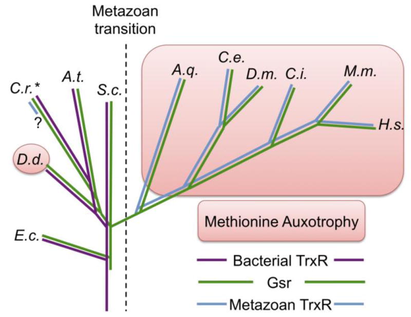
A.t. – Arabidopsis thaliana, A.q. – Amphimedon queenslandica, C.e. – Caenorhabitis elegans, C.i. – Ciona intestinalis, C.r. – Chlamydomonas reinhardtii, D.d. – Dictyostelium discoideum, D.m. – Drosophila melanogaster, E.c. – Escherichia coli, H.s. – Homo sapiens, M.m. – Mus musculus, S.c. – Saccharomyces cerevisiae. The asterisk denotes the single-celled green alga, C. reinhardtii, as representing a sparse but diverse set of non-metazoans that have the metazoan TrxR (blue line with “?”). Predominant others in this group are protozoan intracellular parasites, which might have acquired the gene laterally from a metazoan host. To our knowledge, no similar model yet explains the enigmatic presence of this gene in an alga.
Many non-metazoan species use NADPH to reduce sulfate into sulfide, which is used for de novo biosynthesis of Cys. In these organisms, Cys is a “metabolic hub” that feeds protein synthesis as well as the biosynthesis of all S-containing molecules, including Met, GSH, CoA, SAM, Hcy, LA, and others [112]. Non-sulfate-reducing organisms require an alternative source of Cys. This is provided heterotropically by ingestion of organo-S-compounds, which were produced originally by the sulfate-reducing organisms. Metazoans still use Cys as the metabolic hub for the production of all S-containing molecules, with the notable exception of Met, which was blocked by reversal of the transsulfuration pathway, and Met-cycle intermediates such as SAM and Hcy.
Acquisition of energetic nutrients by early free-living actively-feeding heterotrophs likely provided a collateral abundance of ingested amino acids, including Met and cystine. Indeed, most metazoans require a very similar set of essential amino acids in their diet [137]. This might suggest that the early metazoan lifestyle made many of the costly steps in the biosynthesis of certain amino acids, including sulfate reduction, superfluous. As a consequence, the genes encoding these processes, having become largely unused, could have been lost without substantial adverse consequences (“genetic atrophy”). In non-sulfate-reducing organisms, potential advantages of being able to convert Met → → Cys include the following:
A more demanding role for Cys than Met. Non-metazoans require Met for protein synthesis and synthesis of Met-cycle intermediates. The later are continuously recycled, so loss of protein is the only loss of Met. By contrast Cys is used for protein synthesis and is the precursor for all S-containing metabolites. In sulfate-reducing organisms, Cys is the first organic product of S assimilation and is readily available as the precursor for GSH, CoA, and other S-metabolites. Non-sulfate-reducing organisms, by contrast, obtain S from ingestion of extracellular S-amino acids [129, 138]. Many cells excrete S-containing metabolites, such as GSH (usually as a disulfide), taurine, and H2S, all of which are derived from Cys. Hcy → Cys transsulfuration allows Met, not just cystine, to contribute to secretion of S-metabolites.
Met is a “reductase-efficient” source of Cys; cystine is a more “reductase-demanding” source. In rodents and perhaps all metazoans, dietary Met is available in vast excess over what is needed for incorporation into protein and to support the Met-cycle [118]. Met enters cells ready for use in either protein-synthesis or the Met-cycle. Transsulfuration also allows the thioether-associated S in Met to be transferred to Cys and Cys-based metabolites without disulfide reduction or consumption of NADPH (discussed above). By contrast, cystine requires NADPH-dependent disulfide reduction into Cys before it can be used for protein synthesis or other metabolic pathways. Banerjee and colleagues [115, 116] showed that about half of the S in mammalian cellular GSH pools is Met-derived (discussed above). This supports the idea that the costs of utilizing Met to make Cys must be similar to those of reducing disulfide substrates to make Cys.
Hcy → Cys transsulfuration allows Met to fuel an alternative NADPH-independent disulfide reductase system. The hepatic Met-driven cytosolic disulfide reductase system appears to be sufficiently robust to support liver functions and long-term survival of mice with TrxR1/Gsr-null livers [107, 108]. Having an additional robust NADPH-independent back-up system for the two NADPH-dependent cytosolic disulfide reductase systems could provide survival advantages in cases of severe oxidative or toxic stresses.
Selenium metabolism. Selenoproteins including TrxRs, Gpxs, and others play critical roles in metazoans (see above). Plants do not have selenoproteins, yet they provide the major source of dietary Se for many metazoans in the form of Se-Met and other Se-metabolites, which plants produce as sequestration products [139]. In metazoans, Se-Met transits the Met-cycle and transsulfuration pathway, yielding Sec or selenide for use in the synthesis of selenoproteins [140, 141]. The metazoan transsulfuration direction thus supports selenoproteins.
Signaling. CBS and CSE have not only been flux-reversed, but also partially repurposed [24]. In mammals these two enzymes are the major sources of intracellular H2S production (Figs 4 & 5) [126]. H2S is now recognized as an important signaling molecule in animals, akin to NO and H2O2 [142–144]. It can exert its effects through post- and pre-translational modifications of protein-Cys residues, resulting in protein- or metabolite-S-persulfides (R-SSH; [127, 145, 146]). The potential advantages of advanced signaling by regulated production of H2S, for example to coordinate cellular activities for multicellular life, might have demanded both the reversal and repurposing of the transsulfuration pathway.
Of the five explanations above for why the evolutionary transsulfuration pathway flux-reversal is advantageous for heterotrophs, the last option, Signaling, is the easiest to correlate to unique metazoan needs, and might be the most important attribute that offset potentially detrimental consequences of being Met-auxotrophic. Signaling is important in all cells and many single-celled organisms can communicate (e.g., quorum-sensing). However beyond even the needs in sessile multicellular plants, multicellularity in animals is critically dependent on advanced cell-cell communication, and likely required a quantum leap in signaling. In metazoans, individual cells need to coordinate their differentiation and developmental activities, metabolic activities, reproductive activities, and physical activities. Rapid coordinated motions such as swimming and feeding in primitive metazoans, for example, would require that cellular contractions are synchronized. A classical paradigm is provided by the non-metazoan slime-mold, Dictyostelium, which is considered a ”single-celled social amoeba”. Dictyostelium transits from a free-living single-cell amoeboid stage, to a coordinated motile pseudo-multicellular slug stage, to a pseudo-multicellular anchored stalk stage, in which individual cells differentiate into vegetative or sporulating cell types. Even though this is a more rudimentary coordination of cells than might have been needed in even the earliest metazoans, this life-cycle is dependent on a large number of advanced signaling mechanisms. Indeed, Dictyostelium continues to be an important model for studying signaling mechanisms [147]. In this context, it is intriguing that Dictyostelium is one of the few Met-auxotrophic non-metazoans (see above; Fig. 6). Thus, one might ask: was the loss of Cys → Hcy transsulfuration not simply “genetic atrophy” of an unused function, as suggested above, but rather a selected trait that enhanced signaling capacity and thereby empowered the metazoan transition?
Similar phylogenetic distributions of TrxR-“type” and transsulfuration-“direction”
Intriguingly, metazoans acquired a novel TrxR enzyme via gene duplication and specialization of Gsr [53, 54] apparently coincident with loss of their Cys → Hcy transsulfuration capacity (Fig. 6). Other reviews have been published on the origins and enzymatic differences between the two classes of TrxRs, and the reader is referred to these for such details [48, 52, 54, 62]. Here we will consider why this transition might have occurred.
As the hallmark roles for TrxR are to provide reducing power for RNR and antioxidant enzymes, one might first ask whether the metazoan transition was associated with novel replication- or antioxidant-demands, which might have been better served by the new TrxR. However, there is no obvious reason to suggest that either replication or antioxidant requirements should have shifted at the metazoan transition. As emphasized in the previous section, perhaps the most critical improvement that would have been needed for the transition from protozoan to metazoan was advanced cell-cell communication and signaling.
Roles of the metazoan TrxR1 system in signaling
It is possible that the metazoan TrxR arose specifically for regulation of signaling pathways and not as an improved means of sustaining redox homeostasis. Investigations over the past 15 years have revealed the importance of the mammalian TrxR1 system in redox signaling. Trx1 modulates the oxidation status of protein-Cys residues, and thereby influences the properties or activities of those proteins or enzymes. Included in this, Trx1 plays regulatory roles on many transcription factors, including P53, HIF1, NFκB, AP1, and others [148]. In addition to reducing protein disulfides, however, Trx1 and some Trx-family members, in particular the Trx-related protein of 14 kDa (TRP14), also participate in signaling by regulating protein-S-nitrosylation in NO-signaling and protein-S-persulfidation in H2S-signaling [114, 145, 149]. Trx1 and TRP14 have, in addition to their active site Cys residues, regulatory Cys residues that are subject to posttranslational modification and can alter the activity of the proteins [150–152]. This provides another level of regulation on how Trx1 or TRP14 modulate signaling in cells.
Intracellular signal transduction from many membrane-associated receptors is redox-regulated by mechanisms involving the TrxR1 system. Seminal studies by Finkel’s team and by Rhee’s team showed that ligand binding by the PDGF receptor, EGF receptor, T cell receptor, and other transmembrane receptors was associated with, in addition to auto-phosphorylation of tyrosyl domains on the cytosolic portion of the receptor, activation of a membrane-associated NADPH-oxidase (NOX) complex [153–160]. NOX activation leads to extracellular H2O2 production, which enters the cell through aquaporins [161]. Inside the cell, the H2O2 inactivates ubiquitous protein phosphatases, thereby antagonizing dephosphorylation of the PDGF receptor. Trx1-dependent Prxs counteract this by eliminating the H2O2. Trx1 also re-activates the protein phosphatases, after which the receptor is dephosphorylated and signaling stops [162–166]. This is now considered a general paradigm for signaling by many transmembrane receptors.
More recently, the TrxR1 system has also become recognized as a critical component regulating the catalytic oxidation of some regulatory Cys residues. Winterbourn and colleagues demonstrated that most cellular thiols are poor substrates for oxidation by H2O2. The reasons for this are, first, that the thiols are not reactive enough, so reaction rates with H2O2 are exceptionally slow (~1/mol/sec; [167]). Second, peroxiredoxins (Prx), are highly abundant in the cytosol and have exceptionally nucleophilic active site Cys residues that react with H2O2 at rates ~107-fold faster than most protein-Cys residues [168, 169]. Therefore, it would be nearly impossible for a molecule of H2O2 to evade reacting with a professional peroxidase long enough to oxidize another protein-Cys residue [170]. Dick and colleagues have shown that, for redox regulation of STAT3 protein, Prx2 serves as a catalyst for STAT3 Cys-oxidation. For this, Prx2 rapidly reacts with H2O2 forming an active site sulfenic acid. It then transfers this oxidation-state to regulatory Cys residues on STAT3 [171]. Importantly, the enzymatic activity of the Prxs is regulated by Trx1, and so the TrxR1 system plays a critical role in this regulatory system.
As another example, TrxR1 plays a role in regulating activity of the antioxidant and cytoprotective system driven by nuclear factor erythroid-2-related-2 (Nrf2). Nrf2 provides a robust response to oxidative stress and electrophilic toxins in cells [172]. Either genetic or pharmacologic inhibition of TrxR1 activity induces Nrf2 without inducing oxidative stress, indicating that a part of the stress-sensing regulatory mechanism of Nrf2 is controlled by TrxR1 [49, 173–176]. Finally it should be noted that homozygous disruption of Trx1 or TrxR1 in mouse zygotes results in severe defects in early embryonic patterning without having any detectable impacts on either the level of cellular oxidative stress or cell proliferation [49, 108, 177–181].
The findings above reveal that the mammalian TrxR1 system plays modulatory roles in redox signaling for a diverse spectrum of pathways and responses. Thus, one might ask: was acquisition of these signaling activities critically dependent on the evolution of a new TrxR enzyme, and could this advance in signaling have empowered the metazoan transition?
Closing perspectives: Balancing antioxidant activities and redox signaling
We now know that in hepatocytes and perhaps in many metazoan cell types, cytosolic disulfide reducing power can come from NADPH via TrxR1, from NADPH via Gsr, or from Met catabolism [107]. We also now know that the cytosolic disulfide reducing power required for RNR activity and cell homeostasis can be delivered to its targets by either Trx1 in the absence of GSH or by GSH in the absence of Trx1 [108, 178]. By contrast, a similar TrxR1- Gsr-system redundancy is not evident in signaling. Disruptions of the TrxR1 system, although not substantially impacting either the cellular redox balance or GSH pools, disrupts embryonic patterning, Nrf2 regulation, and other signaling-dependent processes [49]. By contrast, adult animals including humans tolerate severe long-term whole-body pharmacologic depletion of GSH using BSO without evident defects in signaling [182–184]. Perhaps the redox-signaling roles of the TrxR1 system are so crucial in metazoans that its ancestral redox-homeostasis roles must be “excused”, when necessary, for the sake of sustained effective signaling. Gsr, on the other hand, might be more committed to sustaining cellular redox-homeostasis even in the absence of any contribution from TrxR1.
Where would the NADPH-independent system fall in this hypothetical dichotomy? Met delivers its reducing power to the homeostatic machinery via GSH, which argues that it simply provides “back-up support” to the NADPH +Gsr-driven system. On the other hand, for this reducing power to arrive at GSH, the reduced S from Met must transit the metazoan Hcy → Cys transsulfuration pathway, the enzymes, substrates, and products of which supply H2S for signaling activities. Continued studies on the disulfide reductase systems and in the rapidly advancing field of redox signaling will improve our understanding of both the evolution and the physiological roles of each of these systems.
Lastly, these new insights on disulfide reductase systems should be considered in the context of human health and physiology. If the TrxR1-, Gsr-, and Met-driven disulfide reductase systems play distinct roles in signaling and redox homeostasis, then it might be possible to target these pathways to selectively impact specific signaling activities versus homeostatic activities. The implications of this on modulating pathologies including inflammatory diseases, cancers, and neuropathologies could be immense. Almost certainly, there remain other important pathways to be uncovered and there will always be new untested clinical applications arising in the realm of Redox Biology.
Highlights.
All species generate NADPH intracellularly, which fuels disulfide reductase systems
Mammals have an NADPH-independent disulfide reductase system fueled by methionine
Transsulfuation reversal allows Met-fueled reductase system yet makes Met essential
Transsulfuration reversal and a Se-dependent TrxR family co-evolved with metazoans
Metazoan selenoprotein TrxR1 and transsulfuration each support redox signaling
Acknowledgments
Funding to the authors came from the US National Institutes of Health (AG055022 and CA215784); the Montana Agricultural Experiment Station; a stipend from the Wenner-Gren Foundation; The Swedish Cancer Society (Grants CAN 2015/238 and 2015-961), The Swedish Research Council (Grants 2013-03529, 2013-04054, 2014-02603, and 537-2014-360), The Knut and Alice Wallenberg Foundations (Grant KAW 2015.0063), and the Karolinska Institutet.
Abbreviations
- Ahp
alkyl hydroperoxide reductase
- APS
5′-adenylylsulfate
- BSO
buthionine sulfoximine
- CoA
coenzyme A
- Cys
Cysteine
- EGFR
epidermal growth factor receptor
- ER
endoplasmic reticulum
- FTR
ferredoxin-thioredoxin reductase
- Gpx
glutathione peroxidase
- Grx
Glutaredoxin
- GSH
reduced glutathione
- Gsr
glutathione reductase
- GSSG
oxidized glutathione, glutathione-disulfide
- Hcy
Homocysteine
- LA
lipoic Acid
- Lpd
lipoic acid dehydrogenase
- Met
Methionine
- Msr
methionine-sulfoxide reductase
- NAD+
nicotinamide adenine dinucleotide, oxidized
- NADH
nicotinamide adenine dinucleotide, reduced
- NADP+
nicotinamide adenine dinucleotide phosphate, oxidized
- NADPH
nicotinamide adenine dinucleotide phosphate, reduced
- NOX
NADPH-oxidase
- Nrf2
nuclear factor erythroid-2-related-2
- PAPS
3′-phosphoadenosine-5′-phosphosulfate
- PDGFR
platelet-derived growth factor receptor
- PPG
Propargylglycine
- Prx
Peroxiredoxin
- RNR
ribonucleotide reductase
- ROS
reactive oxygen species
- S
Sulfur
- SAH
S-adenosyl homocysteine
- SAM
S-adenosyl methionine
- Se
Selenium
- Sec
Selenocysteine
- TCA
tricarboxylic acid cycle
- Tgr
thioredoxin-glutathione reductase
- TRP14
thioredoxin-related protein of 14 kDa
- Trx
Thioredoxin
- TrxR
thioredoxin reductase
Footnotes
Publisher's Disclaimer: This is a PDF file of an unedited manuscript that has been accepted for publication. As a service to our customers we are providing this early version of the manuscript. The manuscript will undergo copyediting, typesetting, and review of the resulting proof before it is published in its final citable form. Please note that during the production process errors may be discovered which could affect the content, and all legal disclaimers that apply to the journal pertain.
References
- 1.Buchanan BB. Regulation of CO2 assimilation in oxygenic photosynthesis: the ferredoxin/thioredoxin system. Perspective on its discovery, present status, and future development. Arch Biochem Biophys. 1991;288(1):1–9. doi: 10.1016/0003-9861(91)90157-e. [DOI] [PubMed] [Google Scholar]
- 2.Calvin M, Benson AA. The Path of Carbon in Photosynthesis. Science. 1948;107(2784):476–80. doi: 10.1126/science.107.2784.476. [DOI] [PubMed] [Google Scholar]
- 3.Bassham JA, Benson AA, Kay LD, Harris AZ, Wilson AT, Calvin M. The Path of Carbon in Photosynthesis. XXI. The Cyclic Regeneration of Carbon Dioxide Acceptor1. Journal of the American Chemical Society. 1954;76(7):1760–1770. [Google Scholar]
- 4.Fan J, Ye J, Kamphorst JJ, Shlomi T, Thompson CB, Rabinowitz JD. Quantitative flux analysis reveals folate-dependent NADPH production. Nature. 2014;510(7504):298–302. doi: 10.1038/nature13236. [DOI] [PMC free article] [PubMed] [Google Scholar]
- 5.Margittai É, Bánhegyi G. Isocitrate dehydrogenase: A NADPH-generating enzyme in the lumen of the endoplasmic reticulum. Archives of Biochemistry and Biophysics. 2008;471(2):184–190. doi: 10.1016/j.abb.2007.12.017. [DOI] [PubMed] [Google Scholar]
- 6.Gálvez S, Gadal P. On the function of the NADP-dependent isocitrate dehydrogenase isoenzymes in living organisms. Plant Science. 1995;105(1):1–14. [Google Scholar]
- 7.Kruger NJ, von Schaewen A. The oxidative pentose phosphate pathway: structure and organisation. Current Opinion in Plant Biology. 2003;6(3):236–246. doi: 10.1016/s1369-5266(03)00039-6. [DOI] [PubMed] [Google Scholar]
- 8.Ratledge C. The role of malic enzyme as the provider of NADPH in oleaginous microorganisms: a reappraisal and unsolved problems. Biotechnology Letters. 2014;36(8):1557–1568. doi: 10.1007/s10529-014-1532-3. [DOI] [PubMed] [Google Scholar]
- 9.Lewis CA, Parker SJ, Fiske BP, McCloskey D, Gui DY, Green CR, Vokes NI, Feist AM, Vander Heiden MG, Metallo CM. Tracing compartmentalized NADPH metabolism in the cytosol and mitochondria of mammalian cells. Mol Cell. 2014;55(2):253–63. doi: 10.1016/j.molcel.2014.05.008. [DOI] [PMC free article] [PubMed] [Google Scholar]
- 10.Meimaridou E, Kowalczyk J, Guasti L, Hughes CR, Wagner F, Frommolt P, Nurnberg P, Mann NP, Banerjee R, Saka HN, Chapple JP, King PJ, Clark AJ, Metherell LA. Mutations in NNT encoding nicotinamide nucleotide transhydrogenase cause familial glucocorticoid deficiency. Nat Genet. 2012;44(7):740–2. doi: 10.1038/ng.2299. [DOI] [PMC free article] [PubMed] [Google Scholar]
- 11.Yin F, Sancheti H, Cadenas E. Silencing of nicotinamide nucleotide transhydrogenase impairs cellular redox homeostasis and energy metabolism in PC12 cells. Biochim Biophys Acta. 2012;1817(3):401–9. doi: 10.1016/j.bbabio.2011.12.004. [DOI] [PMC free article] [PubMed] [Google Scholar]
- 12.Cody GD. Transition Metal Sulfides and the Origins of Metabolism. Annual Review of Earth and Planetary Sciences. 2004;32(1):569–599. [Google Scholar]
- 13.Wächtershäuser G. Before enzymes and templates: theory of surface metabolism. Microbiological Reviews. 1988;52(4):452–484. doi: 10.1128/mr.52.4.452-484.1988. [DOI] [PMC free article] [PubMed] [Google Scholar]
- 14.McGlynn SE, Kanik I, Russell MJ. Peptide and RNA contributions to iron–sulphur chemical gardens as life’s first inorganic compartments, catalysts, capacitors and condensers. Philosophical Transactions of the Royal Society A: Mathematical, Physical and Engineering Sciences. 2012;370(1969):3007–3022. doi: 10.1098/rsta.2011.0211. [DOI] [PubMed] [Google Scholar]
- 15.Russell MJ, Hall AJ. The emergence of life from iron monosulphide bubbles at a submarine hydrothermal redox and pH front. Journal of the Geological Society. 1997;154(3):377–402. doi: 10.1144/gsjgs.154.3.0377. [DOI] [PubMed] [Google Scholar]
- 16.Summers DP. Ammonia Formation By The Reduction Of Nitrite/Nitrate By Fes: Ammonia Formation Under Acidic Conditions. Origins of Life and Evolution of Biospheres. 2005;35(4):299–312. doi: 10.1007/s11084-005-2040-1. [DOI] [PubMed] [Google Scholar]
- 17.Huber C, Wächtershäuser G. Peptides by Activation of Amino Acids with CO on (Ni,Fe)S Surfaces: Implications for the Origin of Life. Science. 1998;281(5377):670–672. doi: 10.1126/science.281.5377.670. [DOI] [PubMed] [Google Scholar]
- 18.Shipp JA, Gould IR, Shock EL, Williams LB, Hartnett HE. Sphalerite is a geochemical catalyst for carbon hydrogen bond activation. Proceedings of the National Academy of Sciences. 2014;111(32):11642–11645. doi: 10.1073/pnas.1324222111. [DOI] [PMC free article] [PubMed] [Google Scholar]
- 19.Huber C, Wachtershauser G. Activated acetic acid by carbon fixation on (Fe,Ni)S under primordial conditions. Science. 1997;276(5310):245–7. doi: 10.1126/science.276.5310.245. [DOI] [PubMed] [Google Scholar]
- 20.Wächtershäuser G. Pyrite Formation, the First Energy Source for Life: a Hypothesis. Systematic and Applied Microbiology. 1988;10(3):207–210. [Google Scholar]
- 21.Postgate JR. The Sulphate-Reducing Bacteria. 2. Cambridge University Press; Cambridge: 1984. [Google Scholar]
- 22.Archer C, Vance D. Coupled Fe and S isotope evidence for Archean microbial Fe(III) and sulfate reduction. Geology. 2006;34(3):153–156. [Google Scholar]
- 23.Lovley DR. Microbial FE(III) reduction in subsurface environments. 1997 [Google Scholar]
- 24.Olson KR, Straub KD. The Role of Hydrogen Sulfide in Evolution and the Evolution of Hydrogen Sulfide in Metabolism and Signaling. Physiology (Bethesda) 2016;31(1):60–72. doi: 10.1152/physiol.00024.2015. [DOI] [PubMed] [Google Scholar]
- 25.Cortese-Krott MM, Koning A, Kuhnle GGC, Nagy P, Bianco CL, Pasch A, Wink DA, Fukuto JM, Jackson AA, van Goor H, Olson KR, Feelisch M. The Reactive Species Interactome: Evolutionary Emergence, Biological Significance, and Opportunities for Redox Metabolomics and Personalized Medicine. Antioxid Redox Signal. 2017;27(10):684–712. doi: 10.1089/ars.2017.7083. [DOI] [PMC free article] [PubMed] [Google Scholar]
- 26.Bekker A, Holland HD, Wang PL, Rumble D, Iii, Stein HJ, Hannah JL, Coetzee LL, Beukes NJ. Dating the rise of atmospheric oxygen. Nature. 2004;427:117. doi: 10.1038/nature02260. [DOI] [PubMed] [Google Scholar]
- 27.Bergman N, Lenton TM, Watson A. COPSE: A new model of biogeochemical cycling over Phanerozoic time. 2004 [Google Scholar]
- 28.Berner RA. GEOCARBSULF: A combined model for Phanerozoic atmospheric O2 and CO2. Geochimica et Cosmochimica Acta. 2006;70(23):5653–5664. [Google Scholar]
- 29.Hondal RJ, Marino SM, Gladyshev VN. Selenocysteine in thiol/disulfide-like exchange reactions. Antioxid Redox Signal. 2013;18(13):1675–89. doi: 10.1089/ars.2012.5013. [DOI] [PMC free article] [PubMed] [Google Scholar]
- 30.Reich HJ, Hondal RJ. Why Nature Chose Selenium. ACS chemical biology. 2016;11(4):821–41. doi: 10.1021/acschembio.6b00031. [DOI] [PubMed] [Google Scholar]
- 31.Fomenko DE, Gladyshev VN. Comparative genomics of thiol oxidoreductases reveals widespread and essential functions of thiol-based redox control of cellular processes. Antioxid Redox Signal. 2012;16(3):193–201. doi: 10.1089/ars.2011.3980. [DOI] [PMC free article] [PubMed] [Google Scholar]
- 32.Fomenko DE, Marino SM, Gladyshev VN. Functional diversity of cysteine residues in proteins and unique features of catalytic redox-active cysteines in thiol oxidoreductases. Molecules and cells. 2008;26(3):228–35. [PMC free article] [PubMed] [Google Scholar]
- 33.Lazcano A, Guerrero R, Margulis L, Oró J. The evolutionary transition from RNA to DNA in early cells. Journal of molecular evolution. 1988;27(4):283–290. doi: 10.1007/BF02101189. [DOI] [PubMed] [Google Scholar]
- 34.Duncan BK, Miller JH. Mutagenic deamination of cytosine residues in DNA. Nature. 1980;287:560. doi: 10.1038/287560a0. [DOI] [PubMed] [Google Scholar]
- 35.Nordlund P, Reichard P. Ribonucleotide reductases. Annu Rev Biochem. 2006;75:681–706. doi: 10.1146/annurev.biochem.75.103004.142443. [DOI] [PubMed] [Google Scholar]
- 36.Logan DT, Andersson J, Sjoberg BM, Nordlund P. A glycyl radical site in the crystal structure of a class III ribonucleotide reductase. Science. 1999;283(5407):1499–504. doi: 10.1126/science.283.5407.1499. [DOI] [PubMed] [Google Scholar]
- 37.Stubbe J, van Der Donk WA. Protein Radicals in Enzyme Catalysis. Chem Rev. 1998;98(2):705–762. doi: 10.1021/cr9400875. [DOI] [PubMed] [Google Scholar]
- 38.Stubbe J, Ator M, Krenitsky T. Mechanism of ribonucleoside diphosphate reductase from Escherichia coli. Evidence for 3′-C--H bond cleavage. J Biol Chem. 1983;258(3):1625–31. [PubMed] [Google Scholar]
- 39.Kolberg M, Strand KR, Graff P, Andersson KK. Structure, function, and mechanism of ribonucleotide reductases. Biochim Biophys Acta. 2004;1699(1–2):1–34. doi: 10.1016/j.bbapap.2004.02.007. [DOI] [PubMed] [Google Scholar]
- 40.Thelander L, Reichard P. Reduction of ribonucleotides. Annu Rev Biochem. 1979;48:133–58. doi: 10.1146/annurev.bi.48.070179.001025. [DOI] [PubMed] [Google Scholar]
- 41.Zahedi Avval F, Holmgren A. Molecular mechanisms of thioredoxin and glutaredoxin as hydrogen donors for Mammalian s phase ribonucleotide reductase. J Biol Chem. 2009;284(13):8233–40. doi: 10.1074/jbc.M809338200. [DOI] [PMC free article] [PubMed] [Google Scholar]
- 42.Prinz WA, Aslund F, Holmgren A, Beckwith J. The role of the thioredoxin and glutaredoxin pathways in reducing protein disulfide bonds in the Escherichia coli cytoplasm. J Biol Chem. 1997;272(25):15661–7. doi: 10.1074/jbc.272.25.15661. [DOI] [PubMed] [Google Scholar]
- 43.Couto N, Wood J, Barber J. The role of glutathione reductase and related enzymes on cellular redox homoeostasis network. Free Radic Biol Med. 2016;95:27–42. doi: 10.1016/j.freeradbiomed.2016.02.028. [DOI] [PubMed] [Google Scholar]
- 44.Holmgren A, Sengupta R. The use of thiols by ribonucleotide reductase. Free Radic Biol Med. 2010;49(11):1617–28. doi: 10.1016/j.freeradbiomed.2010.09.005. [DOI] [PubMed] [Google Scholar]
- 45.Holmgren A. Hydrogen donor system for Escherichia coli ribonucleoside-diphosphate reductase dependent upon glutathione. Proc Natl Acad Sci U S A. 1976;73(7):2275–9. doi: 10.1073/pnas.73.7.2275. [DOI] [PMC free article] [PubMed] [Google Scholar]
- 46.Sengupta R, Holmgren A. Thioredoxin and glutaredoxin-mediated redox regulation of ribonucleotide reductase. World journal of biological chemistry. 2014;5(1):68–74. doi: 10.4331/wjbc.v5.i1.68. [DOI] [PMC free article] [PubMed] [Google Scholar]
- 47.Laurent TC, Moore EC, Reichard P. Enzymatic Synthesis Of Deoxyribonucleotides. Iv. Isolation And Characterization Of Thioredoxin, The Hydrogen Donor From Escherichia Coli B. J Biol Chem. 1964;239:3436–44. [PubMed] [Google Scholar]
- 48.Arnér ESJ. Focus on mammalian thioredoxin reductases--important selenoproteins with versatile functions. Biochim Biophys Acta. 2009;1790(6):495–526. doi: 10.1016/j.bbagen.2009.01.014. [DOI] [PubMed] [Google Scholar]
- 49.Dagnell M, Schmidt EE, Arner ESJ. The A to Z of modulated cell patterning by mammalian thioredoxin reductases. Free Radic Biol Med. 2018;115:484–496. doi: 10.1016/j.freeradbiomed.2017.12.029. [DOI] [PMC free article] [PubMed] [Google Scholar]
- 50.Dym O, Eisenberg D. Sequence-structure analysis of FAD-containing proteins. Protein Sci. 2001;10(9):1712–28. doi: 10.1110/ps.12801. [DOI] [PMC free article] [PubMed] [Google Scholar]
- 51.Zhong L, Arner ES, Ljung J, Aslund F, Holmgren A. Rat and calf thioredoxin reductase are homologous to glutathione reductase with a carboxyl-terminal elongation containing a conserved catalytically active penultimate selenocysteine residue. J Biol Chem. 1998;273(15):8581–91. doi: 10.1074/jbc.273.15.8581. [DOI] [PubMed] [Google Scholar]
- 52.Williams CH, Arscott LD, Muller S, Lennon BW, Ludwig ML, Wang PF, Veine DM, Becker K, Schirmer RH. Thioredoxin reductase two modes of catalysis have evolved. Eur J Biochem. 2000;267(20):6110–7. doi: 10.1046/j.1432-1327.2000.01702.x. [DOI] [PubMed] [Google Scholar]
- 53.Mariotti M, Ridge PG, Zhang Y, Lobanov AV, Pringle TH, Guigo R, Hatfield DL, Gladyshev VN. Composition and evolution of the vertebrate and mammalian selenoproteomes. PLoS ONE. 2012;7(3):e33066. doi: 10.1371/journal.pone.0033066. [DOI] [PMC free article] [PubMed] [Google Scholar]
- 54.Novoselov SV, Gladyshev VN. Non-animal origin of animal thioredoxin reductases: implications for selenocysteine evolution and evolution of protein function through carboxy-terminal extensions. Protein Sci. 2003;12(2):372–8. doi: 10.1110/ps.0226503. [DOI] [PMC free article] [PubMed] [Google Scholar]
- 55.Novoselov SV, Rao M, Onoshko NV, Zhi H, Kryukov GV, Xiang Y, Weeks DP, Hatfield DL, Gladyshev VN. Selenoproteins and selenocysteine insertion system in the model plant cell system, Chlamydomonas reinhardtii. Embo J. 2002;21(14):3681–93. doi: 10.1093/emboj/cdf372. [DOI] [PMC free article] [PubMed] [Google Scholar]
- 56.Fomenko DE, Xing W, Adair BM, Thomas DJ, Gladyshev VN. High-throughput identification of catalytic redox-active cysteine residues. Science. 2007;315(5810):387–9. doi: 10.1126/science.1133114. [DOI] [PubMed] [Google Scholar]
- 57.Kryukov GV, Castellano S, Novoselov SV, Lobanov AV, Zehtab O, Guigo R, Gladyshev VN. Characterization of mammalian selenoproteomes. Science. 2003;300(5624):1439–43. doi: 10.1126/science.1083516. [DOI] [PubMed] [Google Scholar]
- 58.Steinbrenner H, Sies H. Protection against reactive oxygen species by selenoproteins. Biochim Biophys Acta. 2009;1790(11):1478–85. doi: 10.1016/j.bbagen.2009.02.014. [DOI] [PubMed] [Google Scholar]
- 59.Rao M, Carlson BA, Novoselov SV, Weeks DP, Gladyshev VN, Hatfield DL. Chlamydomonas reinhardtii selenocysteine tRNA[Ser]Sec. RNA. 2003;9(8):923–30. doi: 10.1261/rna.5510503. [DOI] [PMC free article] [PubMed] [Google Scholar]
- 60.Stock T, Rother M. Selenoproteins in Archaea and Gram-positive bacteria. Biochim Biophys Acta. 2009;1790(11):1520–32. doi: 10.1016/j.bbagen.2009.03.022. [DOI] [PubMed] [Google Scholar]
- 61.Ingold I, Berndt C, Schmitt S, Doll S, Poschmann G, Buday K, Roveri A, Peng X, Porto Freitas F, Seibt T, Mehr L, Aichler M, Walch A, Lamp D, Jastroch M, Miyamoto S, Wurst W, Ursini F, Arner ESJ, Fradejas-Villar N, Schweizer U, Zischka H, Friedmann Angeli JP, Conrad M. Selenium Utilization by GPX4 Is Required to Prevent Hydroperoxide-Induced Ferroptosis. Cell. 2018;172(3):409–422. e21. doi: 10.1016/j.cell.2017.11.048. [DOI] [PubMed] [Google Scholar]
- 62.Schmidt EE. DNA Replication in Animal Systems Lacking Thioredoxin Reductase I. In: Seligmann H, editor. DNA Replication-Current Advances. InTechOpen.com; 2011. [Google Scholar]
- 63.Salinas G, Comini MA. Alternative Thiol-Based Redox Systems. Antioxid Redox Signal. 2018;28(6):407–409. doi: 10.1089/ars.2017.7464. [DOI] [PubMed] [Google Scholar]
- 64.Perera VR, Newton GL, Pogliano K. Bacillithiol: a key protective thiol in Staphylococcus aureus. Expert review of anti-infective therapy. 2015;13(9):1089–1107. doi: 10.1586/14787210.2015.1064309. [DOI] [PMC free article] [PubMed] [Google Scholar]
- 65.Imber M, Huyen NTT, Pietrzyk-Brzezinska AJ, Loi VV, Hillion M, Bernhardt J, Tharichen L, Kolsek K, Saleh M, Hamilton CJ, Adrian L, Grater F, Wahl MC, Antelmann H. Protein S-Bacillithiolation Functions in Thiol Protection and Redox Regulation of the Glyceraldehyde-3-Phosphate Dehydrogenase Gap in Staphylococcus aureus Under Hypochlorite Stress. Antioxid Redox Signal. 2018;28(6):410–430. doi: 10.1089/ars.2016.6897. [DOI] [PMC free article] [PubMed] [Google Scholar]
- 66.Chandrangsu P, Loi VV, Antelmann H, Helmann JD. The Role of Bacillithiol in Gram-Positive Firmicutes. Antioxid Redox Signal. 2018;28(6):445–462. doi: 10.1089/ars.2017.7057. [DOI] [PMC free article] [PubMed] [Google Scholar]
- 67.Si M, Zhao C, Zhang B, Wei D, Chen K, Yang X, Xiao H, Shen X. Overexpression of Mycothiol Disulfide Reductase Enhances Corynebacterium glutamicum Robustness by Modulating Cellular Redox Homeostasis and Antioxidant Proteins under Oxidative Stress. Scientific reports. 2016;6:29491. doi: 10.1038/srep29491. [DOI] [PMC free article] [PubMed] [Google Scholar]
- 68.Reyes AM, Pedre B, De Armas MI, Tossounian MA, Radi R, Messens J, Trujillo M. Chemistry and Redox Biology of Mycothiol. Antioxid Redox Signal. 2018;28(6):487–504. doi: 10.1089/ars.2017.7074. [DOI] [PubMed] [Google Scholar]
- 69.delCardayre SB, Stock KP, Newton GL, Fahey RC, Davies JE. Coenzyme A disulfide reductase, the primary low molecular weight disulfide reductase from Staphylococcus aureus. Purification and characterization of the native enzyme. J Biol Chem. 1998;273(10):5744–51. doi: 10.1074/jbc.273.10.5744. [DOI] [PubMed] [Google Scholar]
- 70.Mallett TC, Wallen JR, Karplus PA, Sakai H, Tsukihara T, Claiborne A. Structure of coenzyme A-disulfide reductase from Staphylococcus aureus at 1.54 A resolution. Biochemistry. 2006;45(38):11278–89. doi: 10.1021/bi061139a. [DOI] [PMC free article] [PubMed] [Google Scholar]
- 71.Fairlamb AH, Cerami A. Metabolism and Functions of Trypanothione in the Kinetoplastida. Annual Review of Microbiology. 1992;46(1):695–729. doi: 10.1146/annurev.mi.46.100192.003403. [DOI] [PubMed] [Google Scholar]
- 72.Krauth-Siegel LR, Comini MA, Schlecker T. The Trypanothione System. In: Flohé L, Harris JR, editors. Peroxiredoxin Systems: Structures and Functions. Springer Netherlands; Dordrecht: 2007. pp. 231–251. [DOI] [PubMed] [Google Scholar]
- 73.Manta B, Bonilla M, Fiestas L, Sturlese M, Salinas G, Bellanda M, Comini MA. Polyamine-Based Thiols in Trypanosomatids: Evolution, Protein Structural Adaptations, and Biological Functions. Antioxid Redox Signal. 2018;28(6):463–486. doi: 10.1089/ars.2017.7133. [DOI] [PubMed] [Google Scholar]
- 74.Otero L, Bonilla M, Protasio AV, Fernandez C, Gladyshev VN, Salinas G. Thioredoxin and glutathione systems differ in parasitic and free-living platyhelminths. BMC Genomics. 2010;11:237. doi: 10.1186/1471-2164-11-237. [DOI] [PMC free article] [PubMed] [Google Scholar]
- 75.Jaeger T, Flohe L. The thiol-based redox networks of pathogens: unexploited targets in the search for new drugs. Biofactors. 2006;27(1–4):109–20. doi: 10.1002/biof.5520270110. [DOI] [PubMed] [Google Scholar]
- 76.Mohring F, Pretzel J, Jortzik E, Becker K. The redox systems of Plasmodium falciparum and Plasmodium vivax: comparison, in silico analyses and inhibitor studies. Curr Med Chem. 2014;21(15):1728–56. doi: 10.2174/0929867321666131201144612. [DOI] [PubMed] [Google Scholar]
- 77.Williams DL, Bonilla M, Gladyshev VN, Salinas G. Thioredoxin glutathione reductase-dependent redox networks in platyhelminth parasites. Antioxid Redox Signal. 2013;19(7):735–45. doi: 10.1089/ars.2012.4670. [DOI] [PMC free article] [PubMed] [Google Scholar]
- 78.Lu J, Vlamis-Gardikas A, Kandasamy K, Zhao R, Gustafsson TN, Engstrand L, Hoffner S, Engman L, Holmgren A. Inhibition of bacterial thioredoxin reductase: an antibiotic mechanism targeting bacteria lacking glutathione. Faseb J. 2013;27(4):1394–403. doi: 10.1096/fj.12-223305. [DOI] [PubMed] [Google Scholar]
- 79.Lu J, Vodnala SK, Gustavsson AL, Gustafsson TN, Sjoberg B, Johansson HA, Kumar S, Tjernberg A, Engman L, Rottenberg ME, Holmgren A. Ebsulfur is a benzisothiazolone cytocidal inhibitor targeting the trypanothione reductase of Trypanosoma brucei. J Biol Chem. 2013;288(38):27456–68. doi: 10.1074/jbc.M113.495101. [DOI] [PMC free article] [PubMed] [Google Scholar]
- 80.Moore EC, Reichard P, Thelander L. Enzymatic Synthesis Of Deoxyribonucleotides.V. Purification And Properties Of Thioredoxin Reductase From Escherichia Coli B. J Biol Chem. 1964;239:3445–52. [PubMed] [Google Scholar]
- 81.Holmgren A. Glutathione-dependent synthesis of deoxyribonucleotides. Characterization of the enzymatic mechanism of Escherichia coli glutaredoxin. J Biol Chem. 1979;254(9):3672–8. [PubMed] [Google Scholar]
- 82.Feeney MA, Veeravalli K, Boyd D, Gon S, Faulkner MJ, Georgiou G, Beckwith J. Repurposing lipoic acid changes electron flow in two important metabolic pathways of Escherichia coli. Proc Natl Acad Sci U S A. 2011;108(19):7991–6. doi: 10.1073/pnas.1105429108. [DOI] [PMC free article] [PubMed] [Google Scholar]
- 83.Ritz D, Lim J, Reynolds CM, Poole LB, Beckwith J. Conversion of a Peroxiredoxin into a Disulfide Reductase by a Triplet Repeat Expansion. Science. 2001;294(5540):158–160. doi: 10.1126/science.1063143. [DOI] [PubMed] [Google Scholar]
- 84.Yamamoto Y, Ritz D, Planson AG, Jonsson TJ, Faulkner MJ, Boyd D, Beckwith J, Poole LB. Mutant AhpC peroxiredoxins suppress thiol-disulfide redox deficiencies and acquire deglutathionylating activity. Mol Cell. 2008;29(1):36–45. doi: 10.1016/j.molcel.2007.11.029. [DOI] [PMC free article] [PubMed] [Google Scholar]
- 85.Feeney MA, Veeravalli K, Boyd D, Gon S, Faulkner MJ, Georgiou G, Beckwith J. Repurposing lipoic acid changes electron flow in two important metabolic pathways of Escherichia coli. Proceedings of the National Academy of Sciences. 2011;108(19):7991–7996. doi: 10.1073/pnas.1105429108. [DOI] [PMC free article] [PubMed] [Google Scholar]
- 86.Pontarin G, Fijolek A, Pizzo P, Ferraro P, Rampazzo C, Pozzan T, Thelander L, Reichard PA, Bianchi V. Ribonucleotide reduction is a cytosolic process in mammalian cells independently of DNA damage. Proceedings of the National Academy of Sciences of the United States of America. 2008;105(46):17801–17806. doi: 10.1073/pnas.0808198105. [DOI] [PMC free article] [PubMed] [Google Scholar]
- 87.Mulliez E, Ollagnier S, Fontecave M, Eliasson R, Reichard P. Formate is the hydrogen donor for the anaerobic ribonucleotide reductase from Escherichia coli. Proceedings of the National Academy of Sciences of the United States of America. 1995;92(19):8759–8762. doi: 10.1073/pnas.92.19.8759. [DOI] [PMC free article] [PubMed] [Google Scholar]
- 88.Wei Y, Funk MA, Rosado LA, Baek J, Drennan CL, Stubbe J. The class III ribonucleotide reductase from Neisseria bacilliformis can utilize thioredoxin as a reductant. Proceedings of the National Academy of Sciences. 2014;111(36):E3756–E3765. doi: 10.1073/pnas.1414396111. [DOI] [PMC free article] [PubMed] [Google Scholar]
- 89.Toledano MB, Kumar C, Le Moan N, Spector D, Tacnet F. The system biology of thiol redox system in Escherichia coli and yeast: Differential functions in oxidative stress, iron metabolism and DNA synthesis. FEBS Letters. 2007;581(19):3598–3607. doi: 10.1016/j.febslet.2007.07.002. [DOI] [PubMed] [Google Scholar]
- 90.Trotter EW, Grant CM. Non-reciprocal regulation of the redox state of the glutathione-glutaredoxin and thioredoxin systems. EMBO Rep. 2003;4(2):184–8. doi: 10.1038/sj.embor.embor729. [DOI] [PMC free article] [PubMed] [Google Scholar]
- 91.Muller EG. A glutathione reductase mutant of yeast accumulates high levels of oxidized glutathione and requires thioredoxin for growth. Mol Biol Cell. 1996;7(11):1805–13. doi: 10.1091/mbc.7.11.1805. [DOI] [PMC free article] [PubMed] [Google Scholar]
- 92.Koc A, Mathews CK, Wheeler LJ, Gross MK, Merrill GF. Thioredoxin is required for deoxyribonucleotide pool maintenance during S phase. J Biol Chem. 2006;281(22):15058–63. doi: 10.1074/jbc.M601968200. [DOI] [PubMed] [Google Scholar]
- 93.Camier S, Ma E, Leroy C, Pruvost A, Toledano M, Marsolier-Kergoat MC. Visualization of ribonucleotide reductase catalytic oxidation establishes thioredoxins as its major reductants in yeast. Free Radic Biol Med. 2007;42(7):1008–16. doi: 10.1016/j.freeradbiomed.2006.12.027. [DOI] [PubMed] [Google Scholar]
- 94.Wolosiuk RA, Buchanan BB. Thioredoxin and glutathione regulate photosynthesis in chloroplasts. Nature. 1977;266:565. [Google Scholar]
- 95.Schurmann P, Buchanan BB. The ferredoxin/thioredoxin system of oxygenic photosynthesis. Antioxid Redox Signal. 2008;10(7):1235–74. doi: 10.1089/ars.2007.1931. [DOI] [PubMed] [Google Scholar]
- 96.Buchanan BB, Holmgren A, Jacquot JP, Scheibe R. Fifty years in the thioredoxin field and a bountiful harvest. Biochim Biophys Acta. 2012;1820(11):1822–9. doi: 10.1016/j.bbagen.2012.07.006. [DOI] [PubMed] [Google Scholar]
- 97.Ponsero AJ, Igbaria A, Darch MA, Miled S, Outten CE, Winther JR, Palais G, D’Autreaux B, Delaunay-Moisan A, Toledano MB. Endoplasmic Reticulum Transport of Glutathione by Sec61 Is Regulated by Ero1 and Bip. Mol Cell. 2017;67(6):962–973 e5. doi: 10.1016/j.molcel.2017.08.012. [DOI] [PMC free article] [PubMed] [Google Scholar]
- 98.Martensson J, Lai JC, Meister A. High-affinity transport of glutathione is part of a multicomponent system essential for mitochondrial function. Proc Natl Acad Sci U S A. 1990;87(18):7185–9. doi: 10.1073/pnas.87.18.7185. [DOI] [PMC free article] [PubMed] [Google Scholar]
- 99.Albrecht SC, Barata AG, Grosshans J, Teleman AA, Dick TP. In vivo mapping of hydrogen peroxide and oxidized glutathione reveals chemical and regional specificity of redox homeostasis. Cell Metab. 2011;14(6):819–29. doi: 10.1016/j.cmet.2011.10.010. [DOI] [PubMed] [Google Scholar]
- 100.Cejudo FJ, Ferrandez J, Cano B, Puerto-Galan L, Guinea M. The function of the NADPH thioredoxin reductase C-2-Cys peroxiredoxin system in plastid redox regulation and signalling. FEBS Lett. 2012;586(18):2974–80. doi: 10.1016/j.febslet.2012.07.003. [DOI] [PubMed] [Google Scholar]
- 101.Kelner MJ, Montoya MA. Structural organization of the human glutathione reductase gene: determination of correct cDNA sequence and identification of a mitochondrial leader sequence. Biochem Biophys Res Commun. 2000;269(2):366–8. doi: 10.1006/bbrc.2000.2267. [DOI] [PubMed] [Google Scholar]
- 102.Margittai E, Enyedi B, Csala M, Geiszt M, Banhegyi G. Composition of the redox environment of the endoplasmic reticulum and sources of hydrogen peroxide. Free Radic Biol Med. 2015;83:331–40. doi: 10.1016/j.freeradbiomed.2015.01.032. [DOI] [PubMed] [Google Scholar]
- 103.Kurooka H, Kato K, Minoguchi S, Takahashi Y, Ikeda J, Habu S, Osawa N, Buchberg AM, Moriwaki K, Shisa H, Honjo T. Cloning and characterization of the nucleoredoxin gene that encodes a novel nuclear protein related to thioredoxin. Genomics. 1997;39(3):331–9. doi: 10.1006/geno.1996.4493. [DOI] [PubMed] [Google Scholar]
- 104.Karimpour S, Lou J, Lin LL, Rene LM, Lagunas L, Ma X, Karra S, Bradbury CM, Markovina S, Goswami PC, Spitz DR, Hirota K, Kalvakolanu DV, Yodoi J, Gius D. Thioredoxin reductase regulates AP-1 activity as well as thioredoxin nuclear localization via active cysteines in response to ionizing radiation. Oncogene. 2002;21(41):6317–27. doi: 10.1038/sj.onc.1205749. [DOI] [PubMed] [Google Scholar]
- 105.Hirota K, Matsui M, Murata M, Takashima Y, Cheng FS, Itoh T, Fukuda K, Yodoi J. Nucleoredoxin, glutaredoxin, and thioredoxin differentially regulate NF-kappaB, AP-1, and CREB activation in HEK293 cells. Biochem Biophys Res Commun. 2000;274(1):177–82. doi: 10.1006/bbrc.2000.3106. [DOI] [PubMed] [Google Scholar]
- 106.Pontarin G, Fijolek A, Pizzo P, Ferraro P, Rampazzo C, Pozzan T, Thelander L, Reichard PA, Bianchi V. Ribonucleotide reduction is a cytosolic process in mammalian cells independently of DNA damage. Proc Natl Acad Sci U S A. 2008;105(46):17801–6. doi: 10.1073/pnas.0808198105. [DOI] [PMC free article] [PubMed] [Google Scholar]
- 107.Eriksson S, Prigge JR, Talago EA, Arner ES, Schmidt EE. Dietary methionine can sustain cytosolic redox homeostasis in the mouse liver. Nature communications. 2015;6:6479. doi: 10.1038/ncomms7479. [DOI] [PMC free article] [PubMed] [Google Scholar]
- 108.Prigge JR, Coppo L, Martin SS, Ogata F, Miller CG, Bruschwein MD, Orlicky DJ, Shearn CT, Kundert JA, Lytchier J, Herr AE, Mattsson A, Taylor MP, Gustafsson TN, Arner ESJ, Holmgren A, Schmidt EE. Hepatocyte Hyperproliferation upon Liver-Specific Co-disruption of Thioredoxin-1, Thioredoxin Reductase-1, and Glutathione Reductase. Cell reports. 2017;19(13):2771–2781. doi: 10.1016/j.celrep.2017.06.019. [DOI] [PMC free article] [PubMed] [Google Scholar]
- 109.Su D, Novoselov SV, Sun QA, Moustafa ME, Zhou Y, Oko R, Hatfield DL, Gladyshev VN. Mammalian selenoprotein thioredoxin-glutathione reductase. Roles in disulfide bond formation and sperm maturation. J Biol Chem. 2005;280(28):26491–8. doi: 10.1074/jbc.M503638200. [DOI] [PubMed] [Google Scholar]
- 110.Rogers LK, Bates CM, Welty SE, Smith CV. Diquat induces renal proximal tubule injury in glutathione reductase-deficient mice. Toxicol Appl Pharmacol. 2006;217(3):289–98. doi: 10.1016/j.taap.2006.08.012. [DOI] [PubMed] [Google Scholar]
- 111.Zhang L, Cheng Q, Zhang L, Wang Y, Merrill GF, Ilani T, Fass D, Arner ESJ, Zhang J. Serum thioredoxin reductase is highly increased in mice with hepatocellular carcinoma and its activity is restrained by several mechanisms. Free Radic Biol Med. 2016;99:426–435. doi: 10.1016/j.freeradbiomed.2016.08.028. [DOI] [PubMed] [Google Scholar]
- 112.Lu SC. Glutathione synthesis. Biochim Biophys Acta. 2013;1830(5):3143–53. doi: 10.1016/j.bbagen.2012.09.008. [DOI] [PMC free article] [PubMed] [Google Scholar]
- 113.Lu SC. Regulation of glutathione synthesis. Mol Aspects Med. 2009;30(1–2):42–59. doi: 10.1016/j.mam.2008.05.005. [DOI] [PMC free article] [PubMed] [Google Scholar]
- 114.Pader I, Sengupta R, Cebula M, Xu J, Lundberg JO, Holmgren A, Johansson K, Arner ES. Thioredoxin-related protein of 14 kDa is an efficient L-cystine reductase and S-denitrosylase. Proc Natl Acad Sci U S A. 2014;111(19):6964–9. doi: 10.1073/pnas.1317320111. [DOI] [PMC free article] [PubMed] [Google Scholar]
- 115.Mosharov E, Cranford MR, Banerjee R. The quantitatively important relationship between homocysteine metabolism and glutathione synthesis by the transsulfuration pathway and its regulation by redox changes. Biochemistry. 2000;39(42):13005–11. doi: 10.1021/bi001088w. [DOI] [PubMed] [Google Scholar]
- 116.Vitvitsky V, Mosharov E, Tritt M, Ataullakhanov F, Banerjee R. Redox regulation of homocysteine-dependent glutathione synthesis. Redox Rep. 2003;8(1):57–63. doi: 10.1179/135100003125001260. [DOI] [PubMed] [Google Scholar]
- 117.Stipanuk MH. Role of the liver in regulation of body cysteine and taurine levels: a brief review. Neurochemical research. 2004;29(1):105–10. doi: 10.1023/b:nere.0000010438.40376.c9. [DOI] [PubMed] [Google Scholar]
- 118.Finkelstein JD. Methionine metabolism in mammals. The Journal of nutritional biochemistry. 1990;1(5):228–237. doi: 10.1016/0955-2863(90)90070-2. [DOI] [PubMed] [Google Scholar]
- 119.Finkelstein JD, Martin JJ. Methionine metabolism in mammals. Distribution of homocysteine between competing pathways. Journal of Biological Chemistry. 1984;259(15):9508–13. [PubMed] [Google Scholar]
- 120.Beatty PW, Reed DJ. Involvement of the cystathionine pathway in the biosynthesis of glutathione by isolated rat hepatocytes. Arch Biochem Biophys. 1980;204(1):80–7. doi: 10.1016/0003-9861(80)90009-0. [DOI] [PubMed] [Google Scholar]
- 121.Martinov MV, Vitvitsky VM, Banerjee R, Ataullakhanov FI. The logic of the hepatic methionine metabolic cycle. Biochim Biophys Acta. 2010;1804(1):89–96. doi: 10.1016/j.bbapap.2009.10.004. [DOI] [PMC free article] [PubMed] [Google Scholar]
- 122.Kredich NM. Biosynthesis of Cysteine. EcoSal Plus. 2008;3(1) doi: 10.1128/ecosalplus.3.6.1.11. [DOI] [PubMed] [Google Scholar]
- 123.Stipanuk MH, Ueki I, Dominy JE, Jr, Simmons CR, Hirschberger LL. Cysteine dioxygenase: a robust system for regulation of cellular cysteine levels. Amino Acids. 2009;37(1):55–63. doi: 10.1007/s00726-008-0202-y. [DOI] [PMC free article] [PubMed] [Google Scholar]
- 124.Kabil O, Banerjee R. Enzymology of H2S biogenesis, decay and signaling. Antioxid Redox Signal. 2014;20(5):770–82. doi: 10.1089/ars.2013.5339. [DOI] [PMC free article] [PubMed] [Google Scholar]
- 125.Kabil O, Motl N, Banerjee R. H2S and its role in redox signaling. Biochim Biophys Acta. 2014;1844(8):1355–66. doi: 10.1016/j.bbapap.2014.01.002. [DOI] [PMC free article] [PubMed] [Google Scholar]
- 126.Singh S, Padovani D, Leslie RA, Chiku T, Banerjee R. Relative contributions of cystathionine beta-synthase and gamma-cystathionase to H2S biogenesis via alternative trans-sulfuration reactions. J Biol Chem. 2009;284(33):22457–66. doi: 10.1074/jbc.M109.010868. [DOI] [PMC free article] [PubMed] [Google Scholar]
- 127.Greiner R, Palinkas Z, Basell K, Becher D, Antelmann H, Nagy P, Dick TP. Polysulfides link H2S to protein thiol oxidation. Antioxid Redox Signal. 2013;19(15):1749–65. doi: 10.1089/ars.2012.5041. [DOI] [PMC free article] [PubMed] [Google Scholar]
- 128.Nagy P. Mechanistic chemical perspective of hydrogen sulfide signaling. Methods Enzymol. 2015;554:3–29. doi: 10.1016/bs.mie.2014.11.036. [DOI] [PubMed] [Google Scholar]
- 129.Stipanuk MH. Sulfur amino acid metabolism: pathways for production and removal of homocysteine and cysteine. Annu Rev Nutr. 2004;24:539–77. doi: 10.1146/annurev.nutr.24.012003.132418. [DOI] [PubMed] [Google Scholar]
- 130.Ravanel S, Block MA, Rippert P, Jabrin S, Curien G, Rebeille F, Douce R. Methionine metabolism in plants: chloroplasts are autonomous for de novo methionine synthesis and can import S-adenosylmethionine from the cytosol. J Biol Chem. 2004;279(21):22548–57. doi: 10.1074/jbc.M313250200. [DOI] [PubMed] [Google Scholar]
- 131.Lillig CH, Prior A, Schwenn JD, Aslund F, Ritz D, Vlamis-Gardikas A, Holmgren A. New thioredoxins and glutaredoxins as electron donors of 3′-phosphoadenylylsulfate reductase. J Biol Chem. 1999;274(12):7695–8. doi: 10.1074/jbc.274.12.7695. [DOI] [PubMed] [Google Scholar]
- 132.Nicholas DJD. Biological sulphate reduction. Mineralium Deposita. 1967;2(3):169–180. [Google Scholar]
- 133.Fahey RC, Sundquist AR. Evolution of glutathione metabolism. Advances in enzymology and related areas of molecular biology. 1991;64:1–53. doi: 10.1002/9780470123102.ch1. [DOI] [PubMed] [Google Scholar]
- 134.Ravanel S, Gakiere B, Job D, Douce R. Cystathionine gamma-synthase from Arabidopsis thaliana: purification and biochemical characterization of the recombinant enzyme overexpressed in Escherichia coli. Biochem J. 1998;331(Pt 2):639–48. doi: 10.1042/bj3310639. [DOI] [PMC free article] [PubMed] [Google Scholar]
- 135.Hine C, Mitchell JR. Calorie restriction and methionine restriction in control of endogenous hydrogen sulfide production by the transsulfuration pathway. Experimental gerontology. 2015;68:26–32. doi: 10.1016/j.exger.2014.12.010. [DOI] [PMC free article] [PubMed] [Google Scholar]
- 136.Hansen J, Francke Johannesen P. Cysteine is essential for transcriptional regulation of the sulfur assimilation genes in Saccharomyces cerevisiae. Molecular and General Genetics MGG. 2000;263(3):535–542. doi: 10.1007/s004380051199. [DOI] [PubMed] [Google Scholar]
- 137.Fitzgerald LM, Szmant AM. Biosynthesis of ‘essential’ amino acids by scleractinian corals. Biochem J. 1997;322(Pt 1):213–21. doi: 10.1042/bj3220213. [DOI] [PMC free article] [PubMed] [Google Scholar]
- 138.Stipanuk MH. Metabolism of sulfur-containing amino acids. Annu Rev Nutr. 1986;6:179–209. doi: 10.1146/annurev.nu.06.070186.001143. [DOI] [PubMed] [Google Scholar]
- 139.Pilon–Smits EAH, Quinn CF. Selenium Metabolism in Plants. In: Hell R, Mendel R-R, editors. Cell Biology of Metals and Nutrients. Springer Berlin Heidelberg; Berlin, Heidelberg: 2010. pp. 225–241. [Google Scholar]
- 140.Omi R, Kurokawa S, Mihara H, Hayashi H, Goto M, Miyahara I, Kurihara T, Hirotsu K, Esaki N. Reaction mechanism and molecular basis for selenium/sulfur discrimination of selenocysteine lyase. J Biol Chem. 2010;285(16):12133–9. doi: 10.1074/jbc.M109.084475. [DOI] [PMC free article] [PubMed] [Google Scholar]
- 141.Esaki N, Nakamura T, Tanaka H, Suzuki T, Morino Y, Soda K. Enzymatic synthesis of selenocysteine in rat liver. Biochemistry. 1981;20(15):4492–6. doi: 10.1021/bi00518a039. [DOI] [PubMed] [Google Scholar]
- 142.Kimura H. Signaling molecules: hydrogen sulfide and polysulfide. Antioxid Redox Signal. 2015;22(5):362–76. doi: 10.1089/ars.2014.5869. [DOI] [PMC free article] [PubMed] [Google Scholar]
- 143.Wang R. Physiological implications of hydrogen sulfide: a whiff exploration that blossomed. Physiol Rev. 2012;92(2):791–896. doi: 10.1152/physrev.00017.2011. [DOI] [PubMed] [Google Scholar]
- 144.Bianco CL, Fukuto JM. Examining the reaction of NO and H2S and the possible cross-talk between the two signaling pathways. Proc Natl Acad Sci U S A. 2015;112(34):10573–4. doi: 10.1073/pnas.1513510112. [DOI] [PMC free article] [PubMed] [Google Scholar]
- 145.Doka E, Pader I, Biro A, Johansson K, Cheng Q, Ballago K, Prigge JR, Pastor-Flores D, Dick TP, Schmidt EE, Arner ES, Nagy P. A novel persulfide detection method reveals protein persulfide- and polysulfide-reducing functions of thioredoxin and glutathione systems. Science advances. 2016;2(1):e1500968. doi: 10.1126/sciadv.1500968. [DOI] [PMC free article] [PubMed] [Google Scholar]
- 146.Akaike T, Ida T, Wei FY, Nishida M, Kumagai Y, Alam MM, Ihara H, Sawa T, Matsunaga T, Kasamatsu S, Nishimura A, Morita M, Tomizawa K, Nishimura A, Watanabe S, Inaba K, Shima H, Tanuma N, Jung M, Fujii S, Watanabe Y, Ohmuraya M, Nagy P, Feelisch M, Fukuto JM, Motohashi H. Cysteinyl-tRNA synthetase governs cysteine polysulfidation and mitochondrial bioenergetics. Nature communications. 2017;8(1):1177. doi: 10.1038/s41467-017-01311-y. [DOI] [PMC free article] [PubMed] [Google Scholar]
- 147.Loomis WF. Cell signaling during development of Dictyostelium. Dev Biol. 2014;391(1):1–16. doi: 10.1016/j.ydbio.2014.04.001. [DOI] [PMC free article] [PubMed] [Google Scholar]
- 148.Arner ES, Holmgren A. The thioredoxin system in cancer. Semin Cancer Biol. 2006;16(6):420–6. doi: 10.1016/j.semcancer.2006.10.009. [DOI] [PubMed] [Google Scholar]
- 149.Nagy G, Kardon T, Wunderlich L, Szarka A, Kiss A, Schaff Z, Banhegyi G, Mandl J. Acetaminophen induces ER dependent signaling in mouse liver. Arch Biochem Biophys. 2007;459(2):273–9. doi: 10.1016/j.abb.2006.11.021. [DOI] [PubMed] [Google Scholar]
- 150.Weichsel A, Brailey JL, Montfort WR. Buried S-nitrosocysteine revealed in crystal structures of human thioredoxin. Biochemistry. 2007;46(5):1219–27. doi: 10.1021/bi061878r. [DOI] [PubMed] [Google Scholar]
- 151.Jeong W, Yoon HW, Lee SR, Rhee SG. Identification and characterization of TRP14, a thioredoxin-related protein of 14 kDa. New insights into the specificity of thioredoxin function. J Biol Chem. 2004;279(5):3142–50. doi: 10.1074/jbc.M307932200. [DOI] [PubMed] [Google Scholar]
- 152.Haendeler J, Hoffmann J, Tischler V, Berk BC, Zeiher AM, Dimmeler S. Redox regulatory and anti-apoptotic functions of thioredoxin depend on S-nitrosylation at cysteine 69. Nat Cell Biol. 2002;4(10):743–9. doi: 10.1038/ncb851. [DOI] [PubMed] [Google Scholar]
- 153.Bae YS, Kang SW, Seo MS, Baines IC, Tekle E, Chock PB, Rhee SG. Epidermal growth factor (EGF)-induced generation of hydrogen peroxide. Role in EGF receptor-mediated tyrosine phosphorylation. J Biol Chem. 1997;272(1):217–21. [PubMed] [Google Scholar]
- 154.Choi MH, Lee IK, Kim GW, Kim BU, Han YH, Yu DY, Park HS, Kim KY, Lee JS, Choi C, Bae YS, Lee BI, Rhee SG, Kang SW. Regulation of PDGF signalling and vascular remodelling by peroxiredoxin II. Nature. 2005;435(7040):347–53. doi: 10.1038/nature03587. [DOI] [PubMed] [Google Scholar]
- 155.Devadas S, Zaritskaya L, Rhee SG, Oberley L, Williams MS. Discrete generation of superoxide and hydrogen peroxide by T cell receptor stimulation: selective regulation of mitogen-activated protein kinase activation and fas ligand expression. J Exp Med. 2002;195(1):59–70. doi: 10.1084/jem.20010659. [DOI] [PMC free article] [PubMed] [Google Scholar]
- 156.Kang SW, Rhee SG, Chang TS, Jeong W, Choi MH. 2-Cys peroxiredoxin function in intracellular signal transduction: therapeutic implications. Trends in molecular medicine. 2005;11(12):571–8. doi: 10.1016/j.molmed.2005.10.006. [DOI] [PMC free article] [PubMed] [Google Scholar]
- 157.Rhee SG. Cell signaling. H2O2, a necessary evil for cell signaling. Science. 2006;312(5782):1882–3. doi: 10.1126/science.1130481. [DOI] [PubMed] [Google Scholar]
- 158.Woo HA, Yim SH, Shin DH, Kang D, Yu DY, Rhee SG. Inactivation of peroxiredoxin I by phosphorylation allows localized H(2)O(2) accumulation for cell signaling. Cell. 2010;140(4):517–28. doi: 10.1016/j.cell.2010.01.009. [DOI] [PubMed] [Google Scholar]
- 159.Sundaresan M, Yu ZX, Ferrans VJ, Irani K, Finkel T. Requirement for generation of H2O2 for platelet-derived growth factor signal transduction. Science. 1995;270(5234):296–9. doi: 10.1126/science.270.5234.296. [DOI] [PubMed] [Google Scholar]
- 160.Finkel T. Redox-dependent signal transduction. FEBS Lett. 2000;476(1–2):52–4. doi: 10.1016/s0014-5793(00)01669-0. [DOI] [PubMed] [Google Scholar]
- 161.Bienert GP, Chaumont F. Aquaporin-facilitated transmembrane diffusion of hydrogen peroxide. Biochim Biophys Acta. 2014;1840(5):1596–604. doi: 10.1016/j.bbagen.2013.09.017. [DOI] [PubMed] [Google Scholar]
- 162.Rhee SG. Redox signaling: hydrogen peroxide as intracellular messenger. Exp Mol Med. 1999;31(2):53–9. doi: 10.1038/emm.1999.9. [DOI] [PubMed] [Google Scholar]
- 163.Rhee SG, Bae YS, Lee SR, Kwon J. Hydrogen peroxide: a key messenger that modulates protein phosphorylation through cysteine oxidation. Sci STKE. 2000;2000(53):PE1. doi: 10.1126/stke.2000.53.pe1. [DOI] [PubMed] [Google Scholar]
- 164.Rhee SG, Chang TS, Bae YS, Lee SR, Kang SW. Cellular regulation by hydrogen peroxide. Journal of the American Society of Nephrology: JASN. 2003;14(8 Suppl 3):S211–5. doi: 10.1097/01.asn.0000077404.45564.7e. [DOI] [PubMed] [Google Scholar]
- 165.Rhee SG, Kang SW, Jeong W, Chang TS, Yang KS, Woo HA. Intracellular messenger function of hydrogen peroxide and its regulation by peroxiredoxins. Curr Opin Cell Biol. 2005;17(2):183–9. doi: 10.1016/j.ceb.2005.02.004. [DOI] [PubMed] [Google Scholar]
- 166.Dagnell M, Frijhoff J, Pader I, Augsten M, Boivin B, Xu J, Mandal PK, Tonks NK, Hellberg C, Conrad M, Arner ES, Ostman A. Selective activation of oxidized PTP1B by the thioredoxin system modulates PDGF-beta receptor tyrosine kinase signaling. Proc Natl Acad Sci U S A. 2013;110(33):13398–403. doi: 10.1073/pnas.1302891110. [DOI] [PMC free article] [PubMed] [Google Scholar]
- 167.Winterbourn CC. Comparative reactivities of various biological compounds with myeloperoxidase-hydrogen peroxide-chloride, and similarity of the oxidant to hypochlorite. Biochim Biophys Acta. 1985;840(2):204–10. doi: 10.1016/0304-4165(85)90120-5. [DOI] [PubMed] [Google Scholar]
- 168.Nagy P, Karton A, Betz A, Peskin AV, Pace P, O’Reilly RJ, Hampton MB, Radom L, Winterbourn CC. Model for the exceptional reactivity of peroxiredoxins 2 and 3 with hydrogen peroxide: a kinetic and computational study. J Biol Chem. 2011;286(20):18048–55. doi: 10.1074/jbc.M111.232355. [DOI] [PMC free article] [PubMed] [Google Scholar]
- 169.Nagy P, Winterbourn CC. Redox chemistry of biological thiols. Advances in molecular toxicology. 2010:183–222. [Google Scholar]
- 170.Sies H. Role of metabolic H2O2 generation: redox signaling and oxidative stress. J Biol Chem. 2014;289(13):8735–41. doi: 10.1074/jbc.R113.544635. [DOI] [PMC free article] [PubMed] [Google Scholar]
- 171.Sobotta MC, Liou W, Stocker S, Talwar D, Oehler M, Ruppert T, Scharf AN, Dick TP. Peroxiredoxin-2 and STAT3 form a redox relay for H2O2 signaling. Nature chemical biology. 2015;11(1):64–70. doi: 10.1038/nchembio.1695. [DOI] [PubMed] [Google Scholar]
- 172.Suzuki T, Yamamoto M. Stress-sensing mechanisms and the physiological roles of the Keap1-Nrf2 system during cellular stress. J Biol Chem. 2017 doi: 10.1074/jbc.R117.800169. [DOI] [PMC free article] [PubMed] [Google Scholar]
- 173.Iverson SV, Comstock KM, Kundert JA, Schmidt EE. Contributions of new hepatocyte lineages to liver growth, maintenance, and regeneration in mice. Hepatology. 2011;54:655–63. doi: 10.1002/hep.24398. [DOI] [PMC free article] [PubMed] [Google Scholar]
- 174.Iverson SV, Eriksson S, Xu J, Prigge JR, Talago EA, Meade TA, Meade ES, Capecchi MR, Arner ES, Schmidt EE. A Txnrd1-dependent metabolic switch alters hepatic lipogenesis, glycogen storage, and detoxification. Free Radic Biol Med. 2013;63:369–80. doi: 10.1016/j.freeradbiomed.2013.05.028. [DOI] [PMC free article] [PubMed] [Google Scholar]
- 175.Cebula M, Schmidt EE, Arner ES. TrxR1 as a Potent Regulator of the Nrf2-Keap1 Response System. Antioxid Redox Signal. 2015;23(10):823–53. doi: 10.1089/ars.2015.6378. [DOI] [PMC free article] [PubMed] [Google Scholar]
- 176.Schmidt EE. Interplay between cytosolic disulfide reductase systems and the Nrf2/Keap1 pathway. Biochem Soc Trans. 2015;43(4):632–8. doi: 10.1042/BST20150021. [DOI] [PMC free article] [PubMed] [Google Scholar]
- 177.Bondareva AA, Capecchi MR, Iverson SV, Li Y, Lopez NI, Lucas O, Merrill GF, Prigge JR, Siders AM, Wakamiya M, Wallin SL, Schmidt EE. Effects of thioredoxin reductase-1 deletion on embryogenesis and transcriptome. Free Radic Biol Med. 2007;43(6):911–23. doi: 10.1016/j.freeradbiomed.2007.05.026. [DOI] [PMC free article] [PubMed] [Google Scholar]
- 178.Prigge JR, Eriksson S, Iverson SV, Meade TA, Capecchi MR, Arner ES, Schmidt EE. Hepatocyte DNA replication in growing liver requires either glutathione or a single allele of txnrd1. Free Radic Biol Med. 2012;52(4):803–10. doi: 10.1016/j.freeradbiomed.2011.11.025. [DOI] [PMC free article] [PubMed] [Google Scholar]
- 179.Rollins MF, van der Heide DM, Weisend CM, Kundert JA, Comstock KM, Suvorova ES, Capecchi MR, Merrill GF, Schmidt EE. Hepatocytes lacking thioredoxin reductase 1 have normal replicative potential during development and regeneration. J Cell Sci. 2010;123(Pt 14):2402–12. doi: 10.1242/jcs.068106. [DOI] [PMC free article] [PubMed] [Google Scholar]
- 180.Matsui M, Oshima M, Oshima H, Takaku K, Maruyama T, Yodoi J, Taketo MM. Early embryonic lethality caused by targeted disruption of the mouse thioredoxin gene. Dev Biol. 1996;178(1):179–85. doi: 10.1006/dbio.1996.0208. [DOI] [PubMed] [Google Scholar]
- 181.Jakupoglu C, Przemeck GK, Schneider M, Moreno SG, Mayr N, Hatzopoulos AK, de Angelis MH, Wurst W, Bornkamm GW, Brielmeier M, Conrad M. Cytoplasmic thioredoxin reductase is essential for embryogenesis but dispensable for cardiac development. Mol Cell Biol. 2005;25(5):1980–8. doi: 10.1128/MCB.25.5.1980-1988.2005. [DOI] [PMC free article] [PubMed] [Google Scholar]
- 182.Griffith OW, Meister A. Potent and specific inhibition of glutathione synthesis by buthionine sulfoximine (S-n-butyl homocysteine sulfoximine) J Biol Chem. 1979;254(16):7558–60. [PubMed] [Google Scholar]
- 183.Meister A, Griffith OW, Novogrodsky A, Tate SS. New aspects of glutathione metabolism and translocation in mammals. Ciba Found Symp. 1979;(72):135–61. doi: 10.1002/9780470720554.ch9. [DOI] [PubMed] [Google Scholar]
- 184.Jain A, Martensson J, Stole E, Auld PA, Meister A. Glutathione deficiency leads to mitochondrial damage in brain. Proc Natl Acad Sci U S A. 1991;88(5):1913–7. doi: 10.1073/pnas.88.5.1913. [DOI] [PMC free article] [PubMed] [Google Scholar]