Abstract
Single stranded DNA binding proteins (SSB) are essential to the cell as they stabilize transiently open single stranded DNA (ssDNA) intermediates, recruit appropriate DNA metabolism proteins, and coordinate fundamental processes such as replication, repair and recombination. Escherichia coli single stranded DNA binding protein (EcSSB) has long served as the prototype for the study of SSB function. The structure, functions, and DNA binding properties of EcSSB are well established: The protein is a stable homotetramer with each subunit possessing an N-terminal DNA binding core, a C-terminal protein-protein interaction tail, and an intervening intrinsically disordered linker (IDL). EcSSB wraps ssDNA in multiple DNA binding modes and can diffuse along DNA to remove secondary structures and remodel other protein-DNA complexes. This review provides an update on these features based on recent findings, with special emphasis on the functional and mechanistic relevance of the IDL and DNA binding modes.
Keywords: SSB, ssDNA, RPA, Intersegmental transfer, Diffusion
1. Introduction
The genetic code is encoded and protected within double-stranded DNA (dsDNA). To duplicate DNA, or to repair damage, dsDNA must be unwound by enzymes to expose single-stranded DNA (ssDNA). These transiently exposed ssDNA intermediates are rapidly sequestered and protected by a class of proteins called single-stranded DNA binding (SSB) proteins [1–12]. SSBs play three essential roles in the cell: a) they bind to ssDNA with high affinity in a sequence-independent manner to protect the ssDNA from nucleolytic degradation [2, 10, 13–15], b) through specific protein-protein interactions, they recruit a number of DNA metabolic enzymes to the ssDNA [16], and c) in eukaryotes, they trigger the DNA damage cell cycle checkpoint response [17, 18]. The assembly of SSBs demarcate the nucleoprotein substrates upon which factors that coordinate DNA metabolic processes bind and initiate DNA replication, repair and recombination [19]. Escherichia coli SSB (EcSSB) was one of the early SSB proteins to be functionally and structurally characterized and has long-served as the prototype for mechanistic studies of this class of proteins [1, 4, 6, 13, 20–22]. Comprehensive reviews on the DNA binding properties, structure, and function of EcSSB are available [11, 16]. In this review, we provide an update on the mechanism of action of SSB, with an emphasis on recent studies revealing the dynamic properties of these complexes, the potential roles of the different SSB-ssDNA binding modes, and regulation of SSB activities by the intrinsically disordered C-termini.
2. Structural organization of EcSSB
SSBs are found in all kingdoms of life and while they serve common functional roles, they are structurally divergent (Fig. 1) [23–28]. All SSBs use oligonucleotide/oligosaccharide-binding (OB) domains to bind ssDNA [29]. EcSSB functions as a homotetramer with each subunit containing a single OB-domain (Fig. 1C) [23]. SSBs in thermophilic bacteria such as Deinococcus radiodurans and Thermus aquaticus function as homodimers (Fig. 1B), but each subunit contains two OB-domains each [24, 30, 31]. Several viral and bacteriophage SSBs are known to function as monomers (GP32, Fig. 1A) or dimers (T7 gene 2.5; see the review on the T7 SSB in this volume by Hernandez and Richardson) [25, 32]. The SSB protein from Sulfolobus solfataricus (crenarchaea) also functions as a monomer with a single OB-domain [27]. The eukaryotic SSB, Replication Protein A (RPA), appears to be the most complex and is a heterotrimer with RPA70, RPA32 and RPA14 subunits [33, 34] (see the review on RPA in this volume by Byrne and Oakley). RPA70 harbors three OB-domains and a fourth resides in RPA32 (Fig. 1D) (there are six total OB-domains in RPA with 4 primarily interacting with DNA) [33–35]. hSSB1, another eukaryotic single stranded DNA binding protein that functions in DNA repair, has one OB-fold and functions as a monomer, and in complex with other DNA repair factors. Under conditions of oxidative stress, hSSB1 forms stable higher order oligomers[36–38] (see the review on hSSB1 in this volume by Croft et. al.). The OB-domain interacts with ssDNA through a combination of non-specific base-stacking with aromatic amino acids and electrostatic interactions [23, 26]. While the binding of multiple OB-domains provides the high affinity of SSB to ssDNA, remodeling and displacement is achieved through selective displacement of one or more OB-domains [18, 39–47].
Figure 1. Subunit composition of single strand DNA binding proteins.
Crystal structures of SSB proteins from various organisms and their respective oligomeric states are depicted. Structures were generated from the following PDB IDs: 1GPC, 3UDG, 1EYG and 4GNX.
EcSSB is structurally organized into an N-terminal DNA binding domain, a C-terminal conserved 9 amino acid tip (TIP) that mediates protein-protein interactions, and an intervening non-conserved intrinsically disordered linker (IDL) (Fig. 2A). The OB-domains interact to form the tetrameric DNA binding core around which ssDNA wraps (Fig. 2B) [23]. Among the extensive network of protein-DNA contacts, three Trp residues (W40, W54 and W88) mediate base-stacking interactions with ssDNA and are important for the stability of the EcSSB-ssDNA complex [23, 48]. The IDL region (residues 113–168) is poorly conserved and is not observed in any crystal structure [23, 49, 50]. However, the amino acid composition and the length of the IDL influence the binding mode preferences of EcSSB [51, 52]. Computational analysis of the IDL region predicts it to exist as an ensemble of globular conformations [52], and an overall compaction of these structures has been observed in solution angle X-ray scattering measurements (SAXS) [53]. Precise functional roles for the IDL region have also been elusive as truncation of the IDL or complete deletion of residues 113–168, leaving behind the C-terminal tip fused to the DNA binding core, appear to be sufficient to complement cell survival in vivo [54]. However, the IDL has recently been shown to be crucial for inter-tetramer SSB cooperative binding to ssDNA [51, 52]. The final structural feature of EcSSB is its 9-amino acid C-terminal tip (168–177; TIP). SSB interacting proteins (SIPs) bind to the TIP and are recruited to the ssDNA [16, 55–58]. Short peptides corresponding to the TIP have been crystallized in complex with SIPs, and show the last three amino acids (I175, P176 and F177) docked into a hydrophobic binding pocket of the SIPs [55–61]. The TIP of each SSB subunit represents the dominant site for SSB interaction with other proteins (SIPs). However, it has recently been suggested that the IDL might also mediate protein-protein interactions [62, 63], although direct evidence for this is lacking. More than a dozen SIPs have been identified thus far and these interactions serve as attractive candidates for the development of small molecule inhibitors to perturb SSB function in the cell [16, 59, 61, 64, 65].
Figure 2. Architecture of EcSSB.
A) Schematic of the DNA binding oligonucleotide/oligosaccharide-binding (OB) domain, the C-terminal TIP and the intervening intrinsically disordered loop (IDL) of EcSSB. B) Crystal structure of EcSSB (cartoon) bound to ssDNA (sticks; 1EYG) is shown with each subunit colored. The IDLs are shown extending away from the DNA binding core and the sequence of the TIP are denoted.
3. EcSSB transitions between DNA binding modes
ssDNA can wrap around an EcSSB tetramer with a topology resembling the seams on a tennis ball [23]. Due to the presence of four OB-domains in the tetrameric structure, the number of SSB subunits interacting with ssDNA can vary and this is influenced by solution conditions [7, 22, 66–69]. The variability in the number of ssDNA nucleotides that can interact with an SSB tetramer is exemplified by the observation that SSB can form multiple, distinct binding modes on ssDNA. The population distribution of these binding modes in vitro is sensitive to salt concentration and type, pH, temperature and SSB protein to DNA ratio, as well as Mg2+, and the polyamines, spermidine, and spermine [7, 22, 66–69]. Analysis of EcSSB binding to poly-(dT) revealed the presence of three distinct DNA binding modes: (SSB)35, (SSB)56, and (SSB)65, where the subscript denotes the average number of ssDNA nucleotides occluded by the tetramer [67]. In the (SSB)65 mode, all four SSB subunits are bound to ssDNA forming a “fully wrapped” structure. In the (SSB)35 mode, the ssDNA interacts with an average of only two SSB subunits, while the SSB remains tetrameric. Less is known about the details of the intermediate (SSB)56 structure.
With the exception of the fully wrapped (SSB)65 mode, the precise wrapping of ssDNA in these DNA binding modes and the path of the DNA across the OB-domains during transitions between the modes is not fully understood. Single molecule analysis of binding mode transitions show that EcSSB exists in a dynamic equilibrium between multiple, well-defined structural and functional states [44]. Suksombat et. al. recently examined the energetics of ssDNA unwrapping from a (SSB)65 complex using optical tweezer and fluorescence single molecule approaches [45]. This led to further insights into the topologies of ssDNA wrapping across the four OB-domains. As expected, EcSSB displays the three dominant DNA binding modes (SSB)65, (SSB)56 and (SSB)35. The transitions among the modes occurs without tetramer dissociation, but SSB shows an ability to diffuse along the DNA while releasing segments of ssDNA [43, 44, 70, 71]. Such transitions that free ssDNA from EcSSB-DNA complexes provide opportunities for proteins such as RecA to gain access to the ssDNA and further displace EcSSB. Both the IDL and C-terminal TIP of EcSSB modulate the transitions among the various DNA binding modes [43, 51, 72], and SIPs that interact with the TIP affect the transitions between the DNA binding modes. PriA, PriC, and RecQ, three SIPs, have been shown to interact with SSB in its (SSB)65 mode and facilitate partial unwrapping of the ssDNA [56, 73–76].
4. Conformations of the intrinsically disordered linker (IDL) of EcSSB
While the importance of the DNA binding domain and TIP region for EcSSB function are well established, the role of the IDL is poorly understood. The IDLs are generally conserved in bacteria, but can vary in length (25 – 135 residues) and composition [16, 52, 63]. Interestingly, the human mitochondrial SSB, which is structurally similar to E. coli SSB, is missing an IDL [77]. As we have noted, computational and experimental comparisons of the IDLs from EcSSB and the Plasmodium falciparum SSB (PfSSB) have shed light on its functional roles [51, 52, 78]. The EcSSB IDL is 56 amino acid long and glycine-rich with few charged residues, whereas the PfSSB IDL is 80 amino acid long, asparagine-rich and contains significantly more charged residues. Computational studies of the conformational properties of the IDLs suggest that the EcSSB IDL forms heterogeneous conformations that are globular in nature [51, 52]. In contrast, the IDL from PfSSB is predicted to form more extended structures resembling Flory random coil distributions. These predictions agree with hydrodynamic properties measured in solution for these two proteins [51, 52]. Complete deletion of the IDL of EcSSB eliminates highly cooperative binding of SSB to ssDNA [51, 52]. Interestingly, replacement of the 56 amino acid EcSSB IDL with the 80 amino acid IDL from PfSSB also eliminates cooperative binding, as well as the (SSB)35 DNA binding mode [51]. The current model posits that the globular nature of the EcSSB IDLs promote physical interactions among SSB tetramers when bound to ssDNA and facilitates cooperative binding (Fig. 3) [51]. The IDLs of the majority of the bacterial SSB proteins are homologous in amino acid compositions to that of EcSSB and are also predicted to adopt globular conformations similar to EcSSB. E. coli strains carrying EcSSB variants that lack the IDL region are viable and replicate; however, they show an increased sensitivity to UV irradiation, suggesting that the IDL length and composition is important to recruit DNA repair proteins [51, 52, 79]. One explanation could be that removal of the IDL hinders accessibility of the acidic TIP region to interact with some of the SIP proteins. In support of this explanation, strains carrying SSB with only partial deletions of the IDL respond to UV irradiation with sensitivities similar to wild type [52].
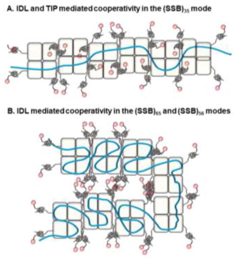
Figure 3. Models of IDL and TIP mediated cooperativity in EcSSB.
A) Cooperative binding of SSB tetramers in the (SSB)35 mode is shown. Proposed interactions between the IDLs of neighboring tetramers along with TIP interactions with free ssDNA binding regions in the OB-domains are denoted. B) Similar cooperative binding to ssDNA in the (SSB)65 and (SSB)56 modes are proposed to be facilitated through interactions between IDLs of multiple tetramers.
5. IDLs mediate cooperativity in SSB-DNA interactions
EcSSB forms cooperative nucleoprotein filaments on long ssDNA substrates that were first visualized by electron microscopy in 1972 [1]. These filaments form under both high and low SSB binding densities, and this cooperative feature was subsequently reproduced in buffers containing low [NaCl] (<10 mM) [66]. Under these conditions, SSB adopts the (SSB)35 binding mode, and hence, it was thought until recently that this binding mode was essential for highly cooperative binding behavior. However, recent evidence shows that at physiological salt concentrations containing either acetate or glutamate, which is the dominant monovalent anion in E. coli, highly cooperative binding is promoted even when SSB is in a fully wrapped (SSB)65 or (SSB)56 mode [51]. This was previously obscured because high [NaCl] had typically been used to selectively populate the (SSB)65 mode and high [Cl−] inhibits cooperativity [7, 22, 68, 80]. The length and composition of the IDL plays a key role in promoting cooperativity. Single molecule studies of SSB-ssDNA interactions in acetate salts show evidence for additional compaction of SSB-DNA complexes beyond that expected from ssDNA wrapping in the (SSB)65 mode [81]. This additional compaction likely reflects cooperative binding that is promoted in acetate salts.
Cooperative binding is not observed for the PfSSB protein which shares a high degree of homology with EcSSB in the DNA binding core [50, 78]. This appears to be due primarily to the very different IDLs of the two SSB proteins. The EcSSB IDL contains only 3 charged residues (2 R and one E) in addition to the 4 negatively charged residues in the TIP region and is predicted to adopt a compact globular conformation [52]. In contrast, the PfSSB IDL contains 26 charged residues in addition to 3 in its different acidic tip and is predicted to form an ensemble of more expanded Flory random coil configurations [52]. The cooperativity observed in EcSSB is stable even under high concentrations of glutamate (0.5 M) indicating that electrostatic interactions are not a major stabilizing factor for cooperativity[51]. In addition, a chimeric version of EcSSB in which the IDL from PfSSB is substituted for the EcSSB IDL no longer shows cooperative DNA binding [51]. Hence, the more globular, uncharged IDL is needed to promote highly cooperative binding indicating a functional role for the IDL in EcSSB.
A role for the IDL and the acidic TIP region was proposed in facilitating cooperative interactions within the (SSB)35 mode [82]. In this model, the TIP from one tetramer interacts with unoccupied DNA binding sites in a neighboring tetramer (Fig. 3A) [83]. However, such a scenario would be prevented in the (SSB)65 mode since all subunits are occupied by ssDNA. Since cooperative binding has now been observed in the fully wrapped binding mode, it is possible that high cooperativity is promoted primarily through direct interactions between IDLs of tetramers (Fig. 3B). It is likely that a combination of these features is utilized during DNA binding mode transitions and further modulated by interactions with SIPs.
6. SSB interacting proteins (SIPs)
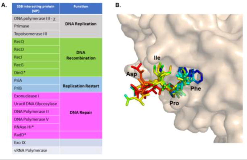
More than one dozen enzymes involved in DNA repair, replication and recombination interact with the TIP of EcSSB. All of the direct SIP:SSB interactions characterized to date are mediated through the TIP, which in EcSSB is – Met-Asp-Phe-Asp-Asp-Asp-Ile-Pro-Phe, with Phe being at the C-terminus (Fig. 2). A detailed list of SIPs, their functions, and structural features have been reviewed previously by Shereda, et. al. [16]. Three additional SIPs (RNaseHI, RadD and DinG) have since been identified and an updated list of SIPs is presented (Fig. 4A) [55, 84, 85]. Briefly, SIPs can be categorized according to their functions in DNA replication, replication restart, recombination and repair. Crystal structures of several SIPs bound to short TIP containing peptides have been solved [55, 57, 58, 60, 75, 86]. In all of these structures, the last three amino acids of the TIP (Ile-Pro-Phe) are well ordered and bind to a hydrophobic pocket in the SIP. An alignment of all the TIPs in these structures is shown in Fig. 4B. The proline and phenylalanine adopt a rigid conformation akin to a hook binding into the active site of the SIPs. The other residues away from the active site are more mobile as evidenced from the deviations in the B-factors upon alignment (Fig. 4B). Direct binding studies suggest that most of the binding free energy for SSB-SIP interactions is due to interactions with the SSB TIP [87].
Figure 4. SSB interactions with SSB interacting proteins (SIPs).
A) An updated list of the SIPs identified to date are categorized according to their cellular function. The asterisks denote newly identified SIPs. B) The crystal structure of exonuclease I in complex with the terminal four residues in the EcSSB TIP (PDB ID: 3C94) is shown. Similar TIP regions from multiple SIP-TIP peptide structures were aligned and shown here colored according to conformational flexibility (B-factors); red and blue denote extremes of high and low flexibility, respectively. The Ile-Pro-Phe residues adopt similar conformations in all these structures, whereas the Asp residue situated away from the active site can adopt multiple conformations.
While almost all SIP interactions to SSB identified to date occur through the TIP region, a recent report suggests that deletions of the IDL regions lead to a loss of SSB interaction with the RecO and RecG proteins in pull down experiments [88]. The authors posit potential roles for three Pro-X-X-Pro motifs in the IDL region being responsible for mediating interactions with RecO and RecG and possibly neighboring SSB tetramers leading to cooperative binding [62, 63, 88]. However, it is known that the TIP region is essential for SSB interactions with RecO and RecG [89–91]. A variant of SSB, SSBΔC8, lacking the TIP does not support RecG- or RecO-related biochemical activities [92, 93]. Further biochemical analysis is required to better understand this potential mode of binding.
7. SSB-SIP interactions affect SSB-DNA binding modes
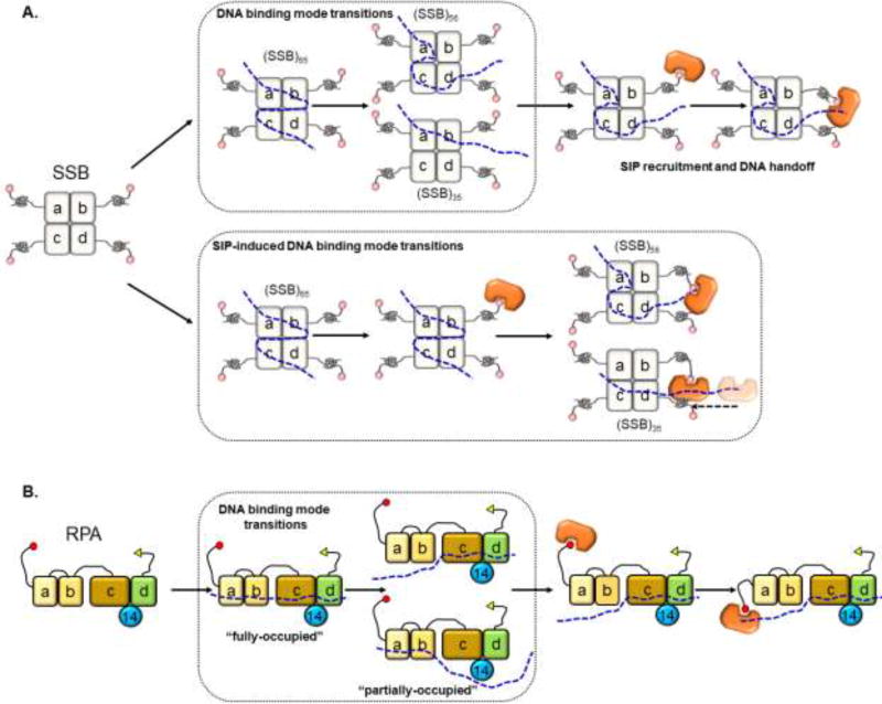
In the (SSB)65 mode, the ssDNA is fully wrapped around the tetramer, yet during DNA repair and recombination, numerous SIP proteins need to gain access to the SSB-bound DNA. Recent studies show that upon interaction with SSB, SIPs can facilitate a partial ssDNA unwrapping that promotes a transition from the (SSB)65 to the (SSB)35 mode (Fig. 5A, B) [43, 56, 74]. One example is the situation when the DNA replication machinery is prematurely displaced when DNA damage is encountered leading to a stalled replication fork. Replication restart under these conditions is coordinated by the PriA DNA helicase, which is recruited to stalled sites through its interaction with SSB [56]. Using FRET to monitor the (SSB)65 versus (SSB)35 modes, Bhattacharyya et. al., showed that PriA binds to SSB and promotes a transition from the fully wrapped (SSB)65 mode to a partially wrapped state, likely similar to the (SSB)35 mode [74]. This transition is necessary for PriA to bind ssDNA and initiate replication restart. PriC, another SIP within this replication restart machinery, also binds to EcSSB and can facilitate formation of the (SSB)35 binding mode [56]. Another example of SIP-induced reordering of the SSB binding mode has been observed with the RecQ helicase. RecQ is a helicase/translocase that catalyzes dsDNA strand separation to resolve complex DNA structures such as double-Holliday junctions, displacement loops (D-loops) and converging replication forks. RecQ is recruited to these structures through its interaction with SSB, and similar to PriA, needs to displace SSB, at least partially, to gain access to the buried ssDNA [76]. In a recent study, Mills et. al. show that RecQ binds to SSB and promotes its transition from the (SSB)65 to the (SSB)35 mode to gain access to the ssDNA, ultimately displacing SSB in the process [76].
Figure 5. DNA binding mode transitions in SSB.
A) SSB can spontaneously transition between the (SSB)35, (SSB)56, and (SSB)65 binding modes, and the transiently open ssDNA allow binding of SIPs. A model where SIPs facilitate transitions between binding modes is also depicted. B) The four-OB domains in RPA that are primarily responsible for ssDNA binding are shown. DNA binding domains (DBD) a, b and c reside in RPA70 and are connected by flexible linkers. DBD c, d and the RPA14 subunit form the trimerization core. RPA is also proposed to transition between multiple binding modes enabling the binding of RPA interacting proteins (orange) to ssDNA vacated by one or more DBDs.
8. Role of SSB-ssDNA dynamics in remodeling of high-affinity SSB-DNA complexes
The rapid and high-affinity binding of EcSSB to ssDNA protects transiently exposed ssDNA during the various DNA metabolic processes. However, the SSB protein must eventually be removed from the ssDNA by other DNA processing enzymes, which have lower affinities for DNA. How do cells displace the high affinity SSB? Three key features of EcSSB help in this matter: a) an SSB tetramer is capable of diffusing along ssDNA even when bound tightly in the fully wrapped (SSB)65 mode [44, 46]. As such it can be moved directionally from a particular site on ssDNA by the action of a nucleic acid motor protein such as an ATP-dependent translocase or a polymerase [94], b) an SSB tetramer can undergo a direct (inter-segment) transfer between sites on ssDNA without dissociation from the ssDNA [42]. This allows the SSB to readily move between sites on a ssDNA molecule and may be important in recycling of SSB between Okazaki fragments on the lagging strand during DNA replication, c) the interaction between the TIP of EcSSB and the SIPs can function to load a specific SIP onto the ssDNA substrate (Fig. 5A).
9. SSB diffusion along ssDNA
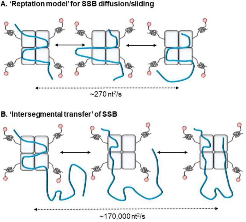
In the (SSB)65 binding mode, all four OB-domains interact with ssDNA. However, an EcSSB tetramer is able to diffuse along ssDNA, with an apparent one-dimensional diffusion coefficient of 270 nt2/s at 37°C [43, 46]. Zhou et. al. proposed a ‘reptation model’, where SSB movement would occur through formation of a transient bulge in the DNA which progressively moves through the four OB-folds (Fig. 6A) [46]. This propensity of EcSSB to diffuse is retained when it is bound to SIPs such as RecO, suggesting that this is a property important for cellular function.
Figure 6. Diffusion of SSB.
A) The reptation model for EcSSB diffusion/sliding is shown where segment(s) of ssDNA-OB domain interactions are perturbed allowing another OB-domain to bind to the free DNA. This enables the entire tetramer to shift or diffuse along the DNA lattice. B) On long ssDNA, SSB achieves rapid diffusion using principles of direct transfer where transiently dissociated ssDNA are replaced by DNA from a distant location. The respective rates for the two processes are denoted.
The ability of SSB proteins to diffuse along ssDNA provides the mechanism by which they can transiently invade a DNA duplex to partially melt a duplex DNA hairpin [43, 95]. Motor proteins can also rectify the movement of SSB along ssDNA. For example, a 5′->3' translocase such as Pif1 can push EcSSB in the same direction, whereas a 3′->5' translocase such as UvrD can push EcSSB in the opposite direction [94]. The diffusional properties of EcSSB can also be utilized by the RecA protein to facilitate formation of a nucleoprotein filament on ssDNA. EcSSB diffusion provides the mechanism by which it can remove secondary structures such as hairpins that hinder RecA filament formation [43].
The ability of an SSB protein to diffuse along ssDNA also is a property of other multi-OB domain proteins such as its eukaryotic homolog replication protein A (RPA; see the review on RPA by Byrne and Oakley in this issue). RPA also harbors four OB-domains, but are arranged within a heterotrimeric subunit architecture (Fig. 5B) [34]. The RPA-70 subunit has three OB-domains and the fourth OB-domain resides in the RPA-32 subunit [26, 28]. RPA also binds to ssDNA in multiple DNA binding modes (occluded site size 18–28 nt/RPA) that are influenced by salt concentration [35]. Similar to EcSSB, RPA also diffuses on ssDNA (diffusional coefficient of ~ 5000 nt2 s−1 at 37°C) [95]. The models proposed for EcSSB diffusion and sliding may also be applicable for RPA (Fig. 5B) and its remodeling by RPA-interacting proteins.
10. Redistribution of SSB on DNA via Direct transfer mechanisms
EcSSB tetramers can also be redistributed along ssDNA via direct transfer or intersegment transfer mechanisms without proceeding through a free (fully dissociated) SSB intermediate (Fig. 6B). Kozlov et. al. showed that this direct transfer proceeds through a doubly ligated SSB intermediate where a SSB tetramer is bound to two ssDNA molecules (or different segments of the same DNA) [47]. One key requirement for direct transfer is the need for multiple DNA binding sites on the SSB and the availability, at least transiently, of free ssDNA binding sites on a DNA bound SSB tetramer. With this in mind, direct transfer occurs much more rapidly for SSB in its (SSB)35 binding mode, with 2 free ssDNA binding sites, than in either the (SSB)65 binding mode [47]. The direct transfer process could be used to recycle SSB during lagging strand DNA synthesis. SSB is thought to bind primarily to the lagging DNA strand during DNA replication, but must then be displaced by DNA PolIII to finish replication [96]. Direct transfer could be used as a mechanism to recycle SSB between Okazaki fragments to promote rapid DNA replication. Consistent with this hypothesis, several SIPs are efficient at shifting SSBs into various DNA binding modes (where one or more OB-domains are free to bind an additional DNA molecule) [56, 74].
An “intersegment” transfer of SSB within a single ssDNA molecule has been hypothesized to explain the roughly 600-fold higher apparent diffusion coefficient of an SSB tetramer on long ssDNA (Fig. 6B) [42]. While one dimensional diffusion of SSB on short ssDNA occurs with an apparent one-dimensional diffusion coefficient of ~270 nt2/s at 37°C (Fig. 6A), a much higher apparent diffusion coefficient of 170,000 nt2/s was measured for much longer ssDNA [42]. This large difference could be explained if SSB can undergo long range intersegmental transfer in addition to its short-range diffusion/sliding. An SSB tetramer bound to one segment of a long ssDNA could transiently unwrap ssDNA from one or more of its OB-domains which could then bind to a more distant segment of the DNA strand (Fig. 6B).
Intersegment transfer would allow SSB to move around and scan a larger span of DNA more efficiently than one-dimensional diffusion [42]. Such long-range movement might be utilized to recruit and position appropriate SIPs at distant locations from where it resides. In addition, when SSB-ssDNA complexes are encountered by a helicase/translocase, the hinderance could be relieved by moving the SSB to a distant spot through intersegment transfer [94]. Such protein relocation mechanisms have been observed for histone repositioning on dsDNA during transcription by RNA polymerase [97]. Local diffusion and intersegment transfer can potentially be coupled as local short-range diffusion/sliding facilitate a transition from a fully wrapped (SSB)65 mode to a partially wrapped (SSB)56 or (SSB)35 mode, making available a ssDNA binding site to participate in intersegment transfer by binding to a distantly located ssDNA site.
11. How many SSB tails are needed for function?
While EcSSB is a homotetramer, the homologous SSB protein in thermophilic organisms function as homodimers (Fig.1). However, a comparison of their structures reveals that they are also composed of four OB-domains with two OB-domains residing in each subunit [24, 31]. This architecture results in a key difference in the number of C-terminal tails and thus two TIP regions per complex. For example, the SSB protein from Deinococcus radiodurans (DrSSB) has two C-terminal tails capable of interacting with up to two SIPs, whereas EcSSB has four C-terminal tails capable of interacting with up to four SIPs. Is there a functional advantage to having four versus two tails? What is the minimum number of tails needed per SSB “tetramer”? We addressed these question by generating linked EcSSB proteins where either two or all four OB-domains were linked using short peptide linkers yielding SSB proteins with four OB-folds, but only either two or one free C-termini [79]. Most of the DNA binding properties of these linked SSB proteins were not greatly affected, although the binding mode transitions were shifted so that the SSBs with fewer tails showed a progressive promotion of the (SSB)35 binding mode. In addition, the one-tailed tetramer showed a reduction in cooperative DNA binding. More significantly, the two-tailed tetramer complemented growth in vivo, whereas the one-tailed tetramer was dominant negative due to decoupling of leading and lagging strand replication [79]. This suggests that SSB must utilize two of its tails simultaneously during some essential process. Strains carrying the two-tailed tetramer grew faster, accumulated spontaneous mutations at a seven-fold faster rate, and were able to survive high doses of UV-irradiation [79]. These features are surprisingly similar to that of Deinococcus radiodurans suggesting that the interaction with the SIPs are a critical component of DNA repair and survival.
12. Cellular roles of DNA binding modes
SSB protein binding to ssDNA using multiple DNA binding modes is not a unique feature of EcSSB. Other bacterial SSB proteins and the eukaryotic RPA complex also display multiple DNA binding modes that are sensitive to solution conditions [35, 95, 98]. In contrast, PfSSB from the apicoplast of Plasmodium falciparum, does not populate an (SSB)35 mode, but rather favors the fully wrapped (SSB)56 and (SSB)65 DNA binding modes [50, 78]. This seems to be a direct consequence of the vastly different and more highly charged PfSSB IDL. The precise cellular functions of these DNA binding modes have been elusive. It is likely that all of the SSB binding modes are populated at least transiently in vivo. This seems obvious since the partially wrapped SSB-DNA structures must be intermediates along the path to forming a fully wrapped structure. In fact, the dynamic transitions among the different SSB-DNA binding modes is likely key to some of its functions. The (SSB)65 binding mode has been proposed to be associated with events in DNA recombination, as conditions that promote this mode in vitro correlate with the propensity of the RecA recombinase to perform strand exchange [43]. Similarly, the (SSB)35 mode has been proposed to be used during DNA replication, and two recent studies lend support to this model. A single molecule investigation conducted under near-physiological buffer conditions, and where the generation of ssDNA is coupled to DNA replication on a fork substrate, has shown that the human mitochondrial SSB (HmSSB) binds primarily using a low site size binding mode, likely the (SSB)35 mode [99]. Using linked EcSSB subunits, Waldman et. al. investigated the effect of preventing the formation of a fully wrapped ssDNA-SSB tetramer on cell viability [72]. By covalently linking two SSB subunits, they generated EcSSB “dimers” in which each subunit contains two OB-domains. This allowed them to make SSB variants in which two OB-domains were selectively rendered defective for ssDNA binding through mutagenesis yielding EcSSB variants unable to form the (SSB)65 or (SSB)56 modes. These variants were able to functionally complement wild type EcSSB, indicating that the SSB protein can carry out its essential functions without the fully wrapped DNA binding mode [72]. These experiments provide the first in vivo evidence that the (SSB)35 binding mode can function in DNA replication. However, these cells do show a hyper-resistance phenotype to DNA damage inducing agents and an accumulation of mutations as a trade-off for survival. These results suggest that adequate DNA repair mechanisms are poorly coordinated and thus a role for the (SSB)65 mode in DNA repair and recombination cannot be ruled out.
13. Conclusions
While recent studies have expanded our knowledge of the dynamics of SSB-ssDNA complexes and how these can be remodeled during DNA metabolism, many questions remain to be addressed: i) what is the molecular basis for how the IDL of EcSSB promotes cooperativity? ii) What is the role of cooperativity in vivo? iii) How and when does SSB interact with its many binding partners (SIPs) to achieve selectivity during DNA replication, repair, and recombination?
Acknowledgments
We thank the members of our laboratories and Dr. Jim Keck (University of Wisconsin, Madison) for their thoughtful suggestions.
FUNDING
This work was supported by grants from the National Institutes of Health [NIGMS 7R15GM110671 to E.A. and GM030498 to TML].
ABBREVIATIONS
- ssDNA
single stranded DNA
- EcSSB
Escherichia coli single stranded DNA binding protein
- OB
oligonucleotide/oligosaccharide
- IDL
intrinsically disordered linker
- SIPs
SSB interacting proteins
- RPA
replication protein A
Footnotes
Publisher's Disclaimer: This is a PDF file of an unedited manuscript that has been accepted for publication. As a service to our customers we are providing this early version of the manuscript. The manuscript will undergo copyediting, typesetting, and review of the resulting proof before it is published in its final citable form. Please note that during the production process errors may be discovered which could affect the content, and all legal disclaimers that apply to the journal pertain.
References
- 1.Sigal N, Delius H, Kornberg T, Gefter ML, Alberts B. A DNA-unwinding protein isolated from Escherichia coli: its interaction with DNA and with DNA polymerases. Proceedings of the National Academy of Sciences of the United States of America. 1972;69:3537–41. doi: 10.1073/pnas.69.12.3537. [DOI] [PMC free article] [PubMed] [Google Scholar]
- 2.Molineux IJ, Gefter ML. Properties of the Escherichia coli DNA-binding (unwinding) protein interaction with nucleolytic enzymes and DNA. Journal of molecular biology. 1975;98:811–25. doi: 10.1016/s0022-2836(75)80012-x. [DOI] [PubMed] [Google Scholar]
- 3.Weiner JH, Bertsch LL, Kornberg A. The deoxyribonucleic acid unwinding protein of Escherichia coli. Properties and functions in replication. The Journal of biological chemistry. 1975;250:1972–80. [PubMed] [Google Scholar]
- 4.Chrysogelos S, Griffith J. Escherichia coli single-strand binding protein organizes single-stranded DNA in nucleosome-like units. Proceedings of the National Academy of Sciences of the United States of America. 1982;79:5803–7. doi: 10.1073/pnas.79.19.5803. [DOI] [PMC free article] [PubMed] [Google Scholar]
- 5.Schneider RJ, Wetmur JG. Kinetics of transfer of Escherichia coli single strand deoxyribonucleic acid binding protein between single-stranded deoxyribonucleic acid molecules. Biochemistry. 1982;21:608–15. doi: 10.1021/bi00533a002. [DOI] [PubMed] [Google Scholar]
- 6.Williams KR, Spicer EK, LoPresti MB, Guggenheimer RA, Chase JW. Limited proteolysis studies on the Escherichia coli single-stranded DNA binding protein. Evidence for a functionally homologous domain in both the Escherichia coli and T4 DNA binding proteins. The Journal of biological chemistry. 1983;258:3346–55. [PubMed] [Google Scholar]
- 7.Lohman TM, Overman LB, Datta S. Salt-dependent changes in the DNA binding co-operativity of Escherichia coli single strand binding protein. Journal of molecular biology. 1986;187:603–15. doi: 10.1016/0022-2836(86)90338-4. [DOI] [PubMed] [Google Scholar]
- 8.Greipel J, Maass G, Mayer F. Complexes of the single-stranded DNA-binding protein from Escherichia coli (Eco SSB) with poly(dT). An investigation of their structure and internal dynamics by means of electron microscopy and NMR. Biophysical chemistry. 1987;26:149–61. doi: 10.1016/0301-4622(87)80018-2. [DOI] [PubMed] [Google Scholar]
- 9.Meyer RR, Laine PS. The single-stranded DNA-binding protein of Escherichia coli. Microbiological reviews. 1990;54:342–80. doi: 10.1128/mr.54.4.342-380.1990. [DOI] [PMC free article] [PubMed] [Google Scholar]
- 10.Kowalczykowski SC, Dixon DA, Eggleston AK, Lauder SD, Rehrauer WM. Biochemistry of homologous recombination in Escherichia coli. Microbiological reviews. 1994;58:401–65. doi: 10.1128/mr.58.3.401-465.1994. [DOI] [PMC free article] [PubMed] [Google Scholar]
- 11.Lohman TM, Ferrari ME. Escherichia coli single-stranded DNA-binding protein: multiple DNA-binding modes and cooperativities. Annual review of biochemistry. 1994;63:527–70. doi: 10.1146/annurev.bi.63.070194.002523. [DOI] [PubMed] [Google Scholar]
- 12.Kuzminov A. Recombinational repair of DNA damage in Escherichia coli and bacteriophage lambda. Microbiology and molecular biology reviews : MMBR. 1999;63:751–813. doi: 10.1128/mmbr.63.4.751-813.1999. table of contents. [DOI] [PMC free article] [PubMed] [Google Scholar]
- 13.Molineux IJ, Gefter ML. Properties of the Escherichia coli in DNA binding (unwinding) protein: interaction with DNA polymerase and DNA. Proceedings of the National Academy of Sciences of the United States of America. 1974;71:3858–62. doi: 10.1073/pnas.71.10.3858. [DOI] [PMC free article] [PubMed] [Google Scholar]
- 14.Mackay V, Linn S. Selective inhibition of the dnase activity of the recBC enzyme by the DNA binding protein from Escherichia coli. The Journal of biological chemistry. 1976;251:3716–9. [PubMed] [Google Scholar]
- 15.Meyer RR, Glassberg J, Scott JV, Kornberg A. A temperature-sensitive single-stranded DNA-binding protein from Escherichia coli. The Journal of biological chemistry. 1980;255:2897–901. [PubMed] [Google Scholar]
- 16.Shereda RD, Kozlov AG, Lohman TM, Cox MM, Keck JL. SSB as an organizer/mobilizer of genome maintenance complexes. Critical reviews in biochemistry and molecular biology. 2008;43:289–318. doi: 10.1080/10409230802341296. [DOI] [PMC free article] [PubMed] [Google Scholar]
- 17.Zou Y, Liu Y, Wu X, Shell SM. Functions of human replication protein A (RPA): from DNA replication to DNA damage and stress responses. Journal of cellular physiology. 2006;208:267–73. doi: 10.1002/jcp.20622. [DOI] [PMC free article] [PubMed] [Google Scholar]
- 18.Fanning E, Klimovich V, Nager AR. A dynamic model for replication protein A (RPA) function in DNA processing pathways. Nucleic acids research. 2006;34:4126–37. doi: 10.1093/nar/gkl550. [DOI] [PMC free article] [PubMed] [Google Scholar]
- 19.von Hippel PH, Delagoutte E. A general model for nucleic acid helicases and their "coupling" within macromolecular machines. Cell. 2001;104:177–90. doi: 10.1016/s0092-8674(01)00203-3. [DOI] [PubMed] [Google Scholar]
- 20.Molineux IJ, Friedman S, Gefter ML. Purification and properties of the Escherichia coli deoxyribonucleic acid-unwinding protein. Effects on deoxyribonucleic acid synthesis in vitro. The Journal of biological chemistry. 1974;249:6090–8. [PubMed] [Google Scholar]
- 21.Molineux IJ, Pauli A, Gefter ML. Physical studies of the interaction between the Escherichia coli DNA binding protein and nucleic acids. Nucleic acids research. 1975;2:1821–37. doi: 10.1093/nar/2.10.1821. [DOI] [PMC free article] [PubMed] [Google Scholar]
- 22.Bujalowski W, Lohman TM. Escherichia coli single-strand binding protein forms multiple, distinct complexes with single-stranded DNA. Biochemistry. 1986;25:7799–802. doi: 10.1021/bi00372a003. [DOI] [PubMed] [Google Scholar]
- 23.Raghunathan S, Kozlov AG, Lohman TM, Waksman G. Structure of the DNA binding domain of E. coli SSB bound to ssDNA. Nature structural biology. 2000;7:648–52. doi: 10.1038/77943. [DOI] [PubMed] [Google Scholar]
- 24.Bernstein DA, Eggington JM, Killoran MP, Misic AM, Cox MM, Keck JL. Crystal structure of the Deinococcus radiodurans single-stranded DNA-binding protein suggests a mechanism for coping with DNA damage. Proceedings of the National Academy of Sciences of the United States of America. 2004;101:8575–80. doi: 10.1073/pnas.0401331101. [DOI] [PMC free article] [PubMed] [Google Scholar]
- 25.Shamoo Y, Friedman AM, Parsons MR, Konigsberg WH, Steitz TA. Crystal structure of a replication fork single-stranded DNA binding protein (T4 gp32) complexed to DNA. Nature. 1995;376:362–6. doi: 10.1038/376362a0. [DOI] [PubMed] [Google Scholar]
- 26.Fan J, Pavletich NP. Structure and conformational change of a replication protein A heterotrimer bound to ssDNA. Genes & development. 2012;26:2337–47. doi: 10.1101/gad.194787.112. [DOI] [PMC free article] [PubMed] [Google Scholar]
- 27.Kerr ID, Wadsworth RI, Cubeddu L, Blankenfeldt W, Naismith JH, White MF. Insights into ssDNA recognition by the OB fold from a structural and thermodynamic study of Sulfolobus SSB protein. The EMBO journal. 2003;22:2561–70. doi: 10.1093/emboj/cdg272. [DOI] [PMC free article] [PubMed] [Google Scholar]
- 28.Bochkareva E, Korolev S, Lees-Miller SP, Bochkarev A. Structure of the RPA trimerization core and its role in the multistep DNA-binding mechanism of RPA. The EMBO journal. 2002;21:1855–63. doi: 10.1093/emboj/21.7.1855. [DOI] [PMC free article] [PubMed] [Google Scholar]
- 29.Bochkarev A, Bochkareva E. From RPA to BRCA2: lessons from single-stranded DNA binding by the OB-fold. Current opinion in structural biology. 2004;14:36–42. doi: 10.1016/j.sbi.2004.01.001. [DOI] [PubMed] [Google Scholar]
- 30.Fedorov R, Witte G, Urbanke C, Manstein DJ, Curth U. 3D structure of Thermus aquaticus single-stranded DNA-binding protein gives insight into the functioning of SSB proteins. Nucleic acids research. 2006;34:6708–17. doi: 10.1093/nar/gkl1002. [DOI] [PMC free article] [PubMed] [Google Scholar]
- 31.Jedrzejczak R, Dauter M, Dauter Z, Olszewski M, Dlugolecka A, Kur J. Structure of the single-stranded DNA-binding protein SSB from Thermus aquaticus. Acta crystallographica Section D, Biological crystallography. 2006;62:1407–12. doi: 10.1107/S0907444906036031. [DOI] [PubMed] [Google Scholar]
- 32.Hollis T, Stattel JM, Walther DS, Richardson CC, Ellenberger T. Structure of the gene 2.5 protein, a single-stranded DNA binding protein encoded by bacteriophage T7. Proceedings of the National Academy of Sciences of the United States of America. 2001;98:9557–62. doi: 10.1073/pnas.171317698. [DOI] [PMC free article] [PubMed] [Google Scholar]
- 33.Chen R, Wold MS. Replication protein A: single-stranded DNA's first responder: dynamic DNA-interactions allow replication protein A to direct single-strand DNA intermediates into different pathways for synthesis or repair. BioEssays : news and reviews in molecular, cellular and developmental biology. 2014;36:1156–61. doi: 10.1002/bies.201400107. [DOI] [PMC free article] [PubMed] [Google Scholar]
- 34.Wold MS. Replication protein A: a heterotrimeric, single-stranded DNA-binding protein required for eukaryotic DNA metabolism. Annual review of biochemistry. 1997;66:61–92. doi: 10.1146/annurev.biochem.66.1.61. [DOI] [PubMed] [Google Scholar]
- 35.Kumaran S, Kozlov AG, Lohman TM. Saccharomyces cerevisiae replication protein A binds to single-stranded DNA in multiple salt-dependent modes. Biochemistry. 2006;45:11958–73. doi: 10.1021/bi060994r. [DOI] [PMC free article] [PubMed] [Google Scholar]
- 36.Touma C, Kariawasam R, Gimenez AX, Bernardo RE, Ashton NW, Adams MN, et al. A structural analysis of DNA binding by hSSB1 (NABP2/OBFC2B) in solution. Nucleic acids research. 2016;44:7963–73. doi: 10.1093/nar/gkw617. [DOI] [PMC free article] [PubMed] [Google Scholar]
- 37.Touma C, Adams MN, Ashton NW, Mizzi M, El-Kamand S, Richard DJ, et al. A data-driven structural model of hSSB1 (NABP2/OBFC2B) self-oligomerization. Nucleic acids research. 2017;45:8609–20. doi: 10.1093/nar/gkx526. [DOI] [PMC free article] [PubMed] [Google Scholar]
- 38.Paquet N, Adams MN, Ashton NW, Touma C, Gamsjaeger R, Cubeddu L, et al. hSSB1 (NABP2/OBFC2B) is regulated by oxidative stress. Scientific reports. 2016;6 doi: 10.1038/srep27446. [DOI] [PMC free article] [PubMed] [Google Scholar]
- 39.Guilliam TA, Brissett NC, Ehlinger A, Keen BA, Kolesar P, Taylor EM, et al. Molecular basis for PrimPol recruitment to replication forks by RPA. Nature communications. 2017;8:15222. doi: 10.1038/ncomms15222. [DOI] [PMC free article] [PubMed] [Google Scholar]
- 40.Brosey CA, Chagot ME, Ehrhardt M, Pretto DI, Weiner BE, Chazin WJ. NMR analysis of the architecture and functional remodeling of a modular multidomain protein, RPA. Journal of the American Chemical Society. 2009;131:6346–7. doi: 10.1021/ja9013634. [DOI] [PMC free article] [PubMed] [Google Scholar]
- 41.Jiang X, Klimovich V, Arunkumar AI, Hysinger EB, Wang Y, Ott RD, et al. Structural mechanism of RPA loading on DNA during activation of a simple pre-replication complex. The EMBO journal. 2006;25:5516–26. doi: 10.1038/sj.emboj.7601432. [DOI] [PMC free article] [PubMed] [Google Scholar]
- 42.Lee KS, Marciel AB, Kozlov AG, Schroeder CM, Lohman TM, Ha T. Ultrafast redistribution of E. coli SSB along long single-stranded DNA via intersegment transfer. Journal of molecular biology. 2014;426:2413–21. doi: 10.1016/j.jmb.2014.04.023. [DOI] [PMC free article] [PubMed] [Google Scholar]
- 43.Roy R, Kozlov AG, Lohman TM, Ha T. SSB protein diffusion on single-stranded DNA stimulates RecA filament formation. Nature. 2009;461:1092–7. doi: 10.1038/nature08442. [DOI] [PMC free article] [PubMed] [Google Scholar]
- 44.Roy R, Kozlov AG, Lohman TM, Ha T. Dynamic structural rearrangements between DNA binding modes of E. coli SSB protein. Journal of molecular biology. 2007;369:1244–57. doi: 10.1016/j.jmb.2007.03.079. [DOI] [PMC free article] [PubMed] [Google Scholar]
- 45.Suksombat S, Khafizov R, Kozlov AG, Lohman TM, Chemla YR. Structural dynamics of E. coli single-stranded DNA binding protein reveal DNA wrapping and unwrapping pathways. eLife. 2015;4:1–23. doi: 10.7554/eLife.08193. [DOI] [PMC free article] [PubMed] [Google Scholar]
- 46.Zhou R, Kozlov AG, Roy R, Zhang J, Korolev S, Lohman TM, et al. SSB functions as a sliding platform that migrates on DNA via reptation. Cell. 2011;146:222–32. doi: 10.1016/j.cell.2011.06.036. [DOI] [PMC free article] [PubMed] [Google Scholar]
- 47.Kozlov AG, Lohman TM. Kinetic mechanism of direct transfer of Escherichia coli SSB tetramers between single-stranded DNA molecules. Biochemistry. 2002;41:11611–27. doi: 10.1021/bi020361m. [DOI] [PubMed] [Google Scholar]
- 48.Ferrari ME, Fang J, Lohman TM. A mutation in E. coli SSB protein (W54S) alters intra-tetramer negative cooperativity and inter-tetramer positive cooperativity for single-stranded DNA binding. Biophysical chemistry. 1997;64:235–51. doi: 10.1016/s0301-4622(96)02223-5. [DOI] [PubMed] [Google Scholar]
- 49.Raghunathan S, Ricard CS, Lohman TM, Waksman G. Crystal structure of the homo-tetrameric DNA binding domain of Escherichia coli single-stranded DNA-binding protein determined by multiwavelength x-ray diffraction on the selenomethionyl protein at 2.9-A resolution. Proceedings of the National Academy of Sciences of the United States of America. 1997;94:6652–7. doi: 10.1073/pnas.94.13.6652. [DOI] [PMC free article] [PubMed] [Google Scholar]
- 50.Antony E, Weiland EA, Korolev S, Lohman TM. Plasmodium falciparum SSB tetramer wraps single-stranded DNA with similar topology but opposite polarity to E. coli SSB. Journal of molecular biology. 2012;420:269–83. doi: 10.1016/j.jmb.2012.04.021. [DOI] [PMC free article] [PubMed] [Google Scholar]
- 51.Kozlov AG, Shinn MK, Weiland EA, Lohman TM. Glutamate promotes SSB protein-protein Interactions via intrinsically disordered regions. Journal of molecular biology. 2017;429:2790–801. doi: 10.1016/j.jmb.2017.07.021. [DOI] [PMC free article] [PubMed] [Google Scholar]
- 52.Kozlov AG, Weiland E, Mittal A, Waldman V, Antony E, Fazio N, et al. Intrinsically disordered C-terminal tails of E. coli single-stranded DNA binding protein regulate cooperative binding to single-stranded DNA. Journal of molecular biology. 2015;427:763–74. doi: 10.1016/j.jmb.2014.12.020. [DOI] [PMC free article] [PubMed] [Google Scholar]
- 53.Green M, Hatter L, Brookes E, Soultanas P, Scott DJ. Defining the Intrinsically Disordered C-Terminal Domain of SSB Reveals DNA-Mediated Compaction. Journal of molecular biology. 2016;428:357–64. doi: 10.1016/j.jmb.2015.12.007. [DOI] [PubMed] [Google Scholar]
- 54.Curth U, Genschel J, Urbanke C, Greipel J. In vitro and in vivo function of the C-terminus of Escherichia coli single-stranded DNA binding protein. Nucleic acids research. 1996;24:2706–11. doi: 10.1093/nar/24.14.2706. [DOI] [PMC free article] [PubMed] [Google Scholar]
- 55.Petzold C, Marceau AH, Miller KH, Marqusee S, Keck JL. Interaction with Single-stranded DNA-binding Protein Stimulates Escherichia coli Ribonuclease HI Enzymatic Activity. The Journal of biological chemistry. 2015;290:14626–36. doi: 10.1074/jbc.M115.655134. [DOI] [PMC free article] [PubMed] [Google Scholar]
- 56.Wessel SR, Marceau AH, Massoni SC, Zhou R, Ha T, Sandler SJ, et al. PriC-mediated DNA replication restart requires PriC complex formation with the single-stranded DNA-binding protein. The Journal of biological chemistry. 2013;288:17569–78. doi: 10.1074/jbc.M113.478156. [DOI] [PMC free article] [PubMed] [Google Scholar]
- 57.Marceau AH, Bahng S, Massoni SC, George NP, Sandler SJ, Marians KJ, et al. Structure of the SSB-DNA polymerase III interface and its role in DNA replication. The EMBO journal. 2011;30:4236–47. doi: 10.1038/emboj.2011.305. [DOI] [PMC free article] [PubMed] [Google Scholar]
- 58.Lu D, Myers AR, George NP, Keck JL. Mechanism of Exonuclease I stimulation by the single-stranded DNA-binding protein. Nucleic acids research. 2011;39:6536–45. doi: 10.1093/nar/gkr315. [DOI] [PMC free article] [PubMed] [Google Scholar]
- 59.Lu D, Bernstein DA, Satyshur KA, Keck JL. Small-molecule tools for dissecting the roles of SSB/protein interactions in genome maintenance. Proceedings of the National Academy of Sciences of the United States of America. 2010;107:633–8. doi: 10.1073/pnas.0909191107. [DOI] [PMC free article] [PubMed] [Google Scholar]
- 60.Shereda RD, Reiter NJ, Butcher SE, Keck JL. Identification of the SSB binding site on E. coli RecQ reveals a conserved surface for binding SSB's C terminus. Journal of molecular biology. 2009;386:612–25. doi: 10.1016/j.jmb.2008.12.065. [DOI] [PMC free article] [PubMed] [Google Scholar]
- 61.Lu D, Windsor MA, Gellman SH, Keck JL. Peptide inhibitors identify roles for SSB C-terminal residues in SSB/exonuclease I complex formation. Biochemistry. 2009;48:6764–71. doi: 10.1021/bi900361r. [DOI] [PMC free article] [PubMed] [Google Scholar]
- 62.Bianco PR, Pottinger S, Tan HY, Nguyenduc T, Rex K, Varshney U. The IDL of E. coli SSB links ssDNA and protein binding by mediating protein-protein interactions. Protein science : a publication of the Protein Society. 2017;26:227–41. doi: 10.1002/pro.3072. [DOI] [PMC free article] [PubMed] [Google Scholar]
- 63.Bianco PR. The tale of SSB. Progress in biophysics and molecular biology. 2017;127:111–8. doi: 10.1016/j.pbiomolbio.2016.11.001. [DOI] [PMC free article] [PubMed] [Google Scholar]
- 64.Marceau AH, Bernstein DA, Walsh BW, Shapiro W, Simmons LA, Keck JL. Protein interactions in genome maintenance as novel antibacterial targets. PloS one. 2013;8:e58765. doi: 10.1371/journal.pone.0058765. [DOI] [PMC free article] [PubMed] [Google Scholar]
- 65.Voter AF, Killoran MP, Ananiev GE, Wildman SA, Hoffmann FM, Keck JL. A High-Throughput Screening Strategy to Identify Inhibitors of SSB Protein-Protein Interactions in an Academic Screening Facility. SLAS discovery : advancing life sciences R & D. 2018;23:94–101. doi: 10.1177/2472555217712001. [DOI] [PMC free article] [PubMed] [Google Scholar]
- 66.Lohman TM, Overman LB. Two binding modes in Escherichia coli single strand binding protein-single stranded DNA complexes. Modulation by NaCl concentration. The Journal of biological chemistry. 1985;260:3594–603. [PubMed] [Google Scholar]
- 67.Bujalowski W, Overman LB, Lohman TM. Binding mode transitions of Escherichia coli single strand binding protein-single-stranded DNA complexes. Cation, anion, pH, binding density effects. The Journal of biological chemistry. 1988;263:4629–40. [PubMed] [Google Scholar]
- 68.Lohman TM, Bujalowski W. Negative cooperativity within individual tetramers of Escherichia coli single strand binding protein is responsible for the transition between the (SSB)35 and (SSB)56 DNA binding modes. Biochemistry. 1988;27:2260–5. doi: 10.1021/bi00407a002. [DOI] [PubMed] [Google Scholar]
- 69.Lohman TM, Bujalowski W, Overman LB, Wei TF. Interactions of the E. coli single strand binding (SSB) protein with ss nucleic acids. Binding mode transitions and equilibrium binding studies. Biochemical pharmacology. 1988;37:1781–2. doi: 10.1016/0006-2952(88)90443-1. [DOI] [PubMed] [Google Scholar]
- 70.Suksombat S, Khafizov R, Kozlov AG, Lohman TM, Chemla YR. Structural dynamics of E. coli single-stranded DNA binding protein reveal DNA wrapping and unwrapping pathways. eLife. 2015;4 doi: 10.7554/eLife.08193. [DOI] [PMC free article] [PubMed] [Google Scholar]
- 71.Ha T, Kozlov AG, Lohman TM. Single-molecule views of protein movement on single-stranded DNA. Annual review of biophysics. 2012;41:295–319. doi: 10.1146/annurev-biophys-042910-155351. [DOI] [PMC free article] [PubMed] [Google Scholar]
- 72.Waldman VM, Weiland E, Kozlov AG, Lohman TM. Is a fully wrapped SSB-DNA complex essential for Escherichia coli survival? Nucleic acids research. 2016;44:4317–29. doi: 10.1093/nar/gkw262. [DOI] [PMC free article] [PubMed] [Google Scholar]
- 73.Wessel SR, Cornilescu CC, Cornilescu G, Metz A, Leroux M, Hu K, et al. Structure and Function of the PriC DNA Replication Restart Protein. The Journal of biological chemistry. 2016;291:18384–96. doi: 10.1074/jbc.M116.738781. [DOI] [PMC free article] [PubMed] [Google Scholar]
- 74.Bhattacharyya B, George NP, Thurmes TM, Zhou R, Jani N, Wessel SR, et al. Structural mechanisms of PriA-mediated DNA replication restart. Proceedings of the National Academy of Sciences of the United States of America. 2014;111:1373–8. doi: 10.1073/pnas.1318001111. [DOI] [PMC free article] [PubMed] [Google Scholar]
- 75.Cadman CJ, Lopper M, Moon PB, Keck JL, McGlynn P. PriB stimulates PriA helicase via an interaction with single-stranded DNA. The Journal of biological chemistry. 2005;280:39693–700. doi: 10.1074/jbc.M508521200. [DOI] [PubMed] [Google Scholar]
- 76.Mills M, Harami GM, Seol Y, Gyimesi M, Martina M, Kovacs ZJ, et al. RecQ helicase triggers a binding mode change in the SSB-DNA complex to efficiently initiate DNA unwinding. Nucleic acids research. 2017;45:11878–90. doi: 10.1093/nar/gkx939. [DOI] [PMC free article] [PubMed] [Google Scholar]
- 77.Yang C, Curth U, Urbanke C, Kang C. Crystal structure of human mitochondrial single-stranded DNA binding protein at 2.4 A resolution. Nature structural biology. 1997;4:153–7. doi: 10.1038/nsb0297-153. [DOI] [PubMed] [Google Scholar]
- 78.Antony E, Kozlov AG, Nguyen B, Lohman TM. Plasmodium falciparum SSB tetramer binds single-stranded DNA only in a fully wrapped mode. Journal of molecular biology. 2012;420:284–95. doi: 10.1016/j.jmb.2012.04.022. [DOI] [PMC free article] [PubMed] [Google Scholar]
- 79.Antony E, Weiland E, Yuan Q, Manhart CM, Nguyen B, Kozlov AG, et al. Multiple C-terminal tails within a single E. coli SSB homotetramer coordinate DNA replication and repair. Journal of molecular biology. 2013;425:4802–19. doi: 10.1016/j.jmb.2013.08.021. [DOI] [PMC free article] [PubMed] [Google Scholar]
- 80.Lohman TM. Kinetics and mechanism of dissociation of cooperatively bound T4 gene 32 protein-single-stranded nucleic acid complexes. 1. Irreversible dissociation induced by sodium chloride concentration jumps. Biochemistry. 1984;23:4656–65. doi: 10.1021/bi00315a022. [DOI] [PubMed] [Google Scholar]
- 81.Bell JC, Liu B, Kowalczykowski SC. Imaging and energetics of single SSB-ssDNA molecules reveal intramolecular condensation and insight into RecOR function. eLife. 2015;4:e08646. doi: 10.7554/eLife.08646. [DOI] [PMC free article] [PubMed] [Google Scholar]
- 82.Kozlov AG, Cox MM, Lohman TM. Regulation of single-stranded DNA binding by the C termini of Escherichia coli single-stranded DNA-binding (SSB) protein. The Journal of biological chemistry. 2010;285:17246–52. doi: 10.1074/jbc.M110.118273. [DOI] [PMC free article] [PubMed] [Google Scholar]
- 83.Su XC, Wang Y, Yagi H, Shishmarev D, Mason CE, Smith PJ, et al. Bound or free: interaction of the C-terminal domain of Escherichia coli single-stranded DNA-binding protein (SSB) with the tetrameric core of SSB. Biochemistry. 2014;53:1925–34. doi: 10.1021/bi5001867. [DOI] [PubMed] [Google Scholar]
- 84.Cheng Z, Caillet A, Ren B, Ding H. Stimulation of Escherichia coli DNA damage inducible DNA helicase DinG by the single-stranded DNA binding protein SSB. FEBS letters. 2012;586:3825–30. doi: 10.1016/j.febslet.2012.09.032. [DOI] [PMC free article] [PubMed] [Google Scholar]
- 85.Chen SH, Byrne-Nash RT, Cox MM. Escherichia coli RadD Protein Functionally Interacts with the Single-stranded DNA-binding Protein. The Journal of biological chemistry. 2016;291:20779–86. doi: 10.1074/jbc.M116.736223. [DOI] [PMC free article] [PubMed] [Google Scholar]
- 86.Lu D, Keck JL. Structural basis of Escherichia coli single-stranded DNA-binding protein stimulation of exonuclease I. Proceedings of the National Academy of Sciences of the United States of America. 2008;105:9169–74. doi: 10.1073/pnas.0800741105. [DOI] [PMC free article] [PubMed] [Google Scholar]
- 87.Kozlov AG, Jezewska MJ, Bujalowski W, Lohman TM. Binding specificity of Escherichia coli single-stranded DNA binding protein for the chi subunit of DNA pol III holoenzyme and PriA helicase. Biochemistry. 2010;49:3555–66. doi: 10.1021/bi100069s. [DOI] [PMC free article] [PubMed] [Google Scholar]
- 88.Tan HY, Wilczek LA, Pottinger S, Manosas M, Yu C, Nguyenduc T, et al. The intrinsically disordered linker of E. coli SSB is critical for the release from single-stranded DNA. Protein science : a publication of the Protein Society. 2017;26:700–17. doi: 10.1002/pro.3115. [DOI] [PMC free article] [PubMed] [Google Scholar]
- 89.Ryzhikov M, Korolev S. Structural studies of SSB interaction with RecO. Methods Mol Biol. 2012;922:123–31. doi: 10.1007/978-1-62703-032-8_7. [DOI] [PubMed] [Google Scholar]
- 90.Ryzhikov M, Koroleva O, Postnov D, Tran A, Korolev S. Mechanism of RecO recruitment to DNA by single-stranded DNA binding protein. Nucleic acids research. 2011;39:6305–14. doi: 10.1093/nar/gkr199. [DOI] [PMC free article] [PubMed] [Google Scholar]
- 91.Slocum SL, Buss JA, Kimura Y, Bianco PR. Characterization of the ATPase activity of the Escherichia coli RecG protein reveals that the preferred cofactor is negatively supercoiled DNA. Journal of molecular biology. 2007;367:647–64. doi: 10.1016/j.jmb.2007.01.007. [DOI] [PMC free article] [PubMed] [Google Scholar]
- 92.Lecointe F, Serena C, Velten M, Costes A, McGovern S, Meile JC, et al. Anticipating chromosomal replication fork arrest: SSB targets repair DNA helicases to active forks. The EMBO journal. 2007;26:4239–51. doi: 10.1038/sj.emboj.7601848. [DOI] [PMC free article] [PubMed] [Google Scholar]
- 93.Hobbs MD, Sakai A, Cox MM. SSB protein limits RecOR binding onto single-stranded DNA. The Journal of biological chemistry. 2007;282:11058–67. doi: 10.1074/jbc.M611007200. [DOI] [PubMed] [Google Scholar]
- 94.Sokoloski JE, Kozlov AG, Galletto R, Lohman TM. Chemo-mechanical pushing of proteins along single-stranded DNA. Proceedings of the National Academy of Sciences of the United States of America. 2016;113:6194–9. doi: 10.1073/pnas.1602878113. [DOI] [PMC free article] [PubMed] [Google Scholar]
- 95.Nguyen B, Sokoloski J, Galletto R, Elson EL, Wold MS, Lohman TM. Diffusion of human replication protein A along single-stranded DNA. Journal of molecular biology. 2014;426:3246–61. doi: 10.1016/j.jmb.2014.07.014. [DOI] [PMC free article] [PubMed] [Google Scholar]
- 96.Kelman Z, O'Donnell M. DNA polymerase III holoenzyme: structure and function of a chromosomal replicating machine. Annual review of biochemistry. 1995;64:171–200. doi: 10.1146/annurev.bi.64.070195.001131. [DOI] [PubMed] [Google Scholar]
- 97.Bintu L, Kopaczynska M, Hodges C, Lubkowska L, Kashlev M, Bustamante C. The elongation rate of RNA polymerase determines the fate of transcribed nucleosomes. Nature structural & molecular biology. 2011;18:1394–9. doi: 10.1038/nsmb.2164. [DOI] [PMC free article] [PubMed] [Google Scholar]
- 98.Kozlov AG, Eggington JM, Cox MM, Lohman TM. Binding of the dimeric Deinococcus radiodurans single-stranded DNA binding protein to single-stranded DNA. Biochemistry. 2010;49:8266–75. doi: 10.1021/bi100920w. [DOI] [PMC free article] [PubMed] [Google Scholar]
- 99.Morin JA, Cerron F, Jarillo J, Beltran-Heredia E, Ciesielski GL, Arias-Gonzalez JR, et al. DNA synthesis determines the binding mode of the human mitochondrial single-stranded DNA-binding protein. Nucleic acids research. 2017;45:7237–48. doi: 10.1093/nar/gkx395. [DOI] [PMC free article] [PubMed] [Google Scholar]


