Abstract
The immunomodulatory drugs, lenalidomide and pomalidomide yield high response rates in multiple myeloma patients, but are associated with a high rate of thrombocytopenia and increased risk of secondary hematologic malignancies. Here, we demonstrate that the immunomodulatory drugs induce self-renewal of hematopoietic progenitors and upregulate megakaryocytic colonies by inhibiting apoptosis and increasing proliferation of early megakaryocytic progenitors via down-regulation of IKZF1. In this process, the immunomodulatory drugs degrade IKZF1 and subsequently down-regulate its binding partner, GATA1. This results in the decrease of GATA1 targets such as ZFPM1 and NFE2, leading to expansion of megakaryocytic progenitors with concomitant inhibition of maturation of megakaryocytes. The down-regulation of GATA1 further decreases CCND1 and increases CDKN2A expression. Overexpression of GATA1 abrogated the effects of the immunomodulatory drugs and restored maturation of megakaryocytic progenitors. Our data not only provide the mechanism for the immunomodulatory drugs induced thrombocytopenia but also help to explain the higher risk of secondary malignancies and long-term cytopenia induced by enhanced cell cycling and subsequent exhaustion of the stem cell pool.
Introduction
Lenalidomide (LEN, CC-5013) and pomalidomide (POM, CC-4047) are immunomodulatory drugs (IMiDs), analogues of thalidomide, which have several cellular effects including immunomodulatory, anti-angiogenic, anti-inflammatory and anti-proliferative effects.1–3 In multiple myeloma (MM) cells, LEN binds to cereblon and thereby, is able to target two specific B-cell transcription factors, Ikaros family zinc finger proteins 1 and 3 (IKZF1 and IKZF3) for proteasomal degradation4,5 and subsequently affect transcription factors critical for multiple myeloma (MM) growth, such as CCAAT-enhancer-binding protein beta (C/EBPβ)6 and IRF4.7 We have shown that IKZF1 is also expressed in CD34+ cells and undergoes degradation after ubiquitination of cereblon when cells are treated with IMiDs.8 LEN is considered a therapeutic breakthrough in the treatment of MM.9 POM is the newest IMiD, and appears to be more potent than LEN in MM.10 However, the use of IMiDs is associated with neutropenia, thrombocytopenia, bone marrow failure and stem cell mobilization.9,11,12 In addition, there is a concern of an increased risk of secondary malignancies such as myelodysplastic syndrome and acute leukemia.13–15
Our laboratory has focused on exploring the effects of IMiDs on different hematopoietic lineages. We showed that IMiDs do not exhibit direct stem cell toxicity, but affect lineage commitment.16,17 Downregulation of GATA1 by IMiDs induces a shift into myeloid lineage commitment at the expense of erythroid commitment.16 The downregulation of SPI1 (PU.1), a critical transcription factor for myeloid maturation, leads to maturational arrest with accumulation of immature myeloid precursors, resulting in neutropenia.17 Nevertheless, IMiD-induced thrombocytopenia, a major adverse side effect, is still not understood.
Here, we investigated the effect of IMiDs on megakaryopoiesis after thrombopoietin (TPO) stimulation. We showed that IMiDs induce self-renewal and proliferation of megakaryocytic progenitors by down-regulating GATA1 as a consequence of the degradation of its binding partner IKZF1. This is accompanied by decreased ZFPM1/FOG-1 and NFE2 expression, leading to inhibition of megakaryocyte maturation. Our data further demonstrated that IMiD induced a decrease in CCND1/cyclin D1 accompanied by an increase in CDKN2A/p16, resulting in the maturational arrest of megakaryocytes (Mks). The effects of IMiDs on megakaryopoiesis could be abrogated by overexpression of GATA1. This study provides for the first-time mechanistic insight into how IMiDs induce thrombocytopenia and potentially contribute to secondary hematologic malignancies by sustained cell proliferation.
Methods
CD34+ cells isolation and culture
Primary CD34+ cells were isolated from discarded peripheral blood leukapheresis products after stem cell mobilization of consenting healthy individuals and MM patients. We tested the CD34+ cells from MM patients or healthy individuals in cell proliferation and colony assays and no difference was observed. Data are not shown. The Institutional Review Boards (IRBs) of the University of Pittsburgh, Pittsburgh, PA and Columbia University, New York, NY approved all studies. Purified CD34+ cells were grown in serum-free hematopoietic growth medium (HPGM) (Lonza) supplemented with 10 ng/mL recombinant human thrombopoietin (rhTPO), 10 ng/mL recombinant human interleukin-3 (rhIL-3), 10 ng/mL recombinant human interleukin-6 (rhIL-6), and 50 ng/mL recombinant human stem cell factor (rhSCF). All cytokines were purchased from PeproTech as described previously.16,17
LEN and POM (Sigma Aldrich) in DMSO were diluted in culture medium and added daily. Cell viability was measured by trypan blue exclusion, and cell proliferation was quantified by manual cell counting every 2 days during culture.
Megakaryocytic colony assays
Megakaryocytic colony forming unit (CFU-Mk) assays were generated using the MegaCult™-C Staining Kit (StemCell Technologies) according to the manufacturer’s instructions. The number of CFU-Mk was determined using an anti-CD41 antibody, an alkaline phosphatase detection system and by counter-staining with Evan’s Blue. The total numbers of colonies were counted on day 12 of culture. The colonies were subdivided by colony size: small (3-20 cells/colony), medium (21-49 cells/colony), or large (≥ 50 cells/colony).
Colony-forming assay
Colony-forming assays were performed as described previously.16,17 For CD34+ cells self-renewal assessment, CD34+ cells were seeded in serum-free HPGM supplemented with rhIL-3, rhIL-6 and rhSCF as mentioned above and cultured in the presence of IMiDs or DMSO. After 14 days in culture, the CD34+ cells of each group (vehicle, LEN and POM) were purified using the CD34+ cell isolation kit and were plated in MethoCult H4434 medium (StemCell Technologies) for 14 days (without vehicle, LEN or POM).
Transmission electron microscopy
To identify megakaryocytic precursors by transmission electron microscopy (TEM), we labeled the precursors with CD61 magnet- beads (Miltenyi Biotec). Treated cells were fixed in 2.5% glutaraldehyde in 0.1 M PBS, pH 7.4, for 1 h and post-fixed in aqueous 1% OsO4, 1% K3Fe(CN)6 for 1 h. The pellet was dehydrated through a graded series of 30–100% ethanol, 100% propylene oxide and then infiltrated in 1:1 mixture of propylene oxide/Polybed 812 epoxy resin (Polysciences) for 1 h. Ultrathin (60 nm) sections were collected and counterstained with uranyl acetate and lead citrate and observed by using a JEOL JEM 1011 transmission electron microscope (JEOL) with a bottom mount AMT 2k digital camera (Advanced Microscopy Techniques).
Statistical analyses
Statistical significance of differences between group means (P<0.05) was established using Student’s t test for two group comparisons. Multiple comparisons were performed using one-way ANOVA with the Bonferroni step down correction of P. Error bars on graphs reflect standard error of the mean.
Results
IMiDs induce self-renewal and expansion of early myeloid and megakaryocytic progenitors by blocking apoptosis and enhancing proliferation
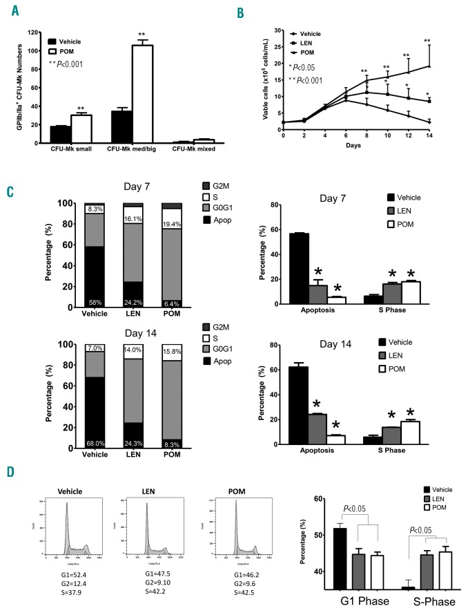
To determine the basis for thrombocytopenia after treatment with IMiDs, we analyzed the effect of POM on megakaryocytic colony formation of CD34+ cells. Under specific conditions allowing the development of CFU-Mk, POM significantly (P<0.001) increased the numbers of CFU-Mk in comparison to vehicle, with an increase from 53.2 (± 2.56) colonies (vehicle) to 144.8 (± 4.74) colonies (POM). The up-regulation of CFU-Mk was especially evident (P<0.001) in medium/large CFU-Mk (34.4 ± 4.03 colonies in vehicle versus 113.8 ± 5.91 colonies in POM), which arose from more primitive progenitors (Figure 1A). We also studied the effects of LEN and POM on proliferation and apoptosis of hematopoietic progenitors. Using CD34+ cells, IMiDs (LEN, P<0.05; POM, P<0.001) increased the absolute cell number in the cultures up to four-fold (LEN) and 10-fold (POM) after 2 weeks (Figure 1B). The cell expansion was not only the result of decreased apoptosis (PI+ cells, day 7: vehicle 58.0%, LEN 24.2%, and POM 6.4%; day 14: vehicle 68.0%, LEN 24.3%, and POM 8.3%; P<0.05), but also of increased mitosis according to the cell cycle analysis results. The proportion of cells in S phase increased (P<0.05) after CD34+ cells were treated with IMiDs on day 7: vehicle 8.3%, LEN 16.1%, POM 19.4% and day 14: vehicle 7.0%, LEN 14.0%, POM 15.8% (Figure 1C). Investigations of the effects of IMiDs on cell cycle regulation of progenitor cells revealed that S-phase cells were increased after IMiDs treatments (day 3: vehicle 37.9%, LEN 42.2%, POM 42.5% in CD34+ cells (Figure 1D).
Figure 1.
IMiDs increase megakaryocytic colony formation and induce self-renewal and expansion of hematopoietic progenitors. (A) Purified CD34+ cells were cultured with POM (pomalidomide) or DMSO as vehicle control using MegaCult-C assay to analyze formation of megakaryocytic colonies (CFU-Mk). Data shown are mean ± SEM from triplicates; Student’s t-test was performed, P-values are two-sided, **P<0.001 (B) Proliferation profiles of CD34+ cells expanded in serum-free HPGM hematopoietic growth medium supplemented with 10 ng/mL TPO with or without 10 μM IMiDs (LEN, lenalidomide or POM). Values shown are mean ± SEM of 7 separate cultures. P-values were calculated via one-way ANOVA with Bonferroni post hoc test. (*P<0.05 compared to vehicle; **P<0.001 compared to vehicle). (C) CD34+ cells cultured as described above were further analyzed by flow cytometry using propidium iodide (PI) staining for apoptosis and cell cycle at days 7 and 14 of culture. Cell pellets were stained for 30 minutes with equal volumes of phosphate-buffered saline (PBS) containing 0.1 mg/mL PI and 0.6% Nonidet P-40 and 2 mg/mL RNAse. Cell cycle analysis was performed on a Beckman Coulter CyAN 9-color High Speed Flowcytometer, and data were analyzed by using Summit 4.3 software (Dako¬Cytomation) (left). P-values were calculated via one-way ANOVA with Bonferroni corrections. Right side is statistic result of triplicates. (D) CD34+ cells were treated with DMSO (0.01%), LEN, or POM (1 μM) for 3 days. The cells gated with CD34+ and analyzed by flow cytometry using propidium iodide (PI) staining for cell cycle. The result shown here is representative of two independent experiment.
IMiDs inhibit megakaryocytic differentiation by blocking endomitosis and maturation
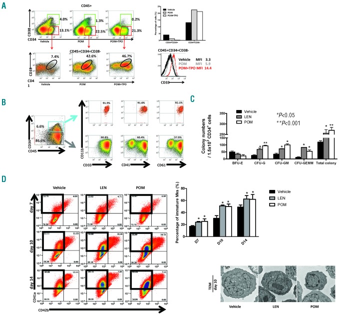
To characterize the CD34+ cells that exhibit increased proliferation and decreased apoptosis in long-term cultures with IMiDs, we performed flow cytometry after 14 days of culture in the presence of IL-3/IL-6/SCF with or without TPO. Flow cytometry revealed a strong induction of early CD34+ cells (CD34+CD38−) after 14 days of treatment with POM compared to vehicle (Figure 2A). Among these early CD34+ cells, more than 40% were double positive for myeloid and megakaryocytic markers, expressing CD33+ and CD41+, after treatment with POM. The development of the immature myeloid/megakaryocytic “hybrids” (CD34+/CD38−/CD41+/CD33+) was independent of TPO (vehicle 7.4%, POM 42.6%, POM+TPO 46.7%). Interestingly, the mean fluorescence intensity (MFI) of CD33 in the CD34+CD38− immature hybrid cells strongly increased when cultured with TPO (MFI: vehicle 3.3, POM 5.9, POM+TPO 14.4). Furthermore, in the presence of POM, we were able to culture and expand CD34+ cells for up to 4 months (Figure 2B). Characterization of cells from long-term cultures by multicolor flow cytometry revealed two cell populations. First, 6.6% of the cells were CD45+34+33+11b+41+61+. These “hybrid” cells maintained CD34+ expression with concomitant expression of myeloid (CD33, CD11b) and megakaryocytic (CD41 and CD61) markers. Second, 86.6% of the cells exhibited a more mature phenotype with loss of CD34 expression, but maintained co-expression of myeloid and megakaryocytic markers, CD45+34-33+11b+41+61+. Despite the culture conditions favoring thrombopoiesis, long-term cultured hematopoietic cells notably still expressed the myeloid markers CD11b and CD33, suggesting that IMiDs induce myeloid development. To address whether IMiDs have a sustained effect on expansion and self-renewal beyond direct exposure, we first cultured CD34+ cells in liquid cultures under the conditions described above. After 14 days, CD34+ cells were selected, and subjected to colony formation assays without adding IMiDs. Even without the continued presence of IMiDs, total colony formation was still significantly increased [vehicle 120, LEN 256 (P<0.05), POM 270 (P<0.001)]. In accordance with prior data,17 we again observed a shift to myeloid lineage commitment with increased myeloid colonies (CFU-G, CFU-GM and CFU-GEMM) at the expense of erythroid colonies (P<0.05; Figure 2C). These data suggested that IMiDs persistently affect self-renewal and lineage commitment of CD34+ cells, resulting in maintenance and expansion of the progenitor cell pool. Our finding showed that IMiDs-induced development of megakaryocytic precursors appears to contrast with the thrombocytopenia in patients caused by IMiDs. Since our previous studies showed that IMiDs are not directly toxic to bone marrow hematopoietic cells,16 we hypothesized that these effects are induced by maturational arrest. To investigate the effects of IMiDs on the maturation of Mks, we evaluated different megakaryocytic cell populations by flow cytometry. CD41a is a lineage marker of Mks throughout all stages of differentiation, whereas CD42b expression is expressed by more mature Mks.18 Hence, CD41a+/CD42b− represents immature Mks, and CD41a+/CD42b+ represents more mature Mks. Flow cytometric analyses showed that the proportion of immature CD41a+/CD42b− cells was significantly increased in the presence of IMiDs (day 7: vehicle 15.8%, LEN 23.0 %, POM 22.9%; day 10: vehicle 32.1%, LEN 50.6%, POM 51.0%; day 14: vehicle 49.7%, LEN 64.7%, POM 66.0%; P<0.05). This difference was visible as soon as day 7 of culture and was maintained throughout the entire culture period (Figure 2D). To confirm the immature and dysplastic morphology, we performed transmission electron microscopy (TEM) after 10 days of IMiD treatment. Mks were identified by CD61-microbead labeling. Again, compared with vehicle, IMiD-treated Mks exhibited immature features, including decreased size, less cytoplasm, and a heterogeneously dilated and abnormally distributed demarcation membrane system (DMS) in the cytoplasm. (Figure 2E)
Figure 2.
IMiDs inhibit megakaryocytic differentiation by blocking endomitosis and maturation. (A) Upper panel: Flow cytometry analysis revealed a strong induction of CD34+CD38−cells (early progenitors) after 14 days of treatment with POM +/− TPO compared to vehicle. Lower panel: Early CD34+CD38− progenitors were further analyzed for CD33+ and CD41+ expressions. Lower right panel: The mean fluorescence intensity (MFI) of CD33 in these CD34+CD38− immature hybrid cells dramatically increased with TPO. (B) In long-term cultures, CD34+ cells were maintained for up to 4 months and multicolor flow cytometry identified 2 cell populations: CD45+34+33+11b+41+61+ hybrid cells (6.6%) and a more mature population of CD45+34-41+61+ cells (86.6%). Without POM, the control cultured cells could be maintained only for up to 3 weeks and were therefore not available for comparison. Data are from one experiment. (C) Purified CD34+ cells were cultured in serum-free HPGM hematopoietic growth medium with DMSO or IMiDs. After culturing for 14 days, CD34+ cells from each group (vehicle, LEN and POM) were selected by immunomagnetic beads and were plated in MethoCult for colony formation assays (with either vehicle, LEN or POM). Data are from one experiment with colonies quantified in triplicate wells. *P<0.05 compared to Vehicle; **P<0.001 compared to Vehicle. Data were compared by one-way ANOVA with Bonferroni post-test. (D) CD34+ cells were cultured in serum-free HPGM hematopoietic growth medium with TPO to induce megakaryopoiesis with or without LEN, POM or DMSO as vehicle. After 7, 10 and 14 days, flow cytometry analysis showed an increase of immature Mks (CD41a+/CD42b−), while more mature Mks (CD41a+/CD42b+) decreased with IMIDs treatment. The result shown here is one representative experiment of triplicates. (E) Morphology of Mks, derived from CD34+ cells grown in serum-free HPGM hematopoietic growth medium with TPO with or without IMiDs at day 7 and 10 of culture. Electron micrographs of three characteristic Mks from different groups (vehicle, LEN and POM) are shown. Mks were identified by CD61+ magnetic bead labeling. Mks in LEN and POM group show features of immaturity, including scant cytoplasm, large nuclei, and a minimal demarcation membrane system. Images are from one experiment representative of two independent experiments each of which included triplicates.
IMiD decreased protein expression of GATA1 and its targets in Mk progenitors
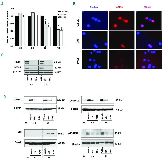
To gain insight into the mechanism of inhibition of megakaryocytic maturation by IMiDs, we evaluated several transcription factors involved in the regulation of megakaryocytic differentiation and maturation. Failure of terminal differentiation and excessive proliferation of Mks have been described to occur in the absence of GATA1.19 By RT-PCR we found significantly (P<0.05) decreased GATA1 levels in Mks treated with IMiDs (Figure 3A). The decrease of GATA1 by IMiDs was confirmed by immunofluorescence of megakaryocytic progenitors (Figure 3B) and Western blot analysis (Figure 3C). We have shown that IMiDs induce cereblone-ubiquitination with subsequent IKZF1 degradation in CD34+ cells8. ZFPM1 (FOG-1), similar to its interacting partner, GATA1, is required for normal differentiation of erythroid precursors and Mks.20 Moreover, most GATA1-regulated events require GATA1 to bind to ZFPM1.21,22 Subsequently, we asked if IMiDs also modulate the expression of ZFPM1. Figure 3D shows that in IMiD-treated CD34+ cells, ZFPM1 is almost completely abrogated after 9 days of treatment with kinetics similar to those of GATA1. GATA1 and ZFPM1 synergistically activate the p45 NFE2 promoter,23,24 which is essential for late maturation of Mks and platelet formation. Accordingly, we found that expression of NFE2 were down-regulated in IMiD-treated cells (Figure 3D). It is known that polyploidy formation in Mks depends on the expression of cyclin D isotypes, and that cyclin D1 is a direct target of GATA1.25 In accordance with this, we found that IMiD-induced downregulation of GATA1 was associated with decreased cyclin D1 (Figure 4D), contributing to the inhibition of maturation. The CDK inhibitor, p16, potently inhibits endomitosis of Mks25 and is decreased upon differentiation of Mks.26 Analysis of p16 by Western blot revealed that p16 was up-regulated by IMiD treatment (Figure 4E), suggesting that the loss of GATA1 induced a cascade of inhibitors of megakaryocytic maturation.
Figure 3.
IMiD decreased protein expression of GATA1 and its targets in Mk progenitors. CD34+ cells were cultured in serum-free HPGM hematopoietic growth medium supplemented with 10 ng/mL TPO to initiate megakaryopoiesis with or without 10 μM IMiDs for the indicated times. (A) Real-time PCR analysis was applied to determine the levels of GATA1 mRNA. The result shown here is one representative experiment of triplicates. (B) Immunofluorescence microscopy was performed to examine the expression of GATA1 on CD34+ cells cultured in serum-free HPGM medium containing 10ng/mL TPO with 10 μM LEN, POM or 0.1% DMSO as vehicle control at day 6. Cells were stained with an antibody that recognize the N terminus of GATA1 (red) and counterstained with DAPI (blue) to visualize the nucleus. Both stainings were merged and revealed a loss of GATA1 when treated with IMiDs. Images shown here is one representative experiment of triplicates. (C) Western blot analysis was applied to determine the levels of GATA1 and IKZF1 protein. β-actin was used as the loading control. Data are representative of three independent experiments. (D) Purified CD34+ cells were cultured in serum-free HPGM hematopoietic growth medium containing 10 ng/mL TPO with 10 μM LEN, POM or 0.1% DMSO as vehicle control for 6 days and 9 days. The expression of the indicated proteins was analyzed by Western blotting. β-actin was used as the loading control. Data are representative of three independent experiments.
Figure 4.
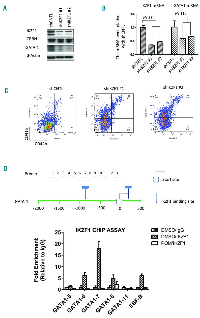
IKZF1 mediates the IMiD-induced inhibition of megakaryocytic maturation. (A) IKZF1 shRNA #1 (shIKZF1-1), #2 (shIKZF1-2) or control shRNA (shCNTL) transduced CD34+cell lysates were analyzed by western blotting to compare the levels of IKZF1, CRBN and GATA1. The result shown here is one representative experiment of triplicates. (B) CD34+ cells were transduced using a lentivirus carrying the control shRNA (shCNTL), IKZF1-shRNA #1 (shIKZF1-1), or IKZF1-shRNA #2 (shIKZF1-2) sequence and cultured in serum-free HPGM hematopoietic growth medium with TPO to induce megakaryopoiesis. At 6 days after transduction, the cells were sorted with GFP and subsequently lysed and analyzed for IKZF1 and GATA1 mRNA levels using qRT-PCR. The result shown here is one representative experiment of triplicates. (C) CD34+ cells were transduced using a lentivirus carrying the control shRNA (shCNTL), IKZF1-shRNA #1 (shIKZF1-1), or IKZF1-shRNA #2 (shIKZF1-2) sequence. CD34+ cells were cultured in serum-free HPGM hematopoietic growth medium with TPO to induce megakaryopoiesis. After 10 days, flow cytometry analyzed the cells gated with GFP and showed an increase of immature Mks (CD41a+/CD42b−) with IKZF1 knockdown. The result shown here is one representative experiment of triplicates. (D) CD34+ cells were treated with DMSO (0.01%) or POM (1 μM) for 24 h and cell lysates were analyzed by chromatin immuno-precipitation (CHIP) using IKZF1 antibody. Control IgG was used as a negative control. The precipitated DNA fragments were subjected to qRT-PCR analysis with primers amplifying the GATA1 promoter. The result shown here is representative of two independent experiments.
IKZF1 mediates the IMiD-induced inhibition of megakaryocytic maturation
Previous studies have demonstrated that lenalidomide induces binding of IKZF1 and IKZF3 to CRBN and promotes their ubiquitination and degradation.27,28 Since IKZF1 is required for the development of the erythroid lineage,29 we were interested in the effects of IMiDs on the interaction between IKZF1 and GATA1 in CD34+cells. So, we examined the role of IKZF1 in POM-induced inhibition of megakaryocytic maturation. First, we confirmed that IKZF1 regulates GATA1 expression in CD34+ cells by knockdown of IKZF1 in CD34+ cells. Knockdown of IKZF1 resulted in decreased GATA-1 protein expression, further suggesting that GATA1 is under IKZF1 regulation (Figure 4A). In IKZF1 knockdown cells, the mRNA level of GATA-1 was significantly decreased, suggesting that GATA-1 expression is regulated by IKZF1 (Figure 4B). To examine the effects of IKZF1 in megakaryopoiesis, we performed flow cytometric analyses. In IKZF1 knockdown cells, the proportion of immature CD41a+/CD42b− cells was significantly increased, suggesting that IKZF1 downregulation is critical for the POM-induced maturational arrest (Figure 4C). To find how GATA1 is regulated at a transcriptional level, we analyzed GATA-1 promoter sequences and several potential IKZF1 binding sites (Figure 4D upper). Indeed, In CHIP assays, we confirmed that IKZF1 binds directly to the GATA1 promoter area. Treatment of CD34+ cells with POM completely inhibited IKZF1 binding to the GATA1 promotor due to the decreased IKZF1 levels (Figure 4D bottom). Our findings showed that IMiDs promote CRBN-dependent degradation of IKZF1 protein in CD34+ cells and decrease GATA1.
Overexpression of GATA1 abrogated the effects of IMiDs on megakaryopoiesis
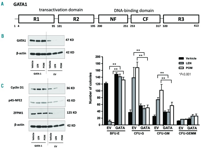
To further confirm the critical role of GATA1 in inhibiting maturation of Mks by IMiDs, we stably overexpressed GATA1 in human CD34+ cells using the pLenti V5 GATA1 expression vector system (Figure 5A). Overexpression (OE) of GATA1 prevented its downregulation by IMiDs compared to control cells transfected with empty vector (EV) alone (Figure 5B). Concomitant with the overexpression of GATA1, GATA1 co-factors and targets, such as ZFPM1, NFE2 and cyclin D1, remained stably expressed despite IMiDs treatment (Figure 5C). Thus, GATA1 appeared to play a critical role in the IMiDs-induced inhibition of megakaryocytic maturation. Most importantly, OE of GATA1 widely abrogated the effects of IMiDs on lineage commitment (Figure 5D). The numbers of BFU-E colonies increased significantly (P<0.001), whereas the numbers of CFU-G/GM colonies in the IMiDs group decreased significantly (P<0.001) when compared to EV-transfected cells. OE of GATA1 abrogated the IMiDs-induced shift of lineage commitment toward myelopoiesis at the expense of erythropoiesis/megakaryopoiesis.
Figure 5.
Overexpression of GATA1 reversed the effects of IMiDs on megakaryopoiesis. (A) Schematic diagram of the structure of GATA1pLenti V5 topo expression constructs. CF and NF represent C- and N-fingers of GATA1; R1, R2, R3 represent three regions of the transactivation domain of GATA1 (modified from reference45). (B) The expression of GATA1 in either EV or GATA1 transfected CD34+ cells were analyzed by Western blotting. CD34+ cells transfected with EV or GATA1 were cultured in serum-free HPGM hematopoietic growth medium containing 10 ng/mL TPO with either vehicle, LEN or POM for 6 days. β-actin was used as the loading control. Data are representative of three independent experiments. (C) Purified CD34+ cells were transfected with GATA1 or EV and cultured in serum-free HPGM hematopoietic growth medium containing 10 ng/mL TPO to induce megakaryocytic development with LEN, POM or DMSO as vehicle control for 6 days. The expression of the indicated proteins was analyzed by Western blotting. β-actin was used as the loading control. Data are representative of three independent experiments. (D) EV and GATA1 transfected CD34+ cells were subjected to colony formation assays using standard MethoCult assays with and without IMiDs. Overexpression of GATA1 abrogated the IMiD-induced upregulation of myeloid lineage commitment and rescued the development of BFU-E. Data shown are mean ± SEM from triplicates; **P<0.001, compared with Vehicle group.
Figure 6.
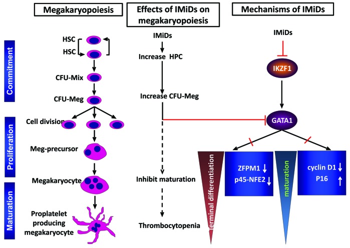
Downregulation of GATA1 by IMiDs induces renewal and expansion of hematopoietic progenitors with a concomitant block of megakaryocytic maturation. Treatment with IMiDs induces self-renewal of hematopoietic progenitors and upregulates megakaryocytic colonies (CFU-Meg) by inhibiting apoptosis and increasing proliferation of early megakaryocytic progenitors. IMiDs down-regulate the transcription factor GATA1 and thereby affect transcription factors and cell cycle regulators controlled by GATA1. The subsequent decrease of ZFPM1 and NFE2 leads to expansion of megakaryocytic progenitors with concomitant inhibition of maturation. A decrease of cyclin D1 and an increase of p16 results in the block of megakaryocytic maturation (Adapted from Figure 7 of reference 25, with permission of Dr. John D. Crispino).
Discussion
Despite the fact that IMiDs are not directly cytotoxic, their use is associated with severe thrombocytopenia (grade 3/4) in up to 25% of patients with MM.10,30 The mechanism for induction of thrombocytopenia is unknown. Here, we determined that IMiDs dramatically expand CD34+ hematopoietic progenitors in liquid cultures and significantly induce megakaryopoiesis and megakaryocytic colony formation. Interestingly, IMiDs generated mainly immature CD41a+/CD42b− megakaryocytes. This was further confirmed by transmission electron microscopy, revealing structural abnormalities of Mks that also suggested a maturational block in megakaryopoiesis.31,32 More strikingly, we were able to maintain a small pool of CD34+ cells for as long as 4 months in liquid culture. Besides CD34+ expression, the long-term cultured cells maintained concomitant expression of myeloid (CD33, CD11b) and megakaryocytic (CD41, CD61) markers, reflecting a phenotype that is similar to acute megakaryoblastic leukemia (AMKL) with GATA1 mutations33,34. GATA1 is a transcription factor critical for development of erythroid and megakaryocytic cells. GATA1 mutations in humans act as a dominant leukemogenic oncogene in megakaryocytic progenitors and cause inherited thrombocytopenia.31 In our studies, the IMiDs-induced proliferation of hematopoietic progenitors and inhibition of maturation of Mks were associated with a loss of GATA1, ZFPM1 (FOG-1) and NFE2 expression. ZFPM1 acts as a cofactor for GATA1 and provides a paradigm for the regulation of cell type-specific gene expression by GATA1.22 GATA1 also drives the expression of another important transcription factor, NFE2 that exists as a heterodimer comprised of p45 and p18 subunits. p45Nfe2−/− mice present with an increase in Mks, a marked defect in maturation and profound thrombocytopenia.35,36 This indicates that terminal maturation of Mks depends heavily on the GATA1/NFE2 axis. In addition to promot ing differentiation of Mks, GATA1 also leads ultimately to cessation of cell proliferation.19,37 This is consistent with our findings showing that IMiDs-induced loss of GATA1 expression in Mks not only inhibits maturation, but also leads to excessive proliferation of megakaryocytic progenitors. GATA1 also regulates numerous CDKs such as cyclin D1 and CDK inhibitors.25 Moreover, overexpression of cyclin D1/CDK4 in GATA1-deficient Mks restored their growth and polyploidization.25 Correspondingly, we observed that downregulating GATA1 in Mks by IMiDs resulted in decreased cyclin D1 expression.
Our findings of the role of IKZF1 in regulating GATA1 expression are in accordance with results by Dijon et al., who reported that lentivirally induced Ik6 (a dominant negative isoform of IKZF1) overexpression resulted in decreased expression of GATA1.29 Cell-cycle regulation is an important mechanism governing the long-term self-renewal potential of HSCs.38 Therefore, long-term treatment of patients with IMiDs might induce a pool of constantly cycling progenitors, leading to premature exhaustion of the stem cell pool. Indeed, patients receiving long-term treatment with IMiDs very often exhibit a hypocellular bone marrow associated with cytopenias.39 Interestingly, Malinge and colleagues reported that IKZF1 knockout mice showed increased megakarayopoiesis. Consistent with this phenotype, studies using mice acute megakaryoblastic leukemia (AMKL) cell line 6133 showed that IKZF1 suppresses megakaryopoiesis by negatively regulating GATA1.40 In contrast, our experiments found that the knockdown of IKZF1 resulted in decreased GATA1 protein expression in human CD34+ cells. The mechanism of IKZF1 in regulating GATA1 in mice malignant cells may be different from that in human hematopoietic progenitor cells.
Interestingly, Kronke et al. reported that Lenalidomide induces ubiquitination and degradation of Casein kinase 1α (CK1α) in del(5q) MDS.41 Furthermore, in a murine model with conditional inactivation of Csnk1a1, Schneider et al. demonstrated that Csnk1a1 haploinsufficiency induces hematopoietic stem cell expansion.42 This is in accordance with our data showing that both pomalidomide and lenlidomide potently downregulate CK1α in hematopoietic progenitors, although pomalidomide exhibits a slightly lower efficiency compared to Lenalidomide (Online Supplementary Figure S1).
GATA1 overexpression allowed development of a more mature Mk phenotype. In addition, it preserved expression of megakaryocyte-specific transcription factors such as NFE2 and ZFPM1 and of cell cycle regulators, including cyclin D1, despite IMiDs treatment, which further indicates that an IMiDs-induced decrease in GATA1 critically affects pathways involved in self-renewal, cell cycle regulation and lineage commitment of CD34+ cells. GATA1 mutations resulting in functional silencing have been found in patients with thrombocytopenia or megakaryocytic acute leukemia.32,43 Therefore, our data suggest that the IMiDs-induced downregulation of IKZF1 and GATA1 favors myeloid lineage commitment and maintains a pool of immature cycling CD34+ cells without maturation, which may lead to stem cell exhaustion. Furthermore, downregulation of GATA1 results in an imbalance of key cellular and molecular regulators that block Mks from continued maturation, likely impeding platelet release and ultimately resulting in thrombocytopenia. sAt this moment, the clinical relevance of our in vitro findings is not entirely clear. However, prolonged treatment with IMiDs has been shown to be associated with an increased risk of MDS/AML and ALL, especially in patients treated with alkylating agents like Melphalan.44 It is therefore possible that the IMiDs-induced increased number of cycling CD34+ cells may enhance the probability of acquiring secondary DNA damage and leukemogenic events induced by other drugs. Hence, in vivo testing of various treatment combinations with IMiDs to explore and potentially predict leukemogenic effects of certain combinations is needed.
Supplementary Material
Acknowledgments
The authors would like to thank Mr. Dale Lewis at the University of Pittsburgh Cell Culture and Cytogenetics Facility for excellent FISH analysis. We would also like to thank Dr. Griffin P. Rodgers at the Molecular and Clinical Hematology Branch, National Heart, Lung, and Blood Institute, Bethesda, MD for providing the pLenti V5 GATA1 expression vectors and empty vectors.
Footnotes
Check the online version for the most updated information on this article, online supplements, and information on authorship & disclosures: www.haematologica.org/content/103/10/1688
Funding
This study was supported by a grant from the LLS and R01CA175313 (SL). MYM, CL, HM were supported in part by RO1 HL093716.
References
- 1.Bartlett JB, Dredge K, Dalgleish AG. The evolution of thalidomide and its IMiD derivatives as anticancer agents. Nat Rev Cancer. 2004;4(4):314–322. [DOI] [PubMed] [Google Scholar]
- 2.Chang DH, Liu N, Klimek V, et al. Enhancement of ligand-dependent activation of human natural killer T cells by lenalidomide: therapeutic implications. Blood. 2006;108(2):618–621. [DOI] [PMC free article] [PubMed] [Google Scholar]
- 3.Quach H, Ritchie D, Stewart AK, et al. Mechanism of action of immunomodulatory drugs (IMiDS) in multiple myeloma. Leukemia. 2010;24(1):22–32. [DOI] [PMC free article] [PubMed] [Google Scholar]
- 4.Kronke J, Udeshi ND, Narla A, et al. Lenalidomide causes selective degradation of IKZF1 and IKZF3 in multiple myeloma cells. Science. 2014;343(6168):301–305. [DOI] [PMC free article] [PubMed] [Google Scholar]
- 5.Chapman MA, Lawrence MS, Keats JJ, et al. Initial genome sequencing and analysis of multiple myeloma. Nature. 2011; 471(7339):467–472. [DOI] [PMC free article] [PubMed] [Google Scholar]
- 6.Li S, Pal R, Monaghan SA, et al. IMiD immunomodulatory compounds block C/EBP{beta} translation through eIF4E down-regulation resulting in inhibition of MM. Blood. 2011;117(19):5157–5165. [DOI] [PMC free article] [PubMed] [Google Scholar]
- 7.Lopez-Girona A, Heintel D, Zhang LH, et al. Lenalidomide downregulates the cell survival factor, interferon regulatory factor-4, providing a potential mechanistic link for predicting response. Br J Haematol. 2011; 154(3):325–336. [DOI] [PubMed] [Google Scholar]
- 8.Li S, Fu J, Mapara M, Lentzsch S. IMiD® compounds affect the hematopoiesis via CRBN dependent degradation of IKZF1 protein in CD34+ Cells. Blood. 2014; 124(21):418–418. [Google Scholar]
- 9.Weber DM, Chen C, Niesvizky R, et al. Lenalidomide plus dexamethasone for relapsed multiple myeloma in North America. N Engl J Med. 2007;357(21):2133–2142. [DOI] [PubMed] [Google Scholar]
- 10.Lacy MQ, Allred JB, Gertz MA, et al. Pomalidomide plus low-dose dexamethasone in myeloma refractory to both bortezomib and lenalidomide: comparison of 2 dosing strategies in dual-refractory disease. Blood. 2011;118(11):2970–2975. [DOI] [PMC free article] [PubMed] [Google Scholar]
- 11.Mazumder A, Kaufman J, Niesvizky R, Lonial S, Vesole D, Jagannath S. Effect of lenalidomide therapy on mobilization of peripheral blood stem cells in previously untreated multiple myeloma patients. Leukemia. 2008;22(6):1280–1281; author reply 1281–1282. [DOI] [PubMed] [Google Scholar]
- 12.Dimopoulos M, Spencer A, Attal M, et al. Lenalidomide plus dexamethasone for relapsed or refractory multiple myeloma. N Engl J Med. 2007;357(21):2123–2132. [DOI] [PubMed] [Google Scholar]
- 13.Attal M, Lauwers-Cances V, Marit G, et al. Lenalidomide maintenance after stem-cell transplantation for multiple myeloma. N Engl J Med. 2012;366(19):1782–1791. [DOI] [PubMed] [Google Scholar]
- 14.McCarthy PL, Owzar K, Hofmeister CC, et al. Lenalidomide after stem-cell transplantation for multiple myeloma. N Engl J Med. 2012;366(19):1770–1781. [DOI] [PMC free article] [PubMed] [Google Scholar]
- 15.Palumbo A, Hajek R, Delforge M, et al. Continuous lenalidomide treatment for newly diagnosed multiple myeloma. N Engl J Med. 2012;366(19):1759–1769. [DOI] [PubMed] [Google Scholar]
- 16.Koh KR, Janz M, Mapara MY, et al. Immunomodulatory derivative of thalidomide (IMiD CC-4047) induces a shift in lineage commitment by suppressing erythropoiesis and promoting myelopoiesis. Blood. 2005;105(10):3833–3840. [DOI] [PubMed] [Google Scholar]
- 17.Pal R, Monaghan SA, Hassett AC, et al. Immunomodulatory derivatives induce PU.1 down-regulation, myeloid maturation arrest, and neutropenia. Blood. 2010; 115(3):605–614. [DOI] [PubMed] [Google Scholar]
- 18.Poirault-Chassac S, Six E, Catelain C, et al. Notch/Delta4 signaling inhibits human megakaryocytic terminal differentiation. Blood. 2010;116(25):5670–5678. [DOI] [PubMed] [Google Scholar]
- 19.Vyas P, Ault K, Jackson CW, Orkin SH, Shivdasani RA. Consequences of GATA-1 deficiency in megakaryocytes and platelets. Blood. 1999;93(9):2867–2875. [PubMed] [Google Scholar]
- 20.Cantor AB, Katz SG, Orkin SH. Distinct domains of the GATA-1 cofactor FOG-1 differentially influence erythroid versus megakaryocytic maturation. Mol Cell Biol. 2002;22(12):4268–4279. [DOI] [PMC free article] [PubMed] [Google Scholar]
- 21.Wilkinson-White L, Gamsjaeger R, Dastmalchi S, et al. Structural basis of simultaneous recruitment of the transcriptional regulators LMO2 and FOG1/ZFPM1 by the transcription factor GATA1. Proc Natl Acad Sci USA. 2011;108(35):14443–14448. [DOI] [PMC free article] [PubMed] [Google Scholar]
- 22.Tsang AP, Visvader JE, Turner CA, et al. FOG, a multitype zinc finger protein, acts as a cofactor for transcription factor GATA-1 in erythroid and megakaryocytic differentiation. Cell. 1997;90(1):109–119. [DOI] [PubMed] [Google Scholar]
- 23.Takayama M, Fujita R, Suzuki M, et al. Genetic analysis of hierarchical regulation for Gata1 and NF-E2 p45 gene expression in megakaryopoiesis. Mol Cell Biol. 2010; 30(11):2668–2680. [DOI] [PMC free article] [PubMed] [Google Scholar]
- 24.Wang X, Crispino JD, Letting DL, Nakazawa M, Poncz M, Blobel GA. Control of megakaryocyte-specific gene expression by GATA-1 and FOG-1: role of Ets transcription factors. EMBO J. 2002;21(19):5225–5234. [DOI] [PMC free article] [PubMed] [Google Scholar]
- 25.Muntean AG, Pang L, Poncz M, Dowdy SF, Blobel GA, Crispino JD. Cyclin D-Cdk4 is regulated by GATA-1 and required for megakaryocyte growth and polyploidization. Blood. 2007;109(12):5199–5207. [DOI] [PMC free article] [PubMed] [Google Scholar]
- 26.Furukawa Y, Kikuchi J, Nakamura M, Iwase S, Yamada H, Matsuda M. Lineage-specific regulation of cell cycle control gene expression during haematopoietic cell differentiation. Br J Haematol. 2000; 110(3):663–673. [DOI] [PubMed] [Google Scholar]
- 27.Kronke J, Udeshi ND, Narla A, et al. Lenalidomide causes selective degradation of IKZF1 and IKZF3 in multiple myeloma cells. Science. 2014;343(6168):301–305. [DOI] [PMC free article] [PubMed] [Google Scholar]
- 28.Lu G, Middleton RE, Sun HH, et al. The myeloma drug lenalidomide promotes the cereblon-dependent destruction of Ikaros proteins. Science. 2014;343(6168):305–309. [DOI] [PMC free article] [PubMed] [Google Scholar]
- 29.Dijon M, Bardin F, Murati A, Batoz M, Chabannon C, Tonnelle C. The role of Ikaros in human erythroid differentiation. Blood. 2008;111(3):1138–1146. [DOI] [PubMed] [Google Scholar]
- 30.Raza A, Reeves JA, Feldman EJ, et al. Phase 2 study of lenalidomide in transfusion-dependent, low-risk, and intermediate-1 risk myelodysplastic syndromes with karyotypes other than deletion 5q. Blood. 2008; 111(1):86–93. [DOI] [PubMed] [Google Scholar]
- 31.Li Z, Godinho FJ, Klusmann JH, Garriga-Canut M, Yu C, Orkin SH. Developmental stage-selective effect of somatically mutated leukemogenic transcription factor GATA1. Nat Genet. 2005;37(6):613–619. [DOI] [PubMed] [Google Scholar]
- 32.Wechsler J, Greene M, McDevitt MA, et al. Acquired mutations in GATA1 in the megakaryoblastic leukemia of Down syndrome. Nat Genet. 2002;32(1):148–152. [DOI] [PubMed] [Google Scholar]
- 33.Choi JK. Hematopoietic disorders in Down syndrome. Int J Clin Exp Pathol. 2008;1(5):387–395. [PMC free article] [PubMed] [Google Scholar]
- 34.Lorsbach RB. Megakaryoblastic disorders in children. Am J Clin Pathol. 2004;122 Suppl(S33–46). [DOI] [PubMed] [Google Scholar]
- 35.Shivdasani RA, Rosenblatt MF, Zucker-Franklin D, et al. Transcription factor NF-E2 is required for platelet formation independent of the actions of thrombopoietin/MGDF in megakaryocyte development. Cell. 1995;81(5):695–704. [DOI] [PubMed] [Google Scholar]
- 36.Lecine P, Villeval JL, Vyas P, Swencki B, Xu Y, Shivdasani RA. Mice lacking transcription factor NF-E2 provide in vivo validation of the proplatelet model of thrombocytopoiesis and show a platelet production defect that is intrinsic to megakaryocytes. Blood. 1998;92(5):1608–1616. [PubMed] [Google Scholar]
- 37.Papetti M, Wontakal SN, Stopka T, Skoultchi AI. GATA-1 directly regulates p21 gene expression during erythroid differentiation. Cell Cycle. 2010;9(10):1972–1980. [DOI] [PMC free article] [PubMed] [Google Scholar]
- 38.Orford KW, Scadden DT. Deconstructing stem cell self-renewal: genetic insights into cell-cycle regulation. Nat Rev Genet. 2008; 9(2):115–128. [DOI] [PubMed] [Google Scholar]
- 39.Fouquet G, Tardy S, Demarquette H, et al. Efficacy and safety profile of long-term exposure to lenalidomide in patients with recurrent multiple myeloma. Cancer. 2013; 119(20):3680–3686. [DOI] [PubMed] [Google Scholar]
- 40.Malinge S, Thiollier C, Chlon TM, et al. Ikaros inhibits megakaryopoiesis through functional interaction with GATA-1 and NOTCH signaling. Blood. 2013; 121(13):2440–2451. [DOI] [PMC free article] [PubMed] [Google Scholar]
- 41.Kronke J, Fink EC, Hollenbach PW, et al. Lenalidomide induces ubiquitination and degradation of CK1alpha in del(5q) MDS. Nature. 2015;523(7559):183–188. [DOI] [PMC free article] [PubMed] [Google Scholar]
- 42.Schneider RK, Adema V, Heckl D, et al. Role of casein kinase 1A1 in the biology and targeted therapy of del(5q) MDS. Cancer Cell. 2014;26(4):509–520. [DOI] [PMC free article] [PubMed] [Google Scholar]
- 43.Nichols KE, Crispino JD, Poncz M, et al. Familial dyserythropoietic anaemia and thrombocytopenia due to an inherited mutation in GATA1. Nat Genet. 2000; 24(3):266–270. [DOI] [PMC free article] [PubMed] [Google Scholar]
- 44.Badros AZ. Lenalidomide in myeloma–a high-maintenance friend. N Engl J Med. 2012;366(19):1836–1838. [DOI] [PubMed] [Google Scholar]
- 45.Zhu J, Chin K, Aerbajinai W, Trainor C, Gao P, Rodgers GP. Recombinant erythroid Kruppel-like factor fused to GATA1 up-regulates delta- and gamma-globin expression in erythroid cells. Blood. 2011;117(11): 3045–3052. [DOI] [PMC free article] [PubMed] [Google Scholar]
Associated Data
This section collects any data citations, data availability statements, or supplementary materials included in this article.


