Abstract
Respiratory inflammation is a frequent and fatal pathologic state encountered in veterinary medicine. Although diluted bee venom (dBV) has potent anti-inflammatory effects, the clinical use of dBV is limited to several chronic inflammatory diseases. The present study was designed to propose an acupoint dBV treatment as a novel therapeutic strategy for respiratory inflammatory disease. Experimental pleurisy was induced by injection of carrageenan into the left pleural space in mouse. The dBV was injected into a specific lung meridian acupoint (LU-5) or into an arbitrary non-acupoint located near the midline of the back in mouse. The inflammatory responses were evaluated by analyzing inflammatory indicators in pleural exudate. The dBV injection into the LU-5 acupoint significantly suppressed the carrageenan-induced increase of pleural exudate volume, leukocyte accumulation, and myeloperoxidase activity. Moreover, dBV acupoint treatment effectively inhibited the production of interleukin 1 beta, but not tumor necrosis factor alpha in the pleural exudate. On the other hand, dBV treatment at non-acupoint did not inhibit the inflammatory responses in carrageenan-induced pleurisy. The present results demonstrate that dBV stimulation in the LU-5 lung meridian acupoint can produce significant anti-inflammatory effects on carrageenan-induced pleurisy suggesting that dBV acupuncture may be a promising alternative medicine therapy for respiratory inflammatory diseases.
Keywords: acupuncture, anti-inflammation, bee venoms, carrageenan-induced pleurisy
Introduction
Inflammation in a respiratory system, such as that associated with pneumonia, pleurisy, and bronchitis, is a frequent and fatal disorder encountered in veterinary medicine. In general, use of steroidal and non-steroidal anti-inflammatory drugs has been a major recommended strategy for the treatment of inflammatory disease [23]. However, though they are effective at relieving inflammation, many reports have demonstrated that the chronic use of these drugs is associated with a significant risk of serious adverse events in gastrointestinal [27], renal [9], and cardiovascular [3] systems. Because of these complications, much attention has been focused on alternative medicine approaches to the treatment of inflammatory diseases [4].
Subcutaneous injection of diluted bee venom (dBV) into an acupoint (termed apipuncture) has been used clinically in traditional oriental medicine, which produces potent anti-inflammatory effects in chronic inflammation patients [32]. Bee venom (BV) is comprised of various water-soluble extracts including peptides, enzymes, and biologically active amines that can stimulate the peripheral nerves and subsequently activate neurons in the brain and spinal cord [16]. Several reports have demonstrated that stimulation of the ST-36 acupoint with dBV significantly activates neurons in the locus coeruleus (LC) [37]. Since LC is the center of the descending noradrenergic system, increased activity of the LC provokes remarkable anti-inflammatory effects through the production of adrenal catecholamine [15]. Nevertheless, the use of dBV apipuncture has been limited to musculoskeletal inflammatory diseases such as arthritis [10], and it has not been considered for use in a therapeutic strategy for the treatment of respiratory inflammatory diseases.
Intrapleural injection of carrageenan induces acute inflammation in the pleural cavity through extravasation of polymorphonuclear leukocytes and macrophages into the pleural space after production of various proinflammatory cytokines or reactive oxygen species [2,8]. Because of these characteristics of carrageenan-induced inflammation, carrageenan-induced pleurisy has been used as a preclinical animal model when studying the mechanisms involved in acute respiratory inflammatory disease or when screening the effects of anti-inflammatory drugs on the respiratory system [34]. On that basis, carrageenan-induced pleurisy is an appropriate animal model for evaluating the anti-inflammatory effects of dBV apipuncture on respiratory inflammatory diseases in veterinary medicine.
By using a carrageenan-induced pleurisy mouse model, the present study aimed to investigate whether (1) stimulation of a specific acupoint with dBV is associated with modulation of the respiratory inflammation; and (2) dBV apipuncture can be an effective alternative therapy in the treatment of respiratory inflammation.
Materials and Methods
Animal preparation
Male Balb/c mice weighing 23 to 25 g and purchased from HanLim Experimental Animal (Korea) were used for this experiment. The animals were housed under controlled conditions: 12 h light/dark cycle, standard temperature (24 ± 2℃), and 40% to 50% humidity. Food and water were provided freely throughout the investigation. All mice were acclimated for one week prior to the beginning of the experiments. The entire experimental procedure and all methods were reviewed and approved by the Seoul National University Animal Care and Use Committee (approval No. SNU-160602-11), following the National Institutes of Health guidelines [33] and were in compliance with the ethical guidelines for investigations of experimental pain in conscious animals [39]. Animals were randomly assigned to experimental groups and subsequent drug treatments and analyses were performed blindly.
Carrageenan-induced pleurisy
Carrageenan-induced pleurisy was performed according to the method described in a previous report but with slight modification [24]. Briefly, mice were anesthetized with 3% isoflurane in a mixture of N2O/O2 gas and submitted to a skin incision at the level of the left sixth intercostal space. Sterile phosphate buffer saline (PBS; vehicle, 200 µL) or PBS containing 2% of λ-carrageenan (Sigma, USA) was injected into the pleural cavity by using a 24-gauge flexible catheter. Twenty-four hours after carrageenan injection, the animals were sacrificed and the chest was carefully opened. The pleural cavity was rinsed with 1 mL of sterile PBS with heparin (20 IU/mL), and the pleural exudate collected by aspiration. Any exudate contaminated with blood was discarded, and the amount of exudate was calculated by subtracting the volume of injected PBS from the total volume of collected fluids. The exudate fluid was diluted with Turk's solution at a ratio of 1:20 and the total number of leukocytes were counted by using an optical microscope and a Neubauer counting chamber. Animals were randomly assigned to experimental groups, and subsequent drug treatment and analyses were performed blindly.
Apipuncture with diluted bee venom
BV from Apis mellifera (Sigma) was diluted to a 0.8 or 0.08 mg/kg concentration in 20 µL of sterile PBS. The dosage of diluted BV (dBV) was selected based on previous reports from our laboratories which showed that a 0.8 mg/kg dose of dBV produced the maximum anti-inflammatory effect without any significant side effects, such as allergic reactions or nociceptive behaviors [11,18]. The dBV was administered subcutaneously into the left Chize acupoint (LU-5; BV-A) located at the depression lateral to the biceps brachii tendon, in the cubital fossa [36] or into an arbitrary non-acupoint located near the midline of the back (BV-NA). The Chize is one of the lung meridians that is anatomically well-defined in mouse and rat, does not affect other acupoints when dBV is injected [36] and is widely used in the treatment of pulmonary diseases [6]. The first dBV treatment was started 5 min before the carrageenan injection and a second dBV treatment was administered 12 h after the carrageenan injection.
Measurement of myeloperoxidase activity
Myeloperoxidase (MPO) activity, a hallmark of neutrophil and macrophage accumulation in the inflammatory process [7], in the pleural exudate was measured at 24 h post-carrageenan injection by using a commercial MPO activity assay kit (Thermo Fisher Scientific, USA). Fluorescence was quantified at excitation/emission wavelengths of 530/590 nm, and the MPO activity of each sample was expressed as relative fluorescence units (RFU) compared to that of a sham group.
Evaluation of proinflammatory cytokines
At 24 h after the carrageenan or vehicle solution injection, the levels of interleukin 1 beta (IL-1β) and tumor necrosis factor alpha (TNF-α) in pleural exudates were evaluated by using a colorimetric, commercial enzyme-linked immunosorbent assay kit (R&D Systems, USA) that included specific monoclonal antibodies for each cytokine. The levels of IL-1β and TNF-α are expressed in pg/mL.
Immunohistochemistry
In order to clarify further the mechanisms associated with the anti-inflammatory effects of the dBV treatment of the LU-5 acupoint, Fos-immunohistochemistry was performed on the hypothalamic paraventricular nucleus (PVN), the brain structure that controls hypothalamic-pituitary-adrenal (HPA) axis reactivity [13]. Briefly, mice were deeply anesthetized 2 h after dBV injection and then perfused transcardially with calcium-free Tyrode's solution followed by a fixative containing 4% paraformaldehyde in 0.1 M phosphate buffer (pH 7.4). The brain was removed from each animal immediately after perfusion, post-fixed for 3 h, and then placed in 30% sucrose in PBS (pH 7.4) 48 h at 4℃. Serial transverse sections (40 µm) were obtained throughout the rostrocaudal extent of the PVN. After elimination of endogenous peroxidase activity with 0.3% hydrogen peroxide and preblocking with 3% normal horse serum in 0.3% Triton X-100 in PBS, the free-floating sections were incubated in rabbit anti-Fos antibody (1:5,000; Calbiochem, USA) overnight at room temperature. Subsequently, the sections were washed and then incubated in biotinylated goat anti-rabbit IgG (1:200; Vector, USA) and the sections were processed by using an avidin–biotin–peroxidase procedure. Finally, visualization was performed using 3, 3′-diaminobenzidine (Sigma) with 0.2% nickel chloride intensification.
Image analysis
Image analysis was conducted as described previously [29]. The PVN sections were examined under a brightfield microscope (Nikon, Japan). PVN sections (from Bregma −0.58 to −0.94 mm) from each animal were scanned with a high-definition camera (DS-Fi3; Nikon). Images were captured to a computer by using NIS element software (Nikon). The number of Fos-immunoreactive (ir) neurons in the PVN were counted bilaterally (both ipsilateral [Ipsi] and contralateral [Contra] to the site of the dBV injection) on each image with a computer-assisted image analysis system (MetaMorph; Universal Imaging, USA). To maintain a constant threshold for each image and to compensate for subtle immunostaining variability, we only counted images that were at least 70% darker than the average gray level of each image after background subtraction and shading correction. All Fos quantitation procedures described above were performed blindly with regard to the experimental condition of each animal.
Statistical analysis
All experimental data are expressed as means ± SEM, and statistical analysis was performed by using Prism 5.0 software (GraphPad Software, USA). Significance of the differences in each experiment was determined by performing one-way ANOVA followed by the Newman-Keuls multiple comparison tests to establish the p value among the experimental groups. An unpaired two-tailed Student's t-test was used to analyze the effects of dBV apipuncture on Fos expression in PVN. Analysis results with p values less than 0.05 were considered statistically significant.
Results
Effect of diluted bee venom on pleural exudate volume in carrageenan-induced pleurisy
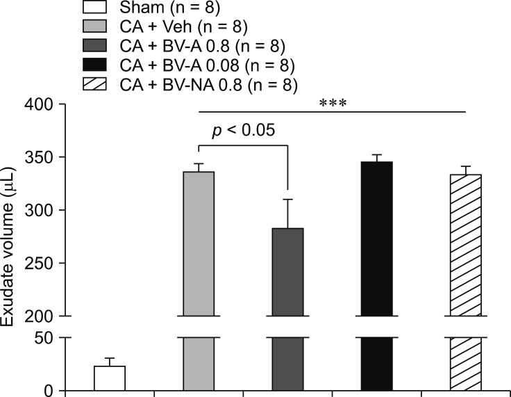
To investigate the potential effect of dBV in carrageenan-induced pleurisy, we examined the volume of pleural exudate from vehicle- or carrageenan-injected animals. Injection of carrageenan into pleural cavity induced an acute inflammatory response characterized by the accumulation of fluid (Fig. 1). Interestingly, carrageenan mice treated with 0.8 mg/kg dBV at the LU-5 acupoint (BV-A 0.8) showed a significant reduction in exudate volume compared to that of vehicle-treated carrageenan mice. However, treatment of 0.08 mg/kg dBV at the LU-5 acupoint (BV-A 0.08) or treatment of 0.8 mg/kg dBV on non-acupoint (BV-NA 0.8) did not induce significant changes compared to that of vehicle-treated carrageenan mouse (Fig. 1).
Fig. 1. Graphs illustrating the effect of diluted bee venom (dBV) treatment on exudate volume in carrageenan (CA)-induced pleurisy. Intrapleural injection of CA significantly increased the exudate volume compared to that of sham animals (Sham). The 0.8 mg/kg dBV apipuncture of LU-5 (CA + BV-A 0.8) significantly blocked the increase of exudate volume compared to that of vehicle-treated CA mouse (CA + Veh) (p < 0.05). However, 0.08 mg/kg dBV apipuncture of LU-5 (CA + BV-A 0.08) or 0.8 mg/kg dBV apipuncture of non-acupoint (CA + BV-NA 0.8) did not change the exudate volume compared to that of vehicle-treated CA mouse (CA + Veh). ***p < 0.001 compared to that of sham animals.
Effect of diluted bee venom on leukocyte accumulation in the pleural cavity
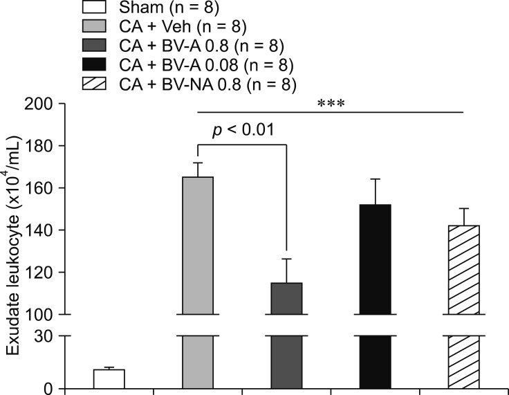
Next, we evaluated the number of leukocyte cells, indicators of inflammation, in pleural exudate. Carrageenan-injected mice showed a significant increase in leukocyte accumulation in the pleural exudate (Fig. 2). However, mice undergoing 0.8 mg/kg dBV treatment of the LU-5 acupoint (BV-A 0.8) showed a potent anti-inflammatory effect (as indicated by a marked reduction in the number of leukocyte cells) when compared with that of vehicle-treated carrageenan mice. Neither 0.08 mg/kg dBV treatment of the LU-5 acupoint (BV-A 0.08) nor 0.8 mg/kg dBV at the non-acupoint (BV-NA 0.8) produced a significant reduction of leukocyte accumulation (Fig. 2).
Fig. 2. Graphs illustrating the effect of diluted bee venom (dBV) treatment on leukocyte accumulation in pleural exudate. Intrapleural injection of carrageenan (CA) markedly induced the accumulation of leukocytes into the CA-injected pleural cavity compared to that of sham animals (Sham). The 0.8 mg/kg dBV apipuncture of LU-5 (CA + BV-A 0.8) significantly inhibited the accumulation of leukocytes in pleural exudate compared with that of vehicle-treated CA mouse (CA + Veh) (p < 0.01). However, 0.08 mg/kg dBV apipuncture of LU-5 (CA + BV-A 0.08) or 0.8 mg/kg dBV apipuncture of non-acupoint (CA + BV-NA 0.8) did not affect the number of leukocytes in pleural exudate compared to that of vehicle-treated CA group (CA + Veh). ***p < 0.001 compared to that of sham animals.
Effect of diluted bee venom on myeloperoxidase activity
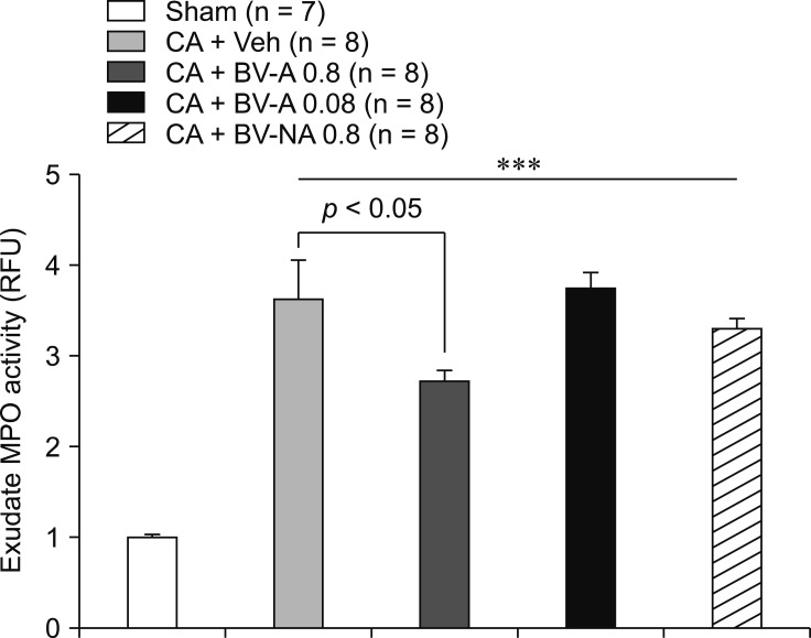
To examine further the effect of dBV apipuncture in the carrageenan-induced leukocyte accumulation, we investigated the activity of MPO in the pleural exudate (Fig. 3). Administration of 0.8 mg/kg dBV at the LU-5 acupoint (BV-A 0.8) significantly inhibited the carrageenan-induced upregulation of MPO activity compared to that of vehicle treatment. However, mice treated with 0.08 mg/kg dBV at the LU-5 acupoint (BV-A 0.08) or 0.8 mg/kg dBV at the non-acupoint (BV-NA 0.8) did not produce significant changes in MPO activity compared to that of the vehicle-treated carrageenan mice (Fig. 3).
Fig. 3. Graphs illustrating the effect of diluted bee venom (dBV) treatment on myeloperoxidase (MPO) activity in pleural exudate. Intrapleural injection of carrageenan (CA) significantly increased the MPO activity in pleural exudate compared to that of sham animals (Sham). The CA mouse treated with 0.8 mg/kg dBV apipuncture of LU-5 (CA + BV-A 0.8) had significantly reduced MPO activity as compared to that of vehicle-treated CA mouse (CA + Veh) (p < 0.05). However, the CA mouse treated with 0.08 mg/kg dBV apipuncture of LU-5 (CA + BV-A 0.08) or 0.8 mg/kg dBV apipuncture of non-acupoint (CA + BV-NA 0.8) did not show significant changes in MPO activity compared to that of vehicle-treated CA mouse (CA + Veh). RFU, relative fluorescence units. ***p < 0.001 compared to that of sham animals.
Effect of diluted bee venom on production of IL-1β and TNF-α
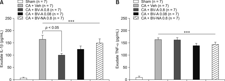
A remarkable increase in IL-1β and TNF-α production were observed in the pleural exudate of carrageenan-injected mice (Fig. 4). Although the increase of IL-1β production due to carrageenan treatment was significantly inhibited by administration of 0.8 mg/kg dBV at the LU-5 acupoint (BV-A 0.8) (panel A in Fig. 4), the same level of dBV treatment did not affect the levels of TNF-α compared to that in vehicle-treated mice (panel B in Fig. 4). No significant changes in IL-1β and TNF-α production were observed following treatment of 0.08 mg/kg dBV at the LU-5 acupoint (BV-A 0.08) or 0.08 mg/kg dBV at the non-acupoint (BV-NA 0.8) compared to that following vehicle treatment (Fig. 4).
Fig. 4. Graphs illustrating the effect of diluted bee venom (dBV) treatment on levels of proinflammatory cytokines in pleural exudate. Intrapleural injection of carrageenan (CA) significantly increased the production of interleukin 1 beta (IL-1β) (A) and tumor necrosis factor alpha (TNF-α) (B) in pleural exudate compared to that of sham animals (Sham). (A) 0.8 mg/kg dBV apipuncture of LU-5 (CA + BV-A 0.8) significantly inhibited the upregulation of IL-1β production compared to that of vehicle-treated CA mouse (CA + Veh) (p < 0.05). However, 0.08 mg/kg dBV apipuncture of LU-5 (CA + BV-A 0.08) or 0.8 mg/kg dBV apipuncture of non-acupoint (CA + BV-NA 0.8) did not affect the level of IL-1β. (B) Treatment of dBV on neither the LU-5 acupoint nor a non-acupoint did not change the level of TNF-α compared to that of vehicle-treated CA mouse (CA + Veh). ***p < 0.001 compared to that of sham animals.
Changes in Fos expression in the hypothalamic paraventricular nucleus following diluted bee venom pretreatment
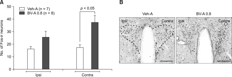
A separate set of Fos-immunohistochemistry-based experiments was performed to clarify whether 0.8 mg/kg dBV administered at the LU-5 acupoint (BV-A 0.8) was associated with activation of PVN neurons. Representative photomicrographs of PVN sections from the dBV-treated mice (panel B in Fig. 5) demonstrate that the number of Fos-ir neurons was significantly increased only in the contralateral side of the PVN in the dBV-treated groups (BV-A 0.8) compared to that of PVN of the vehicle-treated groups (Veh-A) (panel A in Fig. 5).
Fig. 5. Graphs and photomicrographs illustrating the effect of diluted bee venom (dBV) treatment on Fos expression in the ipsilateral (Ipsi) and contralateral (Contra) hypothalamic paraventricular nucleus (PVN). (A) Two hours after 0.8 mg/kg dBV stimulation of the LU-5 acupoint, the number of Fos-immunoreactive (Fos-ir) neurons significantly increased in the contralateral side of PVN in the dBV-treated group (BV-A 0.8) compared to that of the vehicle-treated group (Veh-A) (p < 0.05). (B) Representative PVN sections demonstrating the typical pattern of Fos-ir in the groups. Scale bars = 100 µm.
Discussion
BV apipuncture has been used in oriental medical fields to treat arthritis and rheumatoid diseases and to reduce pain in some Asian countries. Recently Western countries, including the United States, have been using BV treatment as an alternative therapy for multiple sclerosis, arthritis, and chronic inflammation [19]. Nevertheless, BV therapy is still limited to the treatment of musculoskeletal inflammatory diseases, such as arthritis, or neurological diseases [17]. In the present study, we provide novel evidence that dBV treatment of the LU-5 lung meridian acupoint can be an effective approach to alleviating inflammatory responses in the respiratory system. Intrapleural injection of carrageenan evoked acute inflammatory reactions in the pleural cavity, including increases in pleural exudate volume, leukocyte accumulation, MPO activity, and the production of inflammatory cytokines. Interestingly, subcutaneous dBV stimulation into the LU-5 acupoint markedly inhibited the increase of these inflammatory indicators in the carrageenan-induced pleurisy mouse model.
Carrageenan-induced inflammation has been reported to be associated with a biphasic profile of inflammatory responses in which early (within 4 h) and late (after 24 h) phases of inflammation are clearly observed [31]. This distinct profile is caused by infiltration of leukocytes from different lineages; acute neutrophil migration in the early phase and gradual macrophage migration in the late phase [30]. Furthermore, the concentration of cytokine-induced neutrophil chemoattractant-1 (CINC-1) in carrageenan-injected tissue increases to a peak only during the early phase of inflammation and is significantly decreased in the late phase of inflammation [22]. The results in these previous reports support the suggestion that the increased pleural exudate volume and increased production of inflammatory cytokines observed in our current results (24 h post-carrageenan injection) were associated with the accumulation of macrophages.
Interestingly, the present results showed that 0.8 mg/kg dBV apipuncture significantly blocked the increases in carrageenan-induced inflammatory indicators including exudate volume, number of accumulated leukocytes, MPO activity, and IL-1β level compared to the levels in vehicle-treated carrageenan-injected mice. However, the level of TNF-α in the pleural exudate was not affected by dBV treatment. It was previously reported that the expression level of TNF-α significantly increases only during the early phase of carrageenan-induced inflammation, while the production of IL-1β gradually increases during the late phase of inflammation [22,26]. Furthermore, TNF-α has been reported to play a role in regulating the accumulation of macrophages, which are the major source of IL-1β [1]. Therefore, our results suggest that dBV acupoint treatment effectively alleviates the inflammatory responses in carrageenan-induced pleurisy through the suppression of macrophage accumulation and inhibition of IL-1β production in the late phase of inflammation.
BV is composed with various constituents that include the lower molecular weight polypeptides apamin (2 kDa), melittin (3 kDa), mast cell degranulating (MCD) peptide (3 kDa), minimine (6 kDa), and adolapin (11 kDa) as well as a number of higher molecular weight glycoproteins including the enzymes PLA2 (19 kDa), lysophospholipase (22 kDa), and hyaluronidase (38 kDa) [14]. Several studies have demonstrated that direct application of melittin on lipopolysaccharide (LPS)-activated microglia or macrophage cells produces significant anti-inflammatory effects through a nuclear factor-κB–induced JNK pathway [28]. Moreover, purified MCD peptide and adolapin were reported to have anti-inflammatory activity [32], suggesting potent anti-inflammatory effects can be induced by direct BV application. However, in the present study, we did not inject dBV directly into the inflamed area, instead, it was injected into a specific acupoint located on cubital fossa of the left forelimb. Moreover, the amount of MCD peptide and adolapin is reported to present in very small quantities (1–2%) in whole BV [18]. Therefore, it is reasonable to assume that the present anti-inflammatory effects of dBV injection were not produced by the direct anti-inflammatory actions of the BV constituents, rather, they were evoked by another action that occurred following BV stimulation of the LU-5 acupoint.
A number of studies have demonstrated that activation of peripheral afferent nerves by electrical or chemical stimulation can be an effective therapeutic method for improving the inflammatory diseases [12,21]. These reports show the importance of neurological pathways on modulation of inflammatory responses. In this regard, subcutaneous injection of dBV has been considered a possible technique of peripheral nerve stimulation that can modulate inflammatory responses [37]. Our laboratories previously demonstrated that a dBV injection into the ST-36 acupoint (Zusanli) significantly increased the activity of LC neurons in brain stem [37]. Because LC are nuclei containing the cell bodies of noradrenergic neurons [25], the activation of LC neurons facilitates the release of adrenal catecholamines into the bloodstream, which ultimately results in the significant reduction of peripheral inflammation via β-adrenergic receptors [15].
Stimulation of LU-5 has been reported as effective in the improvement of the symptoms and pulmonary functions of patients with respiratory disease by regulating the autonomic nervous system and promoting the release of endogenous peptides at the hypothalamic level [20]. On that basis, we have taken a step toward elucidating some of the mechanistic details underlying the anti-inflammatory effect of dBV stimulation of the LU-5 acupoint. The mice treated with 0.8 mg/kg dBV at LU-5 showed a significantly increased level of Fos-ir in the contralateral PVN neurons. This contralateral PVN activation pattern has been similarly observed in a previous study from our laboratories which demonstrated that subcutaneous dBV injection into the hind limb selectively activates the contralateral LC [38]. Because the PVN is a brain center that activates the HPA axis, it seems plausible that dBV-induced neuronal signaling from the LU-5 acupoint to the contralateral PVN may activate the synthesis and release of adrenal glucocorticoid, a potent anti-inflammatory agent, from the adrenal cortex. Recent studies that support our hypothesis have demonstrated that electroacupuncture on LU-5 notably increases the level of dopamine in the hypothalamus [35], which can activate the HPA axis through dopaminergic neurons in the hypothalamus [5]. Nevertheless, because there is still a lack of research into the direct neurological relationship between the LU-5 acupoint and the PVN, the details of that neurological mechanism need to be investigated in further study.
In conclusion, the results of the current study demonstrate the anti-inflammatory effects of dBV treatment in a carrageenan-induced pleurisy model. The stimulation of the LU-5 acupoint with dBV significantly suppressed the accumulation of pleural exudate, the migration of leukocyte into the pleural cavity, the increase of MPO activity, and the production of IL-1β. These results indicate that dBV apipuncture at the LU-5 acupoint can be a novel alternative therapeutic strategy for respiratory inflammatory disease.
Acknowledgments
This research was supported by a grant (K18181) from the Korea Institute of Oriental Medicine (Daejeon, Republic of Korea).
Footnotes
Conflict of Interest: The authors declare no conflicts of interest.
References
- 1.Authier FJ, Chazaud B, Plonquet A, Eliezer-Vanerot MC, Poron F, Belec L, Barlovatz-Meimon G, Gherardi RK. Differential expression of the IL-1 system components during in vitro myogenesis: implication of IL-1beta in induction of myogenic cell apoptosis. Cell Death Differ. 1999;6:1012–1021. doi: 10.1038/sj.cdd.4400576. [DOI] [PubMed] [Google Scholar]
- 2.Bhattacharyya S, Borthakur A, Anbazhagan AN, Katyal S, Dudeja PK, Tobacman JK. Specific effects of BCL10 Serine mutations on phosphorylations in canonical and noncanonical pathways of NF-κB activation following carrageenan. Am J Physiol Gastrointest Liver Physiol. 2011;301:G475–G486. doi: 10.1152/ajpgi.00071.2011. [DOI] [PMC free article] [PubMed] [Google Scholar]
- 3.Davies NM, Jamali F. COX-2 selective inhibitors cardiac toxicity: getting to the heart of the matter. J Pharm Pharm Sci. 2004;7:332–336. [PubMed] [Google Scholar]
- 4.Efthimiou P, Kukar M. Complementary and alternative medicine use in rheumatoid arthritis: proposed mechanism of action and efficacy of commonly used modalities. Rheumatol Int. 2010;30:571–586. doi: 10.1007/s00296-009-1206-y. [DOI] [PubMed] [Google Scholar]
- 5.Fuertes G, Laorden ML, Milanés MV. Noradrenergic and dopaminergic activity in the hypothalamic paraventricular nucleus after naloxone-induced morphine withdrawal. Neuroendocrinology. 2000;71:60–67. doi: 10.1159/000054521. [DOI] [PubMed] [Google Scholar]
- 6.Gruber W, Eber E, Malle-Scheid D, Pfleger A, Weinhandl E, Dorfer L, Zach MS. Laser acupuncture in children and adolescents with exercise induced asthma. Thorax. 2002;57:222–225. doi: 10.1136/thorax.57.3.222. [DOI] [PMC free article] [PubMed] [Google Scholar]
- 7.Haegens A, Vernooy JH, Heeringa P, Mossman BT, Wouters EF. Myeloperoxidase modulates lung epithelial responses to pro-inflammatory agents. Eur Respir J. 2008;31:252–260. doi: 10.1183/09031936.00029307. [DOI] [PubMed] [Google Scholar]
- 8.Hajhashemi V, Sadeghi H, Minaiyan M, Movahedian A, Talebi A. Central and peripheral anti-inflammatory effects of maprotiline on carrageenan-induced paw edema in rats. Inflamm Res. 2010;59:1053–1059. doi: 10.1007/s00011-010-0225-1. [DOI] [PubMed] [Google Scholar]
- 9.Harirforoosh S, Jamali F. Renal adverse effects of nonsteroidal anti-inflammatory drugs. Expert Opin Drug Saf. 2009;8:669–681. doi: 10.1517/14740330903311023. [DOI] [PubMed] [Google Scholar]
- 10.Kang SS, Pak SC, Choi SH. The effect of whole bee venom on arthritis. Am J Chin Med. 2002;30:73–80. doi: 10.1142/S0192415X02000089. [DOI] [PubMed] [Google Scholar]
- 11.Kang SY, Roh DH, Kim HW, Han HJ, Beitz AJ, Lee JH. Blockade of adrenal medulla-derived epinephrine potentiates bee venom-induced antinociception in the mouse formalin test: involvement of peripheral β-adrenoceptors. Evid Based Complement Alternat Med. 2013;2013:809062. doi: 10.1155/2013/809062. [DOI] [PMC free article] [PubMed] [Google Scholar]
- 12.Kim HW, Uh DK, Yoon SY, Roh DH, Kwon YB, Han HJ, Lee HJ, Beitz AJ, Lee JH. Low-frequency electroacupuncture suppresses carrageenan-induced paw inflammation in mice via sympathetic post-ganglionic neurons, while high-frequency EA suppression is mediated by the sympathoadrenal medullary axis. Brain Res Bull. 2008;75:698–705. doi: 10.1016/j.brainresbull.2007.11.015. [DOI] [PubMed] [Google Scholar]
- 13.Kolber BJ, Muglia LJ. Defining brain region-specific glucocorticoid action during stress by conditional gene disruption in mice. Brain Res. 2009;1293:85–90. doi: 10.1016/j.brainres.2009.03.061. [DOI] [PMC free article] [PubMed] [Google Scholar]
- 14.Kwon YB, Ham TW, Kim HW, Roh DH, Yoon SY, Han HJ, Yang IS, Kim KW, Beitz AJ, Lee JH. Water soluble fraction (<10 kDa) from bee venom reduces visceral pain behavior through spinal α2-adrenergic activity in mice. Pharmacol Biochem Behav. 2005;80:181–187. doi: 10.1016/j.pbb.2004.10.017. [DOI] [PubMed] [Google Scholar]
- 15.Kwon YB, Kim HW, Ham TW, Yoon SY, Roh DH, Han HJ, Beitz AJ, Yang IS, Lee JH. The anti-inflammatory effect of bee venom stimulation in a mouse air pouch model is mediated by adrenal medullary activity. J Neuroendocrinol. 2003;15:93–96. doi: 10.1046/j.1365-2826.2003.00951.x. [DOI] [PubMed] [Google Scholar]
- 16.Kwon YB, Lee HJ, Han HJ, Mar WC, Kang SK, Yoon OB, Beitz AJ, Lee JH. The water-soluble fraction of bee venom produces antinociceptive and anti-inflammatory effects on rheumatoid arthritis in rats. Life Sci. 2002;71:191–204. doi: 10.1016/s0024-3205(02)01617-x. [DOI] [PubMed] [Google Scholar]
- 17.Lee JD, Park HJ, Chae Y, Lim S. An overview of bee venom acupuncture in the treatment of arthritis. Evid Based Complement Alternat Med. 2005;2:79–84. doi: 10.1093/ecam/neh070. [DOI] [PMC free article] [PubMed] [Google Scholar]
- 18.Lee JH, Kwon YB, Han HJ, Mar WC, Lee HJ, Yang IS, Beitz AJ, Kang SK. Bee venom pretreatment has both an antinociceptive and anti-inflammatory effect on carrageenan-induced inflammation. J Vet Med Sci. 2001;63:251–259. doi: 10.1292/jvms.63.251. [DOI] [PubMed] [Google Scholar]
- 19.Lee MS, Pittler MH, Shin BC, Kong JC, Ernst E. Bee venom acupuncture for musculoskeletal pain: a review. J Pain. 2008;9:289–297. doi: 10.1016/j.jpain.2007.11.012. [DOI] [PubMed] [Google Scholar]
- 20.Li L, Yu J, Mu R, Dong S. Clinical effect of electroacupuncture on lung injury patients caused by severe acute pancreatitis. Evid Based Complement Alternat Med. 2017;2017:3162851. doi: 10.1155/2017/3162851. [DOI] [PMC free article] [PubMed] [Google Scholar]
- 21.Liu XY, Zhou HF, Pan YL, Liang XB, Niu DB, Xue B, Li FQ, He QH, Wang XH, Wang XM. Electro-acupuncture stimulation protects dopaminergic neurons from inflammation-mediated damage in medial forebrain bundle-transected rats. Exp Neurol. 2004;189:189–196. doi: 10.1016/j.expneurol.2004.05.028. [DOI] [PubMed] [Google Scholar]
- 22.Loram LC, Fuller A, Fick LG, Cartmell T, Poole S, Mitchell D. Cytokine profiles during carrageenan-induced inflammatory hyperalgesia in rat muscle and hind paw. J Pain. 2007;8:127–136. doi: 10.1016/j.jpain.2006.06.010. [DOI] [PubMed] [Google Scholar]
- 23.Malhotra S, Deshmukh SS, Dastidar SG. COX inhibitors for airway inflammation. Expert Opin Ther Targets. 2012;16:195–207. doi: 10.1517/14728222.2012.661416. [DOI] [PubMed] [Google Scholar]
- 24.Menegazzi M, Di Paola R, Mazzon E, Genovese T, Crisafulli C, Dal Bosco M, Zou Z, Suzuki H, Cuzzocrea S. Glycyrrhizin attenuates the development of carrageenan-induced lung injury in mice. Pharmacol Res. 2008;58:22–31. doi: 10.1016/j.phrs.2008.05.012. [DOI] [PubMed] [Google Scholar]
- 25.Nestor PJ, Scheltens P, Hodges JR. Advances in the early detection of Alzheimer's disease. Nat Med. 2004;10(Suppl):S34–S41. doi: 10.1038/nrn1433. [DOI] [PubMed] [Google Scholar]
- 26.Nicoletti F, Auci DL, Mangano K, Flores-Riveros J, Villegas S, Frincke JM, Reading CL, Offner H. 5-androstenediol ameliorates pleurisy, septic shock, and experimental autoimmune encephalomyelitis in mice. Autoimmune Dis. 2010;2010:757432. doi: 10.4061/2010/757432. [DOI] [PMC free article] [PubMed] [Google Scholar]
- 27.Ong CK, Lirk P, Tan CH, Seymour RA. An evidence-based update on nonsteroidal anti-inflammatory drugs. Clin Med Res. 2007;5:19–34. doi: 10.3121/cmr.2007.698. [DOI] [PMC free article] [PubMed] [Google Scholar]
- 28.Park HJ, Lee HJ, Choi MS, Son DJ, Song HS, Song MJ, Lee JM, Han SB, Kim Y, Hong JT. JNK pathway is involved in the inhibition of inflammatory target gene expression and NF-kappaB activation by melittin. J Inflamm (Lond) 2008;5:7. doi: 10.1186/1476-9255-5-7. [DOI] [PMC free article] [PubMed] [Google Scholar]
- 29.Roh DH, Choi SR, Yoon SY, Kang SY, Moon JY, Kwon SG, Han HJ, Beitz AJ, Lee JH. Spinal neuronal NOS activation mediates sigma-1 receptor-induced mechanical and thermal hypersensitivity in mice: involvement of PKC-dependent GluN1 phosphorylation. Br J Pharmacol. 2011;163:1707–1720. doi: 10.1111/j.1476-5381.2011.01316.x. [DOI] [PMC free article] [PubMed] [Google Scholar]
- 30.Saleh TSF, Calixto JB, Medeiros YS. Anti-inflammatory effects of theophylline, cromolyn and salbutamol in a murine model of pleurisy. Br J Pharmacol. 1996;118:811–819. doi: 10.1111/j.1476-5381.1996.tb15472.x. [DOI] [PMC free article] [PubMed] [Google Scholar]
- 31.Saleh TSF, Calixto JB, Medeiros YS. Effects of anti-inflammatory drugs upon nitrate and myeloperoxidase levels in the mouse pleurisy induced by carrageenan. Peptides. 1999;20:949–956. doi: 10.1016/s0196-9781(99)00086-8. [DOI] [PubMed] [Google Scholar]
- 32.Son DJ, Lee JW, Lee YH, Song HS, Lee CK, Hong JT. Therapeutic application of anti-arthritis, pain-releasing, and anti-cancer effects of bee venom and its constituent compounds. Pharmacol Ther. 2007;115:246–270. doi: 10.1016/j.pharmthera.2007.04.004. [DOI] [PubMed] [Google Scholar]
- 33.U.S. National Institutes of Health. Laboratory animal welfare: public health service policy on humane care and use of laboratory animals by awardee institutions; notice. Fed Regist. 1985;50:19584–19585. [PubMed] [Google Scholar]
- 34.Vazquez E, Navarro M, Salazar Y, Crespo G, Bruges G, Osorio C, Tortorici V, Vanegas H, López M. Systemic changes following carrageenan-induced paw inflammation in rats. Inflamm Res. 2015;64:333–342. doi: 10.1007/s00011-015-0814-0. [DOI] [PubMed] [Google Scholar]
- 35.Wu ZJ, Xu J, Wang J, Gong CP, Cai RL, Hu L. Correlation of electroacupuncture on the heart and lung meridians with dopamine secretion in the hypothalamus of rats in a myocardial ischemia model. Med Acupunct. 2016;28:331–338. [Google Scholar]
- 36.Yin CS, Jeong HS, Park HJ, Baik Y, Yoon MH, Choi CB, Koh HG. A proposed transpositional acupoint system in a mouse and rat model. Res Vet Sci. 2008;84:159–165. doi: 10.1016/j.rvsc.2007.04.004. [DOI] [PubMed] [Google Scholar]
- 37.Yoon SY, Kwon YB, Kim HW, Roh DH, Seo HS, Han HJ, Lee HJ, Beitz AJ, Hwang SW, Lee JH. Peripheral bee venom’s anti-inflammatory effect involves activation of the coeruleospinal pathway and sympathetic preganglionic neurons. Neurosci Res. 2007;59:51–59. doi: 10.1016/j.neures.2007.05.008. [DOI] [PubMed] [Google Scholar]
- 38.Yoon SY, Kwon YB, Kim HW, Roh DH, Seo HS, Han HJ, Lee HJ, Beitz AJ, Lee JH. Bee venom injection produces a peripheral anti-inflammatory effect by activation of a nitric oxide-dependent spinocoeruleus pathway. Neurosci Lett. 2008;430:163–168. doi: 10.1016/j.neulet.2007.10.035. [DOI] [PubMed] [Google Scholar]
- 39.Zimmermann M. Ethical guidelines for investigations of experimental pain in conscious animals. Pain. 1983;16:109–110. doi: 10.1016/0304-3959(83)90201-4. [DOI] [PubMed] [Google Scholar]


