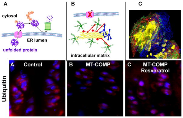
Fig. 4.
MT-COMP promotes the assembly of intracellular ER network in chondrocytes. (A) Schematic model showing how misfolded proteins in the ER are exported to the cytosol by retrotranslocation where ubiquitin (Ub) is added signaling proteasomal degradation [79]. (B) Model depicting how retained MT-COMP interacts with other matrix proteins in the ER generating a matrix network that is likely not able to fit through the retrotranslocation ER pore that leads to the cytosol. (C) Deconvolution microscopy shows intracellular matrix in chondrocyte ER composed of MT-COMP (green), matrilin-3 (blue), types 2 (yellow) and 9 (red) collagens. Adapted from Posey 2009 [69]. Ubiquitin immunohistochemistry of control (D), MT-COMP (E) and resveratrol treated MT-COMP growth plate chondrocytes (F).

