Abstract
We report a case of a 77-year-old man who presented to our shoulder department with a soft tissue mass on his right acromioclavicular (AC) joint. Previously attempted puncture aspiration had revealed serous fluid retention which recurred after each of several drainage attempts. Conventional radiography and MRI of the affected shoulder joint demonstrated a progressed cuff-tear arthropathy with an irreparable tear of the supraspinatus tendon, static superior migration of the humeral head, opening of the AC joint capsule and a superior joint-fluid ‘eruption’ and accumulation called ‘Geyser sign’. Given that the patient’s cuff-tear arthropathy was very well compensated, arthroscopic rotator cuff debridement and open cyst excision were performed. Closure of the superior aspect of the AC joint capsule was performed by the aid of a collagen matrix with additional closure of the deltotrapezial fascia. One year postoperatively, no cyst recurrence was noted.
Keywords: orthopaedics, rotator cuff tears
Background
Despite the fact that acromioclavicular (AC) joint cysts represent a rarely observed condition, their pathogenesis is well described. A distinction of two possible aetiologies and consequently a categorisation into two subtypes was first proposed by Hiller et al.1 Accordingly, type 1 cysts occur due to a disruption of the subacromial bursa in the setting of a degenerated AC joint allowing fluid to escape and accumulate subcutaneously. Type 2 cysts on the other hand are dependent on a cuff-tear arthropathy. In such cases, the full-thickness rotator cuff tear and superior migration of the humeral head lead to a disruption of the inferior AC joint capsule. Consequent pressure of synovial fluid escaping into the subacromial bursa results in a deterioration of the superior AC joint capsule and a cyst-forming subcutaneous joint-fluid accumulation.1
The treatment of type 2 cysts can be challenging, and current literature contains only a few case reports and series on patients affected by this condition. However, several treatment options have been suggested so far, including observation, puncture aspiration, rotator cuff repair or debridement and cyst excision with or without additional acromioplasty, hemiarthroplasty and reverse arthroplasty as well as shoulder arthrodesis.1–14
The aim of this case report is to present a new treatment option for this rarely observed condition.
Case presentation
A 77-year-old right-handed man presented to our shoulder department due to an atraumatic swelling above his right shoulder. Previously attempted puncture had revealed serous fluid retention which recurred after each of several drainage attempts. Medical history revealed the patient had a good general health status. Clinical examination demonstrated a large swelling above the AC joint (figure 1A) without tenderness, as well as a reduced active range of motion particularly in external and internal rotation and a decreased strength in elevation and external rotation. There were no clinical or laboratory findings suggestive of an infection. Conventional radiography (figure 1B) and MRI (figure 1C) depicted a progressed cuff-tear arthropathy (grade IV according to the Hamada classification) with an irreparable tear of the supraspinatus tendon, a partial tear of the subscapularis and infraspinatus tendon, static superior migration of the humeral head with acetabularisation of the acromion, opening of the AC joint capsule and superior joint-fluid ‘eruption’ and accumulation called ‘Geyser sign’ (figure 1C; arrows).1–11 13–15 The patient was not a good candidate for reverse shoulder arthroplasty due to his low pain level and satisfying range of motion. However, he was bothered by a tight sensation associated with the swelling and its appearance, therefore a joint preserving surgical intervention was indicated.
Figure 1.
(A) Preoperative clinical image showing a large swelling over the acromioclavicular joint. (B) Anteroposterior radiograph of the right shoulder revealing a grade IV rotator cuff-tear arthropathy with acetabularisation and narrowing of the glenohumeral joint space. (C) Coronal T2-weighted MRI displaying a superior rotator cuff tear with opening of the acromioclavicular joint capsule, and superior joint-fluid ‘eruption’ (arrows) called Geyser phenomenon.
Differential diagnosis
Important differential diagnoses to be considered are not only infection of the AC joint and haematomas, but also various kinds of benign or malignant bone or soft tissue tumours.1–4 10 11 16 Further, type 1 AC joint cysts should be taken into account. Those appear as a result of a disruption of the subacromial bursa and degeneration of the AC joint. In contrast to the type 2 cysts, the rotator cuff is found to be intact.1
Treatment
Under general anaesthesia, the patient was placed in a beach chair position. The affected shoulder was prepped and draped in a sterile fashion. At first, diagnostic arthroscopy was performed and a massive rotator cuff tear involving the subscapularis, as well as the supraspinatus and infraspinatus tendon, was confirmed. Further, a rupture of the long head of the biceps tendon and depositions of pyrophosphate were noted. The next step involved arthroscopic debridement of the glenohumeral joint surfaces and the rotator cuff. Subsequently, the cyst and its connection to the AC joint were excised through an approximately 7 cm longitudinal incision above the AC joint. Closure of the superior aspect of the AC joint capsule was performed by the aid of a collagen matrix with additional closure of the deltotrapezial fascia.
The histopathological examination proved the structural changes to be a synovial cyst.
Outcome and follow-up
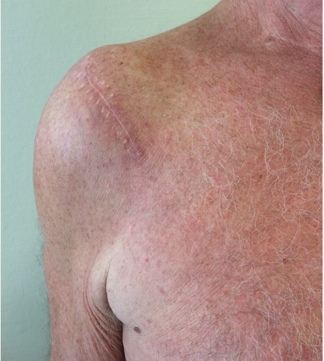
Up to 1 year postoperatively, clinical examination revealed no cyst recurrence (figure 2). The patient’s active range of motion and strength remained unchanged. He achieved 75% in the subjective shoulder value for both shoulders. The Constant score was 79 points for the right and 85 points for the left shoulder.17 X-ray examination revealed no progression of his rotator cuff arthropathy.
Figure 2.

Final follow-up clinical image showing a satisfying result without cyst recurrence.
Discussion
In the present case, the patient suffered only mild symptoms of cuff-tear arthropathy. His main symptom concerned the swelling above his right AC joint. Hence, arthroscopic debridement and open cyst excision were performed. Our decision to support the soft tissue coverage of the AC joint and deltotrapezial fascia with a collagen patch was made in order to reduce the risk of recurrence due to fluid leakage through the open AC joint. As was the case in our patient, the connective tissue above the AC joint can be very thin and barely of sufficient extent to close the AC joint opening. The improved sealing effect was the main reason to use an additional allograft. A similar approach has been presented by Skedros and Knight. They used a human dermis allograft patch to seal the AC region in a patient suffering from a similar condition. Their result concerning cyst recurrence was likewise satisfying, as no recurrence was noted at the 1-year follow-up.18 Assumptions about a possible pinch valve effect have been made in previous reports, and acromioplasty in addition to rotator cuff debridement and cyst excision was suggested to eliminate it, thus preventing cyst recurrence.1 2 4 6 7 10–13 This approach could have been considered. However, evidence is scarce, and a case of cyst recurrence after excision and acromioplasty has been reported previously.18 19
Overall, a trend towards surgical treatment can be found in current literature.
There is an agreement that repetitive puncture aspiration of these types of cysts should be avoided due to a high recurrence rate.3–6 8 10 11 In addition to cyst excision, rotator cuff repair is recommended whenever possible. Arthroscopic irrigation and debridement with or without additional acromioplasty can be effective as a salvage procedure in cases of irreparable rotator cuff defects—particularly in elderly patients with low functional shoulder demands.1 2 4–7 11 13 17 Severely symptomatic cuff-tear arthropathy on the other hand should be addressed by reverse shoulder arthroplasty.1 2 5 12 Furthermore, some authors reported spontaneous cyst resolution and thus suggested observation as another possible option.2 11
Learning points.
Repetitive puncture aspiration for evacuation of type 2 cysts in rotator cuff-tear arthropathy should be avoided due to the high recurrence rate.
In cases of well-compensated rotator cuff arthropathy, good results can be achieved through excision of the cysts.
Sealing of the acromioclavicular joint capsule by means of an allograft patch might reduce the risk of recurrence of the cyst.
Reverse total shoulder arthroplasty and cyst excision are indicated in patients who suffer from advanced symptomatic rotator cuff arthropathy and cyst formation.
Footnotes
Contributors: PM planned and performed the surgery with support from MS. NM wrote the manuscript with input from PM, FP and MS. All authors provided critical feedback. All authors read and approved the final manuscript.
Funding: The authors have not declared a specific grant for this research from any funding agency in the public, commercial or not-for-profit sectors.
Competing interests: None declared.
Patient consent: Obtained.
Provenance and peer review: Not commissioned; externally peer reviewed.
References
- 1.Hiller AD, Miller JD, Zeller JL. Acromioclavicular joint cyst formation. Clin Anat 2010;23:NA–52. 10.1002/ca.20918 [DOI] [PubMed] [Google Scholar]
- 2.Singh RA, Hay BA, Hay SM. Management of a massive acromioclavicular joint cyst: The geyser sign revisited. Shoulder Elbow 2013;5:62–4. 10.1111/j.1758-5740.2012.00221.x [DOI] [Google Scholar]
- 3.Craig EV. The geyser sign and torn rotator cuff: clinical significance and pathomechanics. Clin Orthop Relat Res 1984;191:213–5. [PubMed] [Google Scholar]
- 4.Ockert B, Mutschler W, Biberthaler P, et al. Die Akromioklavikulargelenkzyste. Der Orthopäde 2009;38:974–80. 10.1007/s00132-009-1468-9 [DOI] [PubMed] [Google Scholar]
- 5.Cooper HJ, Milillo R, Klein DA, et al. The MRI geyser sign: acromioclavicular joint cysts in the setting of a chronic rotator cuff tear. Am J Orthop 2011;40:E118–21. [PubMed] [Google Scholar]
- 6.Cvitanic O, Schimandle J, Cruse A, et al. The acromioclavicular joint cyst: glenohumeral joint communication revealed by MR arthrography. J Comput Assist Tomogr 1999;23:141–3. 10.1097/00004728-199901000-00029 [DOI] [PubMed] [Google Scholar]
- 7.Lizaur Utrilla A, Marco Gomez L, Perez Aznar A, et al. Rotator cuff tear and acromioclavicular joint cyst. Acta Orthop Belg 1995;61:144–6. [PubMed] [Google Scholar]
- 8.Groh GI, Badwey TM, Rockwood CA. Treatment of cysts of the acromioclavicular joint with shoulder hemiarthroplasty. J Bone Joint Surg Am 1993;75:1790–4. 10.2106/00004623-199312000-00008 [DOI] [PubMed] [Google Scholar]
- 9.Selvi E, De Stefano R, Frati E, et al. Rotator cuff tear associated with an acromioclavicular cyst in rheumatoid arthritis. Clin Rheumatol 1998;17:170–1. 10.1007/BF01452269 [DOI] [PubMed] [Google Scholar]
- 10.Nowak DD, Covey AS, Grant RT, et al. Massive acromioclavicular joint cyst. J Shoulder Elbow Surg 2009;18:e12–e14. 10.1016/j.jse.2008.11.009 [DOI] [PubMed] [Google Scholar]
- 11.Tanaka S, Gotoh M, Mitsui Y, et al. A Case Report of an Acromioclavicular Joint Ganglion Associated with a Rotator Cuff Tear. Kurume Med J 2017;63:29–32. 10.2739/kurumemedj.MS6300002 [DOI] [PubMed] [Google Scholar]
- 12.Feeley BT, Gallo RA, Craig EV. Cuff tear arthropathy: current trends in diagnosis and surgical management. J Shoulder Elbow Surg 2009;18:484–94. 10.1016/j.jse.2008.11.003 [DOI] [PubMed] [Google Scholar]
- 13.Mullett H, Benson R, Levy O. Arthroscopic treatment of a massive acromioclavicular joint cyst. Arthroscopy 2007;23:446.e1–446.e4. 10.1016/j.arthro.2005.12.037 [DOI] [PubMed] [Google Scholar]
- 14.Shaarani SR, Mullett H. Reverse Total Shoulder Replacement with Minimal ACJ Excision Arthroplasty for Management of Massive ACJ Cyst - A Case Report. Open Orthop J 2014;8:298–301. 10.2174/1874325001408010298 [DOI] [PMC free article] [PubMed] [Google Scholar]
- 15.Hamada K, Fukuda H, Mikasa M, et al. Roentgenographic findings in massive rotator cuff tears. A long-term observation. Clin Orthop Relat Res 1990;254:92–6. 10.1097/00003086-199005000-00014 [DOI] [PubMed] [Google Scholar]
- 16.Echols PG, Omer GE, Crawford MK. Juxta-articular myxoma of the shoulder presenting as a cyst of the acromioclavicular joint: a case report. J Shoulder Elbow Surg 2000;9:157–9. 10.1067/mse.2000.104148 [DOI] [PubMed] [Google Scholar]
- 17.Constant CR, Murley AH. A clinical method of functional assessment of the shoulder. Clin Orthop Relat Res 1987;214:160–4. [PubMed] [Google Scholar]
- 18.Skedros JG, Knight AN. Massive acromioclavicular ganglionic cyst treated with excision and allograft patch of acromioclavicular region. J Shoulder Elbow Surg 2012;21:e1–e5. 10.1016/j.jse.2011.07.033 [DOI] [PubMed] [Google Scholar]
- 19.Postacchini F, Perugia D, Gumina S. Acromioclavicular joint cyst associated with rotator cuff tear. A report of three cases. Clin Orthop Relat Res 1993;294:111–3. [PubMed] [Google Scholar]


