Abstract
The chromatin state is finely tuned to regulate function and specificity for transcription factors such as Estrogen-Receptor alpha (ER) which contributes to cell growth in breast cancer. ER transcriptional potential is mediated, in large part, by the specific associated proteins and co-factors that interact with it. Despite the identification and characterisation of several ER coregulators, a complete and systematic view of ER regulating chromatin modifiers is lacking. By exploiting a focused siRNA screen that investigated the requirement for a library of 330 chromatin regulators in ER-mediated cell growth, we find that the NuRD and coREST histone deacetylation complexes are critical for breast cancer cell proliferation. Further, by proteomic and genomics approaches, we discover the transcription factor TRPS1 to be a key interactor of the NuRD and coREST complexes. Interestingly, TRPS1 gene amplification occurs in 28% of human breast tumours and is associated with poor prognosis. We propose that TRPS1 is required to repress spurious binding of ER, where it contributes to the removal of histone acetylation. Our data suggests that TRPS1 is an important ER associated transcriptional repressor that regulates cell proliferation, chromatin acetylation and ER binding at the chromatin of cis-regulatory elements.
Introduction
The transcription factor ER is known to be a primary driver in several breast cancer subtypes and is present in more than 75% of all diagnosed cases (Johnston & Dowsett, 2003). Activation of ER by its ligand estrogen leads to receptor dimerization and this subsequently elicits transcriptional change by directly binding to chromatin (Thomas & Gustafsson, 2011). ER activation in breast cancer models lead to both activation and repression of genes (Thomas & Gustafsson, 2015). In breast cancer, ER is routinely targeted therapeutically, using direct ER antagonists such as Tamoxifen or by inhibiting estrogen synthesis using Aromatase Inhibitors (AI) (Johnston & Dowsett, 2003). Although these treatments that target ER activity have dramatically improved patient survival, response to therapy is variable and resistance to hormonal therapy is common. Understanding the fundamental components of the ER complex and the factors involved in mediating gene expression or repression is essential for understanding treatment response differences and for developing newer treatment strategies.
Recent studies have highlighted the importance of epigenetic regulation in cancer (Rodríguez-Paredes & Esteller, 2011; Dawson & Kouzarides, 2012). Chromatin modifiers can affect post-translational modifications of histones that are associated with regulating chromatin accessibility in active regions. Chromatin accessibility at ER binding regions is thought to be regulated prior to ER activation through the help of pioneering transcription factors such as FOXA1 and others (Jozwik & Carroll, 2012). This suggests that chromatin remodelling and epigenetic mechanisms regulate and predetermine ER binding capabilities. This is particularly important given that ER binding patterns are known to vary substantially within tumours from women that have distinct treatment responsiveness and clinical outcome (Ross-Innes et al, 2012).
Aberrant transcriptional activity in cancer is often linked with mutations in chromatin regulators (Plass et al, 2013). For example, the H3K4 histone methyltransferase MLL2 and MLL3 are mutated in 7.4% and 12.3% of breast cancers respectively (Curtis et al, 2012). We have recently identified a role for MLL3 in directly regulating ER activity (Serandour et al, 2011; Jozwik et al, 2016), suggesting that MLL3 mutations may be directly influencing the transcriptional activity of ER activity in cancer. Indeed, several chromatin regulators have been shown to influence ER activity. Components of the SWI/SNF complex such as BRG1 and BRM have been shown to regulate ER activity (DiRenzo et al, 2000; Belandia et al, 2002) and are often deregulated in cancer (Hohmann & Vakoc, 2014). Chromatin modifiers and readers are becoming viable drug targets and are currently the subject of intensive and promising pharmacological and clinical studies (Helin & Dhanak, 2013). In vitro, HDAC inhibition or altering the methylation status of cancer lines have been shown to reverse resistance to Tamoxifen (Fortunati et al, 2010). Recent studies targeting the ER cofactor BRD4, have demonstrated promising results in both ER-negative and relapsed ER-positive breast cancer (Alluri et al, 2014; Shu et al, 2016; Ladd et al, 2016). The basis of ER gene regulation is a dynamic and complex one and an understanding of the epigenetic mechanisms underpinning ER function is essential for exploiting chromatin regulatory proteins as putative drug targets.
In this study, we take a multifaceted approach, involving siRNA screens, genomic and proteomics approaches to identify the key chromatin regulators involved in modulating histone acetylation and ER function at enhancer elements within breast cancer cells. Our findings reveal a critical nexus involving the NuRD and co-REST complex, which results in the discovery of TRPS1 as a direct modulator of enhancer potential.
Results
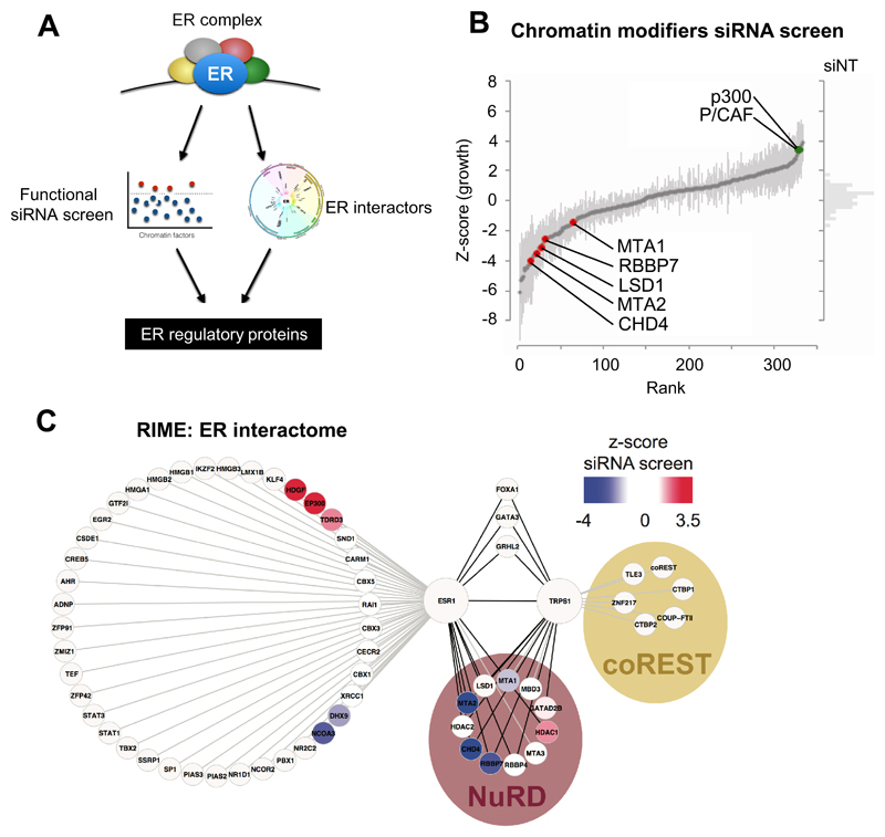
We employed a high-throughput siRNA-screen assay to identify chromatin regulators implicated in ER-positive breast cancer cell growth. Coupled with this genetic approach, we also undertook a proteomic screening approach called RIME (Mohammed et al, 2013), allowing us to identify ER interactors by pulldown and proteomics analysis (Figure 1A) whilst simultaneously revealing insight into which of these factors have a functional role in breast cancer cell growth.
Figure 1. siRNA and proteomics screen to identify ER repressors.
(A) Outline of screening approach for identifying novel ER regulatory proteins. (B) Graph representing changes in growth following siRNA knock-down for each chromatin modifiers in MCF7 cells. Change in cell viability is represented on the Y-axis as Z-score deviations from the mean. (C) Graph representing the RIME proteomic interactors of ER and TRPS1. Proteins identified as both a RIME interactor and a hit based on the siRNA screen have been coloured by their (siRNA screen) Z-score values.
We systematically performed a siRNA-based genetic screen for 332 chromatin regulators using the ER-positive breast cancer cell line MCF7. This screen has been previously validated and used to identify chromatin regulators in human epidermal stem cells (Mulder et al, 2012). Asynchronous MCF7 cells were transfected and subsequently grown for five days after which cell viability was assessed using a cellular ATP assay. A pool of three individual SiRNAs were used against each target factor and four replicates were performed. Transfection plates were verified to have no position based artefacts. Analysis of results indicated that siRNA treatments resulted in both increased and decreased cell proliferation (Figure 1B) when compared to non-targeting control siRNA. BRCA1, BRD4, CHAF1A, EP400, MLL2, SMNDC1 and TDRD7 were among the top 10% of genes where siRNA knockdown led to growth inhibition. As such, these represent genes that are required for cell viability and proliferation. Several of these factors such as BRCA1 and BRD4 have been previously linked with the direct regulation of ER, strengthening the validity of our screen (Kawai et al, 2002; Nagarajan et al, 2014). More recently, MLL2 has also been implicated as an important factor in shaping the chromatin landscape for ER binding (Toska et al, 2017). Interestingly, siRNA knockdown of several transcriptional repressors such as the members of the NuRD and coREST complexes: CHD3, CHD4, LSD1, MTA1, MTA2, RBBP7 also led to decreased growth (Figures 1B). This is in contrast to their perceived role as inhibitors of transcription and cell growth and shows that each of these putative co-repressor proteins are required for cell proliferation. Given that multiple components of the NuRD and coREST complex were identified, there is a clear need for these complexes in ER-driven cancer cell growth.
We performed parallel proteomics experiments with the aim of differentiating direct ER linked chromatin regulators from secondary or ER independent regulatory complexes. Immuno-precipitation based mass spectrometry experiments (RIME) of ER (Mohammed et al, 2013; 2016) were performed and ER linked chromatin complexes were compared to factors identified in the siRNA screen (Figure 1C). We found CHD4, LSD1, MTA2 and RBBP7 to be interacting with ER and required for sustained cell growth, as assessed in the siRNA library screening approach. Thus, members of NuRD or coREST complexes were identified from two independent screens to be both essential factors for cell proliferation and components of the ER complex (Figure 1, Table S1 and Figure S1A).
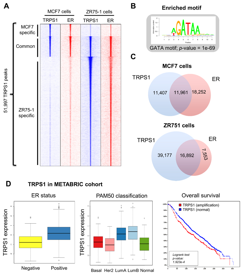
We hypothesised that for ER to initiate both activation and repression of transcription, sequence specific transcriptional repressors may be involved in effecting this distinction. Since canonical components of the NuRD/coREST complexes do not contain sequence specific transcription factors, we analysed the ER RIME dataset for potential ER interacting transcriptional repressors that could potentially mediate the activity elicited by the NuRD/coREST complex. As a result, we identified the transcription factor TRPS1 (Trichorhinophalangeal Syndrome I) to be a novel interactor of ER. TRPS1 is a repressive transcription factor possessing a GATA-like DNA binding domain similar to GATA3, which has been extensively characterised as an important ER coregulator (Eeckhoute et al, 2007; Theodorou et al, 2013). Furthermore, TRPS1 interacts with CTBP2 (Perdomo & Crossley, 2002) and with HDAC1 and HDAC2 (Wu et al, 2014), suggesting that TRPS1 is part of transcriptional repression complex. In breast cancer, TRPS1 is expressed in the luminal compartment of breast mammary gland (Chen et al, 2011) and as such, suggests that it may play a functional role with the ER complex in vivo. Analysis of the METABRIC dataset (997 breast cancers), indicated that TRPS1 tends to be more expressed in ER-positive breast cancer and in the luminal A and B subtypes (Figure 3D). The TRPS1 genomic region is amplified in 575 tumours out of 2051 (28%) and this gene amplification is significantly associated with poor survival (log rank test p-value: 1.923e-4). Moreover, a previous in silico analysis combining a siRNA screen several cancer genomics datasets predicted TRPS1 as an oncogene in breast cancer and TRPS1 overexpression increases colony formation in non-tumorigenic mammary gland cells MCF10A (Sanchez-Garcia et al, 2014).
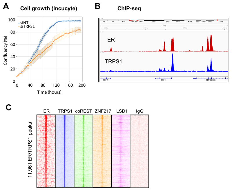
To explore the putative functional role of TRPS1 in breast cancer cells, we assessed whether TRPS1 is required for cell proliferation. We performed Incucyte analysis of cell growth in MCF7 cells after TRPS1 depletion. We found that TRPS1 depletion slowed the growth rate of MCF7 cells, suggesting that TRPS1 is required for optimal cell proliferation (Figure 2A).
Figure 2. Cell growth in TRPS1 depletion condition and ChIP-seq of TRPS1 and novel TRPS1 interactors.
(A) MCF7 cell growth assessed by Incucyte following knock-down of TRPS1. Average cell confluence was assessed every 3 hours for a total of 200 hours in siRNA control (blue dots) and siRNA TRPS1 (orange dots) conditions. Cells depleted of TRPS1 grow slower than control transfected cells. (B) Genome browser snapshot of ChIP-seq peaks for ER and TRPS1 in MCF7 cells at the TFF1 gene locus. (C) Heatmaps representing the ChIP-seq signals of ER, TRPS1, coREST, ZNF217, LSD1 and an IgG control at TRPS1 peaks in MCF7 cells.
To explore the role of TRPS1 in breast cancer cells we performed TRPS1 RIME experiments to explore the interactome and regulatory networks associated with this protein (Mohammed et al, 2013). We conducted TRPS1 RIME experiments in MCF7, T47D and ZR75-1 ER-positive breast cancer cells and used a commercial anti-TRPS1 antibody. TRPS1 was successfully immunoprecipitated in all three cell lines, confirming the validity of the experiment and the specificity of the TRPS1 antibody. TRPS1 was identified to interact with several components of the NuRD and coREST complexes and this result was consistent in all three cell lines and between replicates. TRPS1 was found to physically interact with CHD4, coREST, CTBP1, CTBP2, GATAD2B, HDAC2, LSD1, MBD3, MTA1, MTA2, RBBP4, RBBP7 and ZNF217 (Figure 1C). We also identified transcription factors such as COUP-TFII, FOXA1 and GATA3, which are key ER complex components (Theodorou et al, 2013; Hurtado et al, 2011; Kurihara et al, 2007) to be TRPS1 interactors. Although the interaction between TRPS1 Ikaros domain and CTBP2 has been characterized previously (Perdomo & Crossley, 2002), we confirmed this interaction with the coREST complex by a reciprocal RIME of the core coREST component ZNF217. The ZNF217 RIME identified ZNF217 and several coREST members (coREST, CTBP1, CTBP2, HDAC1, HDAC2, LSD1), along with TRPS1, which was the second most enriched protein after ZNF217 (Figure Supplementary 1C).
Based on the RIME data, we reasoned that the transcription factor TRPS1 recruits the HDAC complexes coREST and NuRD to the chromatin. Given that coREST and NuRD components were essential for cell proliferation based on the siRNA screen, the transcriptional targeting role of TRPS1 potentially renders it a key component of ER biology. In order to identify the genome-wide binding pattern of TRPS1 and to assess potential chromatin interplay with the ER complex, we performed ChIP-sequencing of TRPS1 in MCF7 and ZR75-1 cells (two replicates for each cell line). Uniquely mapped reads were used to call peaks using MACS2 (Zhang et al, 2008). In total, we found 23,368 TRPS1 binding sites in MCF7 cells and 56,069 binding sites in ZR751 cells. By motif analysis, we found the GATA motif as the top motif enriched in TRPS1 peaks (Figure 3B and Supplementary Figure S2). This is in good agreement with the fact that TRPS1 binds DNA through its GATA-like DNA binding domain. Strikingly, we found that TRPS1 co-occupies the chromatin with ER in a cell-type specific manner (Figures 2B and 3A). In MCF7 cells, ER bound 51 % of TRPS1 peaks whilst the reverse overlap comparison found 40 % of all ER peaks were occupied by TRPS1 (Figure 3C).
Figure 3. TRPS1 and ER share binding regions in breast cancer.
(A) Heatmaps representing the ChIP-seq signals of ER and TRPS1 in MCF7 and ZR-751 cells showing common binding sites and cell line specific binding sites. (B) Motif analysis of TRPS1 bound regions reveals GATA as the most enriched motif. (C) Overlap between ER and TRPS1 binding sites in MCF7 cells. (D) TRPS1 data in METABRIC cohort: TRPS1 expression level in 997 breast tumours based on ER positivity and PAM50 classification and breast-specific survival.
As such, ChIP-seq analysis suggests a global functional connection between TRPS1 and the ER complex in breast cancer cells. To further explore the putative role of components discovered in our RIME and siRNA screens, we performed ChIP-seq of coREST, LSD1 and ZNF217 in MCF7 cells. IgG control ChIP-seq were included as negative controls. We could not validate CHD4 antibodies by RIME and therefore did not proceed with this specific factor any further. We found binding signal of coREST, LSD1 and ZNF217 at ER/TRPS1 binding sites and at TRPS1-only binding sites (Figure 2C and Supplementary Figure 3). This result supports our hypothesis that TRPS1 is involved in the recruitment of transcription repressive-proteins at regions that are bound by the ER complex.
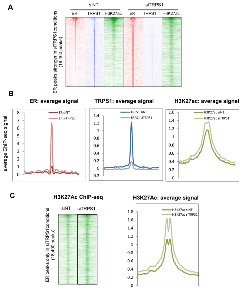
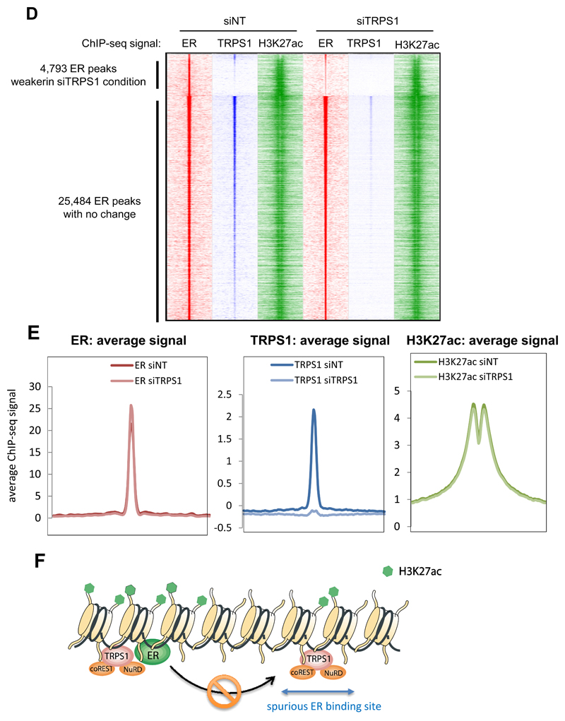
As the coREST and NuRD complexes have histone deacetylation activity, we assessed whether TRPS1 and its interacting chromatin regulators contribute to histone acetylation. As TRPS1 is potentially facilitating the HDAC activity of the coREST and NuRD complexes, we hypothesised that inhibition of TRPS1 would result in increased histone acetylation at the enhancer elements bound by ER, due to impaired deacetylation. We depleted TRPS1 by siRNA (Western blot validation is shown in Supplementary Figure S5) and mapped TRPS1 binding and H3K27ac enrichment by ChIP-seq in MCF7 cells, each of which was conducted in triplicate. This data was integrated with ER ChIP-seq analysis following TRPS1 depletion. As expected, the depletion of TRPS1 by siRNA results in substantially reduced TRPS1 binding (replicate 1: 11,254 peaks in siRNA control versus 4,386 peaks in siRNA TRPS1; replicate 2: 36,547 peaks in siRNA control versus 583 peaks in siRNA TRPS1; replicate 3: 26,054 peaks in siRNA control versus 1,997 peaks in siRNA TRPS1). Importantly, we found that upon TRPS1 depletion, 4793 ER peaks were weaker, 25,484 ER peaks did not change and interestingly, 18,400 ER peaks were stronger (Figure 4A). Furthermore, we found that the stronger ER peaks following TRPS1 depletion were also TRPS1 binding sites under control condition (i.e. in siRNA Non-Target transfected conditions). At these regions where ER is stronger following TRPS1 silencing, TRPS1 was lost as expected and H3K27ac signal was increased (Figure 4B and 4C). As such, at a subset of TRPS1 binding sites, the presence of TRPS1 decreases ER binding and histone acetylation. There was no significant change in H3K27ac at the ER peaks that are conserved in control and TRPS1 depletion conditions (Figures 4D and 4E), implying that these regions are not influenced by TRPS1. This suggests that TRPS1 functions to restrict ER binding as removal of TRPS1 results in increased number of ER binding sites as well as a failure to deacetylate the H3K27ac chromatin mark at spurious regions. Overall, these data support the hypothesis that the presence of TRPS1 mediates histone deacetylation and that decreases in TRPS1 levels relieve this repression and culminate in increased ER binding and enhancer acetylation.
Figure 4. TRPS1 is an ER repressor.
(A) Heatmaps representing siTRPS1-specific ER peaks and the ChIP-seq signal of ER, TRPS1 and H3K27ac in siNT (siRNA Non-Target) and siTRPS1 conditions in MCF7 cells. (B) Average signal at siTRPS1-specific ER peaks. (C) Second replicate of ChIP-seq H3K27ac showing the gain of H3K27ac at ER gained peaks after TRPS1 depletion. (D) Heatmaps representing the ChIP-seq signal of ER, TRPS1 and H3K27ac at ER lost peaks (siNT specific) and ER conserved peaks (siNT/siTRPS1 common) in control and TRPS1 depletion conditions. (E) Average ChIP-seq signal of ER, TRPS1 and H3K27ac at ER siNT/siTRPS1 common sites. (F) Proposed model for ER regulation by TRPS1: TRPS1 limits ER binding through chromatin deacetylation via NuRD and coREST complex.
In order to characterise global transcriptional consequences of TRPS1 inhibition, we performed RNA-sequencing experiments in MCF7 cells transfected with siRNA against TRPS1 or a non-targeting (NT) control siRNA for 48 hours. The cells were also co-treated with estradiol (E2) or control (vehicle) for the last 12 hours, 4 replicates were performed. Using BETA analysis combining siNT/siTRPS1 differential gene expression (expression fold-change under 0.5 or above 1.5) and TRPS1 ChIP-seq data, we could identify 213 direct gene targets repressed by TRPS1 and 10 direct gene targets activated by TRPS1 (Supplementary Table 3). This suggests that TRPS1 is mostly a transcriptional repressor (examples of genes repressed by TRPS1 in Supplementary Figure 9). With DESeq2, we identified 779 E2-up-regulated genes and 570 E2-down-regulated genes in control condition (expression fold change under 0.5 or above 1.5; adjusted p value < 0.001). Among these genes, only 569 and 406 genes were still respectively E2-up and E2-down-regulated after TRPS1 depletion (Supplementary Figure 6A). Overall, 27% of E2 up-regulated genes were not activated in the absence of TRPS1, while 29% of E2 down-regulated genes were no longer repressed upon TRPS1 silencing. Furthermore, the genes whilst expression is still E2-regulated upon TRPS1 depletion are also significantly less E2-activated or E2-repressed (Supplementary Figure 6B). This suggests that TRPS1 depletion directly influences ER activity and importantly, reorganises ER binding events, characterised by a substantial increase in spurious ER binding sites. We then investigated potential transcriptional activity linked to the 18,400 newly enriched ER binding sites formed upon TRPS1 depletion. We identified 4036 genes’ promoters located at less than 30 kb from these stronger peaks. Among these genes, 75 are E2-regulated in control, 61 are E2-regulated upon TRPS1 depletion and 86 are E2-regulated in both conditions. Thus, although TRPS1 prevents ER binding at several thousand sites, depletion to TRPS1 alone is not sufficient to alter transcriptional activity.
The TRPS1 gene is very often amplified in breast cancer (28% of 2051 cases in the METABRIC cohort) (20% of 963 patients in the TCGA cohort) and its amplification is associated with worse prognosis (Figure 3D). The TRPS1 gene is located in the 8q arm, a chromosomal region which is often amplified in breast cancer. Therefore, we cannot exclude that the worse prognosis of TRPS1 gene amplification is due to a more global effect of the 8q arm amplification. In the METABRIC dataset, we found that the TRPS1 lower expression is associated with worse prognosis in all breast cancer (p-value = 0.005; Supplementary Figure 3) but this observation is not significant when considering ER-negative and ER-positive tumours separately. This might be because the TRPS1 gene is more likely to be expressed in ER-positive breast cancer that are of better prognosis than ER-negative breast cancer (Figure 3D).
Discussion
Taken together, our data suggests that transcriptional repressors like NuRD and coREST complexes are needed for gene regulation by ER and for ER-mediated cell growth. At ER binding sites, TRPS1 might participate in maintaining histone deacetylation through the recruitment of the repressing complexes together with other transcription factors (Figure 4F). We hypothesise that this chromatin resetting is necessary to allow the recruitment of ER at the appropriate time and location within the genome (Perissi et al, 2010; Cerase et al, 2015). At other genomic regions, TRPS1-mediated histone deacetylation would prevent spurious ER binding and therefore reduces the number of ER binding regions. Our mechanistic insight, along with known consequences of TRPS1 genomic alterations in cancer, suggests TRPS1 to be an important factor regulating ER and breast cancer progression.
Materials and Methods
Biological material
MCF7, T47D and ZR75-1 human cell lines were obtained from ATCC and grown in DMEM or RPMI supplemented with 10 % FBS at 37 °C in 5% CO2 in a humidified incubator. For proliferation assay, cells were seeded into 24-well dishes at a cell density of 2 × 10⁴ per well and growth was monitored using the Essen Bioscience INCUCYTE machine (Ann Arbor, MI, USA). The cell lines were authenticated and tested negative for mycoplasma contamination by the CRUK CI Biorepository Core.
siRNA screen
The siRNA screen was conducted as previously published (Mulder et al, 2012). 332 siRNAs were custom made from Ambion and targeted factors containing HD, BROMO, CHROMO, PWWP, tandem BRCT, TUDOR, BAH, MBT, SET (including DOT1L), JMJC, JMJN, PRMT, HAT, HDAC, SIRT, DNMT, MBD and SNF2 ATP-dependent remodelers.
For siRNA transfections, 22 million cells were harvested and resuspended in 1.98 mL of buffer SF (Amaxa nucleofection kit). 165 uL of this was aliquoted into round bottom 96 well plates along with 2 uL of siRNA at 10 mM concentration. The mixture was then electroporated (Amaxa 96 well-plate electroporater) and 180 uL of warm media was added to each well. To another round bottom 96 well plate contaning 190 uL of media, 60 uL of the electroporated mixture was added. 10 uL of this was then transferred to flat bottom 96 well plates with an additional 100 uL of media. A final of 2400 cells were seeded in each well. Media was changed the day after transfection for all wells and every alternate day thereafter for a total of 6 days. Cells were then analysed using a cell viability assay (CellTitre-glo, Promega).
Four replicates of each experiment were performed and data obtained from the assay was initially screened for consistency between replicates. A Grubbs’ test was performed to identify and subsequently remove outliers among the four replicates.
siRNA assay for ChIP-seq and RNA-seq
Cells were transfected with siRNA using Lipofectamine2000 (Invitrogen). TRPS1 was silenced using a siRNA pool ON-TargetplusSMARTpool purchased from Dharmacon (catalogue number M-009644-01-0005). AllStars Negative Control siRNA (Qiagen) was used as a negative control.
ChIP-seq library preparation
The antibodies used for the ChIP-seq were anti-coREST (Abcam, ab32631), anti-ER (Santa-Cruz, ref. sc-543), anti-H3K27ac (Abcam, ref. ab4729), anti-LSD1 (Abcam, ab17721), anti-TRPS1 (R&D Systems, ref. AF4838), anti-ZNF217 (Santa-Cruz, sc-55351) and rabbit serum (sc-2027). ChIP were performed as previously described in Schmidt et al. (Methods, 2010), using 10 ug of antibody and 60 million cells. The ChIP-seq and the input libraries were prepared using the TruSeq ChIP Sample Prep Kit (Illumina, ref. IP-202-1012).
RNA-seq library preparation
For hormonal deprivation, MCF7 cells were cultured in phenol-red free DMEM supplemented with 5% charcoal-treated FBS. After 24 hours, the cells were transfected with siRNA against TRPS1 or a control siRNA for 48 hours. The cells were also co-treated with 100 nM estradiol or vehicle for the last 12 hours. 4 biological replicates were performed.
Western-blots
Western-blots were processed as previously described in Ross-Innes et al (Ross-Innes et al, 2010). The antibodies used were anti-TRPS1 (R&D Systems, ref. AF4838) and anti-histone H3 (ab1791).
Rapid IP-mass spectrometry of endogenous protein (RIME)
Rapid immunoprecipitation-mass spectrometry experiments were performed as previously described (Mohammed et al., 2013). The antibodies used were anti-TRPS1 (R&D Systems, ref. AF4838) and anti-ZNF217 (Abcam, ab48133). MS data were submitted to PRIDE Archive (Project PXD xxxxxx).
Bioinformatics
For ChIP-seq libraries, reads were mapped with Bowtie2 to the indexed reference genome UCSC hg19 downloaded from Bowtie2 website. Sam files were then sorted and converted in bam files with Samtools. Sorted bam files were the used for peak calling using MACS2 (Zhang et al, 2008) with default parameters. Common peaks between two replicates were used for the Venn diagrams. Sequencing depth-normalized heatmaps were generated as described previously (Ross-Innes et al, 2012). For screen-shots, normalized bgr files were made thanks to MACS2 with the command spmr (fragment pileup per million reads) and visualized on Integrative Genomics Viewer. Motif analysis was performed using the Cistrome SeqPos motif tool (version 1.0.0; Liu et al., 2011). The de novo motif search and the scan the TRANSFAC motifs was done on the 2,000 strongest TRPS1 peaks in MCF7 cells in terms of MACS2 fold enrichment, 200 bp around the summits. RNA-seq libraries were made in 4 biological replicates. Reads were mapped with Tophat and processed with DESeq2. Significant differentially expressed genes were filtered with adjusted p-value < 0.001 and fold change > 1,5 or < 0.5. To predict the direct gene targets of TRPS1, a BETA analysis was performed using the siNT/siTRPS1 differential gene expression and the TRPS1 ChIP-seq peaks (Wang et al., 2013). Sequencing data were submitted to GEO Dataset (GSE xxxxxx).
Supplementary Material
Acknowledgements
We would like to thank Clive D’Santos and Chris Taylor from the CRUK CI Proteomics core, James Hadfield and the other members of the Genomics core, and Suraj Menon from the Bioinformatics core. Jason Carroll is supported by an ERC Consolidator grant and a Komen Scholar Award. We would like to acknowledge the support of the University of Cambridge, Cancer Research UK and Hutchison Whampoa Limited.
Footnotes
Author Contributions
A.S., H.M., K.M., J.C designed the project and wrote the manuscript. A.S., H.M. and A.M. performed the experiments and analysed the data.
Conflict of Interest
The authors declare that they have no conflict of interest.
References
- Alluri PG, Asangani IA, Chinnaiyan AM. BETs abet Tam-R in ER-positive breast cancer. Cell Res. 2014;24:899–900. doi: 10.1038/cr.2014.90. [DOI] [PMC free article] [PubMed] [Google Scholar]
- Belandia B, Orford RL, Hurst HC, Parker MG. Targeting of SWI/SNF chromatin remodelling complexes to estrogen-responsive genes. EMBO J. 2002;21:4094–4103. doi: 10.1093/emboj/cdf412. [DOI] [PMC free article] [PubMed] [Google Scholar]
- Cerase A, Pintacuda G, Tattermusch A, Avner P. Xist localization and function: new insights from multiple levels. Genome Biol. 2015;16:166. doi: 10.1186/s13059-015-0733-y. [DOI] [PMC free article] [PubMed] [Google Scholar]
- Chen JQ, Bao Y, Litton J, Xiao L, Zhang H-Z, Warneke CL, Wu Y, Shen X, Wu S, Katz RL, Sahin A, et al. Expression and relevance of TRPS-1: a new GATA transcription factor in breast cancer. Horm Cancer. 2011;2:132–143. doi: 10.1007/s12672-011-0067-5. [DOI] [PMC free article] [PubMed] [Google Scholar]
- Curtis C, Shah SP, Chin S-F, Turashvili G, Rueda OM, Dunning MJ, Speed D, Lynch AG, Samarajiwa S, Yuan Y, Gräf S, et al. The genomic and transcriptomic architecture of 2,000 breast tumours reveals novel subgroups. Nature. 2012;486:346–352. doi: 10.1038/nature10983. [DOI] [PMC free article] [PubMed] [Google Scholar]
- Dawson MA, Kouzarides T. Cancer Epigenetics: From Mechanism to Therapy. Cell. 2012;150:12–27. doi: 10.1016/j.cell.2012.06.013. [DOI] [PubMed] [Google Scholar]
- DiRenzo J, Shang Y, Phelan M, Sif S, Myers M, Kingston R, Brown M. BRG-1 is recruited to estrogen-responsive promoters and cooperates with factors involved in histone acetylation. Mol Cell Biol. 2000;20:7541–7549. doi: 10.1128/mcb.20.20.7541-7549.2000. [DOI] [PMC free article] [PubMed] [Google Scholar]
- Eeckhoute J, Keeton EK, Lupien M, Krum SA, Carroll JS, Brown M. Positive cross-regulatory loop ties GATA-3 to estrogen receptor alpha expression in breast cancer. Cancer Res. 2007;67:6477–6483. doi: 10.1158/0008-5472.CAN-07-0746. [DOI] [PubMed] [Google Scholar]
- Fortunati N, Bertino S, Costantino L, De Bortoli M, Compagnone A, Bandino A, Catalano MG, Boccuzzi G. Valproic acid restores ER alpha and antiestrogen sensitivity to ER alpha-negative breast cancer cells. Mol Cell Endocrinol. 2010;314:17–22. doi: 10.1016/j.mce.2009.09.011. [DOI] [PubMed] [Google Scholar]
- Helin K, Dhanak D. Chromatin proteins and modifications as drug targets. Nature. 2013;502:480–488. doi: 10.1038/nature12751. [DOI] [PubMed] [Google Scholar]
- Hohmann AF, Vakoc CR. A rationale to target the SWI/SNF complex for cancer therapy. Trends Genet. 2014;30:356–363. doi: 10.1016/j.tig.2014.05.001. [DOI] [PMC free article] [PubMed] [Google Scholar]
- Hurtado A, Holmes KA, Ross-Innes CS, Schmidt D, Carroll JS. FOXA1 is a key determinant of estrogen receptor function and endocrine response. Nat Genet. 2011;43:27–33. doi: 10.1038/ng.730. [DOI] [PMC free article] [PubMed] [Google Scholar]
- Johnston SRD, Dowsett M. Aromatase inhibitors for breast cancer: lessons from the laboratory. Nat Rev Cancer. 2003;3:821–831. doi: 10.1038/nrc1211. [DOI] [PubMed] [Google Scholar]
- Jozwik KM, Carroll JS. Pioneer factors in hormone-dependent cancers. Nat Rev Cancer. 2012;12:381–385. doi: 10.1038/nrc3263. [DOI] [PubMed] [Google Scholar]
- Jozwik KM, Chernukhin I, Serandour AA, Nagarajan S, Carroll JS. FOXA1 Directs H3K4 Monomethylation at Enhancers via Recruitment of the Methyltransferase MLL3. Cell Rep. 2016;17:2715–2723. doi: 10.1016/j.celrep.2016.11.028. [DOI] [PMC free article] [PubMed] [Google Scholar]
- Kawai H, Li H, Chun P, Avraham S, Avraham HK. Direct interaction between BRCA1 and the estrogen receptor regulates vascular endothelial growth factor (VEGF) transcription and secretion in breast cancer cells. Oncogene. 2002;21:7730–7739. doi: 10.1038/sj.onc.1205971. [DOI] [PubMed] [Google Scholar]
- Kurihara I, Lee D-K, Petit FG, Jeong J, Lee K, Lydon JP, DeMayo FJ, Tsai M-J, Tsai SY. COUP-TFII mediates progesterone regulation of uterine implantation by controlling ER activity. PLoS Genet. 2007;3:e102. doi: 10.1371/journal.pgen.0030102. [DOI] [PMC free article] [PubMed] [Google Scholar]
- Ladd B, Mazzola AM, Bihani T, Lai Z, Bradford J, Collins M, Barry E, Goeppert AU, Weir HM, Hearne K, Renshaw JG, et al. Effective combination therapies in preclinical endocrine resistant breast cancer models harboring ER mutations. Oncotarget. 2016;7:54120–54136. doi: 10.18632/oncotarget.10852. [DOI] [PMC free article] [PubMed] [Google Scholar]
- Mohammed H, D'Santos C, Serandour AA, Ali HR, Brown GD, Atkins A, Rueda OM, Holmes KA, Theodorou V, Robinson JLL, Zwart W, et al. Endogenous purification reveals GREB1 as a key estrogen receptor regulatory factor. Cell Rep. 2013;3:342–349. doi: 10.1016/j.celrep.2013.01.010. [DOI] [PMC free article] [PubMed] [Google Scholar]
- Mohammed H, Taylor C, Brown GD, Papachristou EK, Carroll JS, D'Santos CS. Rapid immunoprecipitation mass spectrometry of endogenous proteins (RIME) for analysis of chromatin complexes. Nat Protoc. 2016;11:316–326. doi: 10.1038/nprot.2016.020. [DOI] [PubMed] [Google Scholar]
- Mulder KW, Wang X, Escriu C, Ito Y, Schwarz RF, Gillis J, Sirokmány G, Donati G, Uribe-Lewis S, Pavlidis P, Murrell A, et al. Diverse epigenetic strategies interact to control epidermal differentiation. Nat Cell Biol. 2012;14:753–763. doi: 10.1038/ncb2520. [DOI] [PubMed] [Google Scholar]
- Nagarajan S, Hossan T, Alawi M, Najafova Z, Indenbirken D, Bedi U, Taipaleenmäki H, Ben-Batalla I, Scheller M, Loges S, Knapp S, et al. Bromodomain protein BRD4 is required for estrogen receptor-dependent enhancer activation and gene transcription. Cell Rep. 2014;8:460–469. doi: 10.1016/j.celrep.2014.06.016. [DOI] [PMC free article] [PubMed] [Google Scholar]
- Perdomo J, Crossley M. The Ikaros family protein Eos associates with C-terminal-binding protein corepressors. European Journal of Biochemistry. 2002;269:5885–5892. doi: 10.1046/j.1432-1033.2002.03313.x. [DOI] [PubMed] [Google Scholar]
- Perissi V, Jepsen K, Glass CK, Rosenfeld MG. Deconstructing repression: evolving models of co-repressor action. Nat Rev Genet. 2010;11:109–123. doi: 10.1038/nrg2736. [DOI] [PubMed] [Google Scholar]
- Plass C, Pfister SM, Lindroth AM, Bogatyrova O, Claus R, Lichter P. Mutations in regulators of the epigenome and their connections to global chromatin patterns in cancer. Nat Rev Genet. 2013;14:765–780. doi: 10.1038/nrg3554. [DOI] [PubMed] [Google Scholar]
- Rodríguez-Paredes M, Esteller M. Cancer epigenetics reaches mainstream oncology. Nat Med. 2011;17:330–339. doi: 10.1038/nm.2305. [DOI] [PubMed] [Google Scholar]
- Ross-Innes CS, Stark R, Holmes KA, Schmidt D, Spyrou C, Russell R, Massie CE, Vowler SL, Eldridge M, Carroll JS. Cooperative interaction between retinoic acid receptor-alpha and estrogen receptor in breast cancer. Genes Dev. 2010;24:171–182. doi: 10.1101/gad.552910. [DOI] [PMC free article] [PubMed] [Google Scholar]
- Ross-Innes CS, Stark R, Teschendorff AE, Holmes KA, Ali HR, Dunning MJ, Brown GD, Gojis O, Ellis IO, Green AR, Ali S, et al. Differential oestrogen receptor binding is associated with clinical outcome in breast cancer. Nature. 2012;481:389–393. doi: 10.1038/nature10730. [DOI] [PMC free article] [PubMed] [Google Scholar]
- Sanchez-Garcia FL, Villagrasa P, Matsui J, Kotliar D, Castro VN, Akavia U-D, Chen B-J, Saucedo-Cuevas L, Rodriguez Barrueco R, Llobet-Navas D, Silva JM, et al. Integration of Genomic Data Enables Selective Discovery of Breast Cancer Drivers. Cell. 2014;159:1461–1475. doi: 10.1016/j.cell.2014.10.048. [DOI] [PMC free article] [PubMed] [Google Scholar]
- Serandour AA, Avner S, Percevault F, Demay F, Bizot M, Lucchetti-Miganeh C, Barloy-Hubler F, Brown M, Lupien M, Métivier R, Salbert G, et al. Epigenetic switch involved in activation of pioneer factor FOXA1-dependent enhancers. Genome Res. 2011;21:555–565. doi: 10.1101/gr.111534.110. [DOI] [PMC free article] [PubMed] [Google Scholar]
- Shu S, Lin CY, He HH, Witwicki RM, Tabassum DP, Roberts JM, Janiszewska M, Huh SJ, Liang Y, Ryan J, Doherty E, et al. Response and resistance to BET bromodomain inhibitors in triple-negative breast cancer. Nature. 2016;529:413–417. doi: 10.1038/nature16508. [DOI] [PMC free article] [PubMed] [Google Scholar]
- Theodorou V, Stark R, Menon S, Carroll JG. GATA3 acts upstream of FOXA1 in mediating ESR1 binding by shaping enhancer accessibility. Genome Res. 2013;23:12–22. doi: 10.1101/gr.139469.112. [DOI] [PMC free article] [PubMed] [Google Scholar]
- Thomas C, Gustafsson J-Å. The different roles of ER subtypes in cancer biology and therapy. Nat Rev Cancer. 2011;11:597–608. doi: 10.1038/nrc3093. [DOI] [PubMed] [Google Scholar]
- Thomas C, Gustafsson J-Å. Progesterone receptor-estrogen receptor crosstalk: a novel insight. Trends Endocrinol Metab. 2015;26:453–454. doi: 10.1016/j.tem.2015.08.002. [DOI] [PubMed] [Google Scholar]
- Toska E, Osmanbeyoglu HU, Castel P, Chan C, Hendrickson RC, Elkabets M, Dickler MN, Scaltriti M, Leslie CS, Armstrong SA, Baselga J. PI3K pathway regulates ER-dependent transcription in breast cancer through the epigenetic regulator KMT2D. Science. 2017;355:1324–1330. doi: 10.1126/science.aah6893. [DOI] [PMC free article] [PubMed] [Google Scholar]
- Wu L, Wang Y, Liu Y, Yu S, Xie H, Shi X, Qin S, Ma F, Tan TZ, Thiery JP, Chen L. A central role for TRPS1 in the control of cell cycle and cancer development. Oncotarget. 2014;5:7677–7690. doi: 10.18632/oncotarget.2291. [DOI] [PMC free article] [PubMed] [Google Scholar]
- Zhang Y, Liu T, Meyer CA, Eeckhoute J, Johnson DS, Bernstein BE, Nusbaum C, Myers RM, Brown M, Li W, Liu JS. Model-based analysis of ChIP-Seq (MACS) Genome Biol. 2008;9:R137. doi: 10.1186/gb-2008-9-9-r137. [DOI] [PMC free article] [PubMed] [Google Scholar]
Associated Data
This section collects any data citations, data availability statements, or supplementary materials included in this article.


