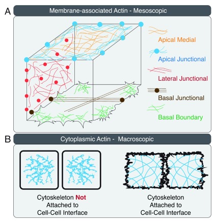
Figure 1. Actin organization in a three-dimensional epithelial cell.
( A) Actin arrangement on the apical, lateral, and basal membranes of the epithelial cell is illustrated at the mesoscopic cellular level. ( B) Connectivity between cell–cell interface and the actin cytoskeleton is illustrated at the macroscopic multi-cellular level.

