Abstract
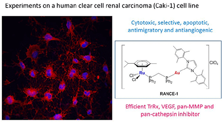
Heterobimetallic compounds are designed to harness chemotherapeutic traits of distinct metal species into a single molecule. The ruthenium-gold (Ru-Au) family of compounds based on Au-N-heterocyclic carbene (NHC) fragments [Cl2(p-cymene)Ru(μ-dppm)Au(NHC)]ClO4 was conceived to combine the known antiproliferative and cytotoxic properties of Au-NHC-based compounds and the antimigratory, antimetastatic and antiangiogenic characteristic of specific Ru-based compounds. Following recent studies of the anticancer efficacies of these Ru–Au-NHC complexes with promising potential as chemotherapeutics against colorectal, and renal cancers in vitro, we report here on the mechanism of a selected compound, [Cl2(p-cymene)Ru(μ-dppm)Au(IMes)]ClO4 (RANCE-1, 1). The studies were carried out in vitro using a human clear cell renal carcinoma cell line (Caki-1). These studies indicate that bimetallic compound RANCE-1 (1) is significantly more cytotoxic than the Ru (2) or Au (3) monometallic derivatives. RANCE-1 significantly inhibits migration, invasion and angiogenesis, which are essential for metastasis. RANCE-1 was found to disturb pericellular proteolysis by inhibiting cathepsins, and the metalloproteases MMP and ADAM which play key roles in the etiopathogenesis of cancer. RANCE-1 also inhibits the mitochondrial protein TrxR that is often overexpressed in cancer cells and facilitates apoptosis evasion. We found that while Auranofin perturbed migration and invasion to similar degrees as RANCE-1 (1) in Caki-1 renal cancer cells, RANCE-1 (1) inhibited antiangiogenic formation and VEGF expression. We found that Auranofin and RANCE-1 (1) have distinct proteolytic profiles. In summary, RANCE-1 constitutes a very promising candidate for further preclinical evaluations in renal cancer.
Keywords: Bimetallic, ruthenium-gold, renal cancer, mechanisms, Auranofin
Table of Contents
Introduction
Heterometallic compounds have been developed to enhance the anti-cancer properties of single metallodrugs. The hypothesis is that the incorporation of two different biologically active metals in the same molecule may improve their anti-tumor activity as a result of metal specific interactions with distinct biological targets (cooperative effect) or by the improved chemicophysical properties of the resulting heterometallic compound (synergism).1 Recent reports have highlighted the success of this approach2–28 although there are very few articles documenting comparisons of the heteronuclear compounds with the monometallic fragments (alone or in combination).1,3,15,23–27 Our group has developed heterometallic complexes containing Au(I) phosphane and Au(I) N-heterocyclic carbene moieties as potential cancer chemotherapeutics.23–28 We have established a number of titanocene-Au derivatives with high efficacy against ovarian and prostate cancers in vitro23,26 and renal cancer both in vitro24 and in vivo.25 We and others have also recently reported on the design of heterometallic Ru-Au complexes with in vitro efficacy against HCT 116 colon cancer cell lines.27 We found that in most cases, a synergistic effect of the heterometallic compound when compared to its monometallic counterparts (either alone or in combination).23–27 Most recently, we reported on the synthesis and anticancer activity of family of Ru-Au cationic complexes incorporating Au-N-heterocyclic carbene ligands of the type Cl2(p-cymene)Ru(μ-dppm)Au(NHC)]ClO4.28 These compounds displayed high stability in physiological conditions, notable cytotoxicity in renal and colon cancer cell lines, and cytotoxic selectivity.28 Preliminary mechanistic studies indicated that these compounds behave unlike cisplatin and more like other Au(I) derivatives containing lipophilic ligands such as phosphanes (e.g. Auranofin) and N-heterocyclic carbenes.29 The compounds did not exhibit interaction with DNA but inhibited mitochondrial thioredoxin reductase in Caki-1 renal cancer cell lines.28
Here we report on further mechanistic studies of the selected compound [Cl2(p-cymene)Ru(μ-dppm)Au(IMes)]ClO4 (RANCE-1, 1), and two monometallic derivatives [Ru(p-cymene)Cl2(dppm-κP)] R (2),30 and [AuCl(IMes)] ANCE-1 (3)31 in a human clear cell renal carcinoma (Caki-1) cell line. We demonstrate that these compounds decrease viability of Caki-1 cells by a mechanism consistent with apoptosis induction, the compounds also inhibit of migration, invasion and angiogenesis and markers associated with those pathologies.
Material and Methods
Metallic compounds
Auranofin was purchased from Strem and used without further purification. [Cl2(p-cymene)Ru(μ-dppm)Au(IMes)]ClO4 (RANCE-1, 1),27 monometallic [Ru(p-cymene)Cl2(dppm-κP)] (2)30 and [AuCl(IMes)] ANCE-1 (3)31 were prepared as described previously.
Cell lines
Human renal cell carcinoma line Caki-1 was newly obtained for these studies from the American Type Culture Collection (ATCC) (Manassas, Virginia, USA) and cultured in Roswell Park Memorial Institute (RPMI-1640) (Mediatech Inc., Manassas, VA) media containing 10% Fetal Bovine Serum, certified, heat inactivated, US origin (FBS) (Gibco, Life Technologies, US), 1% Minimum Essential Media (MEM) nonessential amino acids (NEAA, Mediatech) and 1% penicillin–streptomycin (PenStrep, Mediatech). IMR90 (human foetal lung fibroblast) cells were purchased from ATCC (Manassas, Virginia, USA) and maintained in Dulbecco’s modified Eagle’s medium (DMEM) (Mediatech) supplemented with 10% FBS, 1% NEAA and 1% PenStrep. HUVEC (human umbilical vein endothelial) cells were obtained from American Type Culture Collection (ATCC) and cultured in Medium 200PRF (Gibco, Life Technologies, US) and Low Serum Growth Supplement (LSGS) (Gibco, Life Technologies, US).
Cell Viability Analysis
The cytotoxic profile (IC50) of RANCE-1 (1) was determined by assessing the viability of Caki-1 and IMR90 control cells treated with the appropriate cultured medium containing 0.1 μM, 1 μM, 10 μM and 100 μM of RANCE-1 (1) or the monometallic compounds R (2) and ANCE-1 (3) alone and in combination for 72h using the colorimetric cell viability assay PrestoBlue (Invitrogen). The cytotoxic profile of Auranofin and cisplatin (for comparative purposes) was also determined. All compounds were dissolved in DMSO except Cisplatin that was dissolved in H2O with a final DMSO concentration of 1%.
Cell Death Assay
The cell cycle profile in Caki-1 cancerous cells cultured in the appropriate medium containing IC50 concentrations of RANCE-1 (1), and Auranofin for 24h was analysed by flow cytometry stained with Annexin V (FITC) dye (Invitrogen). Staurosporine and Ionomycin treated cells were also stained with Annexin V dye and served as positive controls for apoptosis and cell death. FITC fluorescence intensity was detected with a flow cytometry analysis was conducted using a BD LSR II flow cytometer. 10*105 events per sample were recorded. The flow cytometer was calibrated prior to use.
Cell Cycle Profile
The cell cycle profile in Caki-1 cancerous cells cultured in the complete RPMI medium containing IC20 concentrations of RANCE-1 (1) and Auranofin for 24h was analysed by flow cytometry wherein total DNA was stained with FxCycle Violet (FCV; DAPI) dye (Invitrogen). DAPI fluorescence intensity was detected with a BD LSR II flow cytometer and flow cytometry analysis was conducted using BD FACSDiva 8.0.2 10*105 events per sample were recorded. The flow cytometer was calibrated prior to use.
Cell Migration and Invasion Analysis
The antimigratory profile of RANCE-1 (1), the monometallic compounds R (2) and ANCE-1 (3) and Auranofin was assessed by in vitro scratch assay using Caki-1 cells treated with the appropriate cultured medium containing IC20 concentration of the compounds. The diluting agent (0.1% DMSO) served as positive control. Briefly, using 6-well collagen-coated plate cells were allowed to seed for 24h. After which, cells were serum starved for 24 hours, scratched by 200 μL tips. 24 hours after injury, cells were photographed using a Moticam camera mounted on a Zeiss microscope inverted microscope at 20×. The area invaded was measured in 5 randomly selected segments from each photo then averaged. Data were collected from two independent experiments performed.
The anti-invasion profile of RANCE-1 (1), the monometallic compounds R (2) and ANCE-1 (3) and Auranofin was assessed by transwell assay by determining the invasion of Caki-1 cancerous cells treated with the appropriate cultured medium containing IC20 concentration of the compounds. The diluting agent (0.1% DMSO) served as positive control. Briefly, Geltrex® a Reduced Growth Factor Basement Membrane Matrix (Invitrogen) is thawed and added to a 24-well transwell insert and solidified in a 37 °C incubator for 30 minutes to form a thin gel layer. Cell solution of 5*105 cells in serum-free media is added on top of the Geltrex® coating to simulate invasion through the extracellular matrix. In insert was mounted into well containing complete media. Invasion of cells toward the chemotactic gradient membrane to the underside was assessed after 24 hours when the membrane is fixed and stained with haematoxylin and eosin. Then the invaded cells were captured with a Moticam camera mounted on a Zeiss microscope inverted microscope at 20×. Cell numbers were counted from 5 randomly selected fields of view from each photo then averaged. Data were collected from at least two independent experiments performed.
Angiogenesis Analysis
The antiangiogenic profile of RANCE-1 (1) and Auranofin were determined by assessing the endothelial tube formation of Human umbilical vein endothelial cells (HUVECs) treated with the appropriate cultured medium containing the above mentioned compounds. Briefly, 96 well plates were coated with Geltrex®, Reduced Growth Factor Basement Membrane Matrix (Invitrogen) (50μl/well) and incubated at 37°C for 30 minutes to allow gelation to occur. HUVECs were added to the top of the gel at a density of 6,000 cells/well in the presence or absence of the RANCE-1 or its monometallic controls (1 μM). The diluting agent (1% DMSO) served as positive control. Cells were incubated at 37°C with 5% CO2 for 24h and pictures were captured with a Moticam camera mounted on a Zeiss microscope inverted microscope at 10×. Quantification of tube formation was assisted by SCORE, a web based image analysis system (S.CO BioLifescience). Tube formation quantified by Number of branching points (tube nodes, TN) and total length skeleton (tube length, TL). The data was obtained from the average of three wells per treatment condition.
Thioredoxin reductase activity assay
Caki-1 cells treated in vitro with IC50 concentrations of either RANCE-1 (1), the monometallic compounds R (2) and ANCE-1 (3) and Auranofin or 0.1% DMSO were lysed after 24 hours of treatment. The lysate was then mixed with Thioredoxin Reductase Assay buffer, after 20 minutes incubation DTNB (3,3’-Disulfanediylbis(6-nitrobenzoic acid)) was added TrxR levels were detected according to the manufacturer’s instructions (Abcam kit, ab83463) using the BioTek Microplate Reader (BioTek U.S., Winooski, VT) at λ = 412 nm. Tests were done in duplicate. TrxR activity was calculated based on the linear amount of TNB produced per mg of total protein and adjusted for background activity from enzymes other than TrxR in the lysates.
Protease array
Caki-1 cells treated in vitro with either IC20 concentration of RANCE-1 (1), Auranofin or 0.1% DMSO were lysed after 72 hours of treatment. Before application to the array, protein concentration was determined by BCA. Then 150 μg of lysate was incubated for 24 h with the Proteome Profiler Human Protease Array Kit (ARY025, R&D Systems). The relative expression levels of the proteases were determined according to the manufacturer’s protocol, and signal intensities were compared using HLImage++ software (R&D).
Interleukin array
Caki-1 cells treated in vitro with either IC20 concentrations of RANCE-1 (1), Auranofin or 0.1% DMSO were lysed after 72 hours of treatment, cell culture supernatant was collected and interleukin expression was determined by Multi-Target ELISA array kit (PathScan Cytokine Antibody Array Kit, Cell Signalling). The relative expression levels of the proteases were determined according to the manufacturer’s protocol, and signal intensities were compared using HLImage++ software (R&D).
VEGF Assay
Caki-1 cells treated in vitro with either IC20 concentrations of RANCE-1 (1), Auranofin or 0.1% DMSO were lysed after 72 hours of treatment, cell culture supernatant was collected and VEGF expression was assessed by a VEGF human ELISA kit (100663 Abcam).
Results and Discussion
Cytotoxicity, Selectivity, Cell Death and Cell Cycle Arrest
The cytotoxicity of the bimetallic Ru-Au compound RANCE-1 (1), and monometallic ruthenium R (2) and gold ANCE-1 (3) compounds was evaluated. The cytotoxic profile of Auranofin and cisplatin (for comparative purposes) was also determined. In this assay, human renal Caki-1 and non-tumorigenic human foetal lung fibroblast (IRM-90) were incubated with the indicated compound for 72 hours and compared for sensitivity to cisplatin, and Auranofin. The results are summarized in Table 1.
Table 1.
Cell viability IC50 values (μM) in Caki-1 cells and IMR-90 fibroblasts for bimetallic RANCE-1 (1) and monometallic Ru R (2) Au ANCE-1 (3) compounds. Auranofin and cisplatin were used as controls.a The values are also plotted in a bar graph for a better visualization of the selectivity profiles.
| Caki-1 | IMR90 | 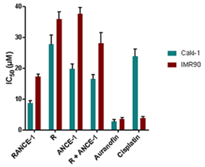 |
|
| RANCE-1 (1) | 8.7 ± 0.9 | 17.3 ± 0.8 | |
| R (2) | 27.8 ± 2.9 | 35.9 ± 2.3 | |
| ANCE-1 (3) | 19.8 ± 1.6 | 37.6 ± 2.1 | |
| R (2) + ANCE-1 (3) | 16.6 ± 1.3 | 28.1± 3.4 | |
| Auranofin | 2.8 ± 0.6 | 3.7 ± 0.4 | |
| Cisplatin | 23.9 ± 2.4 | 3.9 ± 0.5 | |
All compounds were dissolved in 1% of DMSO and diluted with media before addition to cell culture medium for a 72 hour incubation period. Cisplatin was dissolved in H2O·
RANCE-1 (1) derivative is more cytotoxic to the Caki-1 cells than cisplatin, monometallic ruthenium R (2) and gold ANCE-1 (3) compounds. Importantly, RANCE-1 (1) is considerably less toxic to the non-tumorigenic IRM-90 than cisplatin and ruthenium R (2) which makes RANCE-1 more selective. While Auranofin is also cytotoxic with a low IC50 value in Caki-1 cells it is only moderately selective. We studied the effect of the combination of monometallic ruthenium R (2) and gold ANCE-1 (3) compounds (1:1 equivalents) on Caki-1 using the same conditions as for the bimetallic compound. The resulting IC50 values were larger than those of RANCE-1 (1), while remaining selective. This fact supports the idea that there is indeed a synergistic effect for RANCE-1 (1) on the renal cancer cells as described for other Ru-Au and Ti-Au compounds described by our group.25–28 In view of the results obtained, we decided not to explore the effects of cisplatin in Caki-1 cells any further.
Following the evaluation of the cytotoxicity of bimetallic RANCE-1 and monometallic ruthenium R (2), gold ANCE-1 (3) and Auranofin, we proceed to evaluate cell death mechanisms for RANCE-1 and Auranofin. Caki-1 renal cancer cells were incubated with the indicated compound at the IC50 concentration for 72 hours. We observed that RANCE-1 (1) induce apoptosis in 82% of cells killed and Auranofin (86%) of cells (Figure 1). Auranofin and other gold(I) compounds are known to be apoptotic in several cancer cell lines,32,33 and p-cymene Ru derivatives (as the classic RAED described by Sadler and RAPTA-C described by Dyson) are also mainly apoptotic (ca 80%) on the ovarian cancer cell line A2780.34 Thus the apoptotic behavior of bimetallic Ru-Au RANCE-1 in terms of cell death may be due to the presence of both the Ru and Au fragments.
Figure 1.
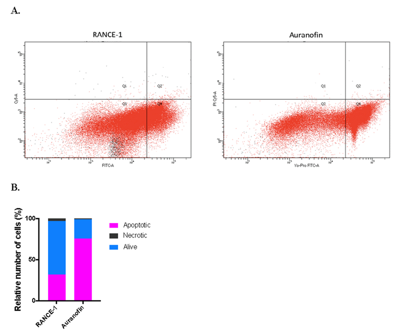
Cell death assays on Caki-1 cells induced by IC50 concentrations of RANCE-1 (1), and Auranofin measured by using two-colour flow cytometric analysis, after 24 h of incubation. (A) Flow cytometry histogram of RANCE-1 (1) induced apoptosis in 32% while Auranofin induced apoptosis in 76% of cell population (B) Bar-graph representation of from flow cytometry histogram quantifying cell death.
Next we evaluated the effects of most cytotoxic RANCE-1 (1), and Auranofin for cell cycle arrest. We observed that cells treated with RANCE-1 (1) had the greatest percentage of cells in Sub G1 (26%) and more G1/G0 (37%) the fewest cells in S phase (16%), and similarly few cells in G2/M (20%). Similarly for other p-cymene Ru derivatives such as RAED and RAPTA-C it was reported that greatest percentage of cells was in G1/G0 (60%) and fewest cells in S phase (ca. 15%) and G2/M (5%) for ovarian cancer A278 cell lines.34 Auranofin treated cells were almost exclusively in G0/G1 (82%), suggesting that the compound induced complete G1/G0 arrest, as has already been reported for AF in other cell lines,35–38 and also in cancer cells treated with anti-inflammatory drugs.39
Inhibition of migration and invasion
In advanced tumors, increased cell migration and invasion is a hallmark of metastasis40,41. We therefore evaluated the anti-invasive properties of RANCE-1 (1), the monometallic Ru R (2), and Au ANCE-1 (3) compounds. The effect of the compounds on migration was determined using a wound-healing 2D scratch assay (Figure 3A). RANCE-1 and Auranofin significantly reduced migration by 82% and 88% respectively. While the cytotoxicity and apoptotic properties of Auranofin on different cancer cell lines are well known,33 the efficacy of Auranofin was unexpected and has not been previously reported. The monometallic Au compound ANCE-1 (3) reduced migration by 26% while the monometallic Ru derivative R (2) reduced migration by 68%. The antimetastatic attributes of R (2) as it prevents in vitro cell invasion had been previously described for the triple-negative MDA-MB231 cancer cells30 and this potential metastatic phenotype was one of the reasons we choose the Ru fragment of the RANCE-1 heterometallic compound.
Figure 3.
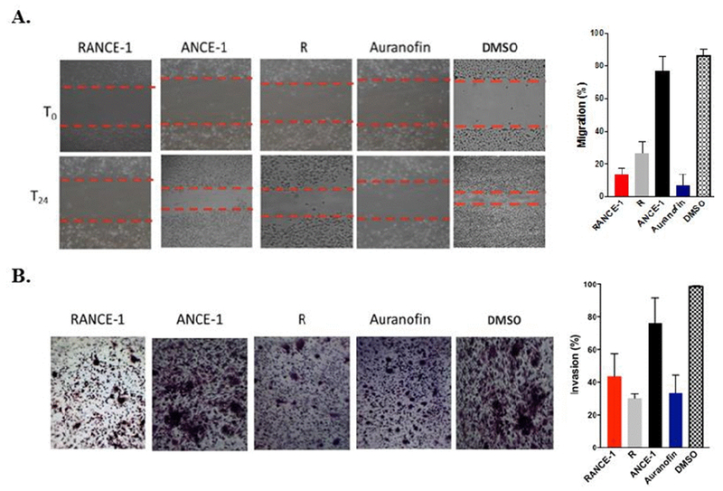
Cell migration and Invasion Inhibition Assays for bimetallic Ru-Au compound RANCE-1 (1), the monometallic Ru R (2) and Au ANCE-1 (3) compounds and Auranofin. A) Inhibition of migration (2D wound-healing scratch assay). Scratch assay showing that RANCE-1, R and Auranofin interfere with Caki-1 migration. Panels show representative images of untreated cells at time points T0 (top row) when the compound is added to the assay up to 24 hours (bottom row). B) Inhibition of invasion (3D Transwell Assay). Cells were seeded onto a transwell migration chambers containing filters coated on with Geltrex® matrix, then incubated for 24h with IC20 concentrations of RANCE-1 (1), R (2), ANCE-1 (3) or Auranofin. The transwell assay shows that RANCE-1 (1), R (2) and Auranofin interfere with Caki-1 invasion. Panels show representative images of treated cells at time points T24. Error bars indicate standard deviations.
RANCE-1 was observed to inhibit invasion in a 3D Transwell assay fitted with Geltrex® matrix an extracellular matrix analogue (Figure 3B). RANCE-1 (1) reduced invasion by 66%, as we observed with migration the monometallic Auranofin was an excellent inhibitor of invasion (54% of invasion is inhibited). The inhibition of invasion by the Ru monometallic compound R (2) at 30% was quite robust (as previously described for another cancer cell line)30, higher even than that of RANCE-1 or Auranofin. The monometallic Au compound ANCE-1 on the other hand only inhibited 18% of migration in agreement with the data obtained in the 2D scratch assay.
Inhibition of angiogenesis
Neovascularization plays an essential role in the pathology of tumor growth. We chose the formation tube-like structures by HUVEC cells on an extracellular matrix, as a mean to assess in angiogenesis of RANCE-1 (1), R (2), ANCE-1 (3) and Auranofin. The assay chosen consists in assessing the endothelial tube formation of Human umbilical vein endothelial cells (HUVECs) (Figure 4). The number of tubes and nodes was counted as described by DeCicco‐Skinner et al.42 The greater the inhibition of tube formation, that is the lowest length of tube (TL) and the lowers number of nodes (TN) the higher the antiangiogenic properties of a compound.
Figure 4.
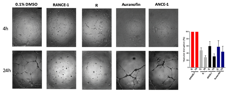
Induction of endothelial cell reorganization into 3D vessel structures. Human umbilical vein endothelial cells (HUVEC) were seeded in plate coated with Geltrex® matrix using completer HUVEC media and incubated at 37°C and 5% CO2. At post-seeding IC10 concentrations of RANCE-1, A, ANCE-1, Auranofin or 0.1% DMSO was added. (A) representative phase-contrast images captured 24h after the compounds were added. (B) Quantitation of tube formation.
The ruthenium containing derivatives RANCE-1 (1) and R (2) were observed to inhibit all tube formation in a 3D assay on Geltrex® matrix an extracellular matrix analogue (Figure 4). The gold monometallic compounds ANCE-1 (3) and Auranofin cause mild disturbances in tube arrangement but there is viable tube formation. This indicates that the ruthenium component of the compound may be responsible for the antiangiogenic properties. However RANCE-1 displays an impressive antiangiogenic effect (higher than that of the monometallic compound R (2)).
Inhibition of targets associated to cancer progression, cisplatin resistance, metastasis and angiogenesis
Inhibition of Thioredoxin Reductase
Changes in cell anti-oxidant capacity are a characteristic of many chemo-resistant cancers. Overexpression of thioredoxin reductase (TrxR) is a critical part of cisplatin-resistant cancer cell survival, thus making this enzyme an important anti-cancer target.40,43–46 We have reported on the relevant inhibition of TrxR in Caki-1 cells by Auranofin25 and heterometallic titanocene-Au25,26 and Ru-Au complexes.28 Therefore, we measured the activity of thioredoxin reductase in Caki-1 cells, following incubation with compounds RANCE-1 (1), Ru R (2) and Au ANCE-1 (3) and Auranofin as a positive control (Figure 5A). We observed that the TrxR inhibition activity of all the compounds did not improve much between 24h and 72h. As we have already reported,25 after 72 h of incubation Caki-1 TrxR activity is significantly reduced by Auranofin (86%). The inhibition of TrxR by RANCE-1 (1) and monometallic Au(I) compound ANCE-1 (3) was almost identical (RANCE-1, 47%; ANCE-1 (40%). While R (2) inhibited TrxR by less than 10% in that same time range. This indicates that the Au(I) fragment present in heterometallic RANCE-1 compound is most likely responsible for the TrxR inhibition effect.
Figure 5.
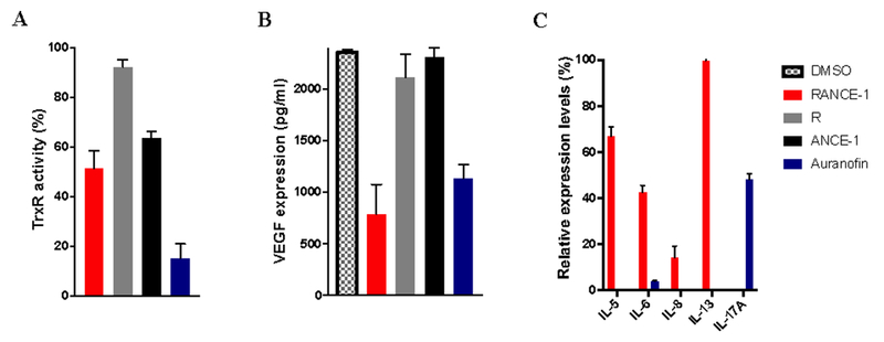
Inhibition of prometastatic protumorigenic factors in in Caki-1 renal cancer cells A. Inhibition of mitochondrial protein TrxR by bimetallic Ru-Au compound RANCE-1 (1), the monometallic ruthenium R (2) and gold ANCE-1 (3) compounds and Auranofin (1 μM) for 72 hours. The values for indicate the percentage of reduction of TrxR activity relative to 0.1% DMSO treated control. B. Inhibition of pro-angiogenic proteins VEGF by bimetallic Ru-Au compound RANCE-1 (1), the monometallic ruthenium R (2) and gold ANCE-1 (3) compounds and Auranofin (IC 20 concentrations) for 24 hours. The values for indicate the absolute concentration (pg/ml) of VEGF following treatment, 0.1% DMSO control. C. RANCE-1 (1) and Auranofin induced changes in the expression levels of Interleukins. Analysis of 150ng of protein extracted from cell lysate collected from RANCE-1 or Auranofin (IC20) treated cells. All the data shown are representative of triplicates.
Inhibition of VEGF
Vascular endothelial growth factor (VEGF) is the key mediator of angiogenesis in cancer, where it is up-regulated by oncogene expression, a variety of growth factors.47–49 Before most solid tumors can grow beyond past the 0.5 cm diameter, they require blood vessels for nutrients and oxygen.39,48,49 The production of VEGF and other growth factors by the tumor results in the ‘angiogenic switch’, where new vasculature is formed in and around the tumor, promoting exponential growth.50 Also, ruthenium compounds have been reported to inhibit VEGF.51 Given the key tumor promoting properties of VEGF we set out to evaluate the inhibitory effects of the RANCE-1 (1), the monometallic ruthenium R (2) and gold ANCE-1 (3) compounds and Auranofin on VEGF, an attractive targets in cancer therapy. We found that VEGF secretion is significantly inhibited by both RANCE-1 (1) and Auranofin after 72h of incubation (70% and 55% reduction respectively), while R (2) and ANCE-1 (3) lead to no notable inhibition of VEGF secretion (Figure 5B). Angiogenesis is a complex process driven by diverse activities and an intricate sequence of factors. While VEGF is known to be a key regulator of angiogenesis and its downregulation is often correlated with reduced angiogenesis, such correlation is not absolute. As our data indicate while there is evident inhibition of tube formation by RANCE-1 and significantly less inhibition of tube formation by Auranofin, they are both potent inhibitors of VEGF, this suggest that there might be more at play in RANCE-1 inhibition of VEGF and its effect on tube formation.
The inhibition of VEGF-2 has been recently reported for a ruthenium (III) macrocyclic cationic compound described by Che et al. which inhibited MS-1 endothelial and HUVEC tube formation (a model for angiogenesis) and inhibited angiogenesis in chicken chlorioallantoic membrane (CAM).51
Inhibition of Interleukins
Inflammatory cytokines such as interleukins (IL) are known to be associated with malignant progression of breast and lung cancers.50,52,53 IL-5, IL-6, IL-8, IL-12 and IL-17A are of particular relevance as their elevated expression has been detected in multiple epithelial tumors and has been shown to be associated with tumor metastasis.52,53 ILs also play an important role in tumor promotion and metastasis through various proteolytic interaction and through control of matrix metalloproteinases (MMP) expression and the expression of angiogenic proteins growth factors such as VEGF50. We studied the inhibition of ILs by selected most active compounds bimetallic RANCE-1 (1) and Auranofin after 72 hour of incubation at IC20 concentrations (Figure 5C). We found that RANCE-1 (1) inhibited IL-6 expression by 60%. RANCE-1 (1) inhibited IL-5 expression by 37% and had no effect on IL-13 expression, and resulted in a complete inhibition of IL-17A expression. The inhibition of IL-17 is of great clinical interest because increased IL-17A expression is associated with ER(−) and triple negative tumor hyper proliferation and poor prognosis in breast cancer. IL-17 is also known to drives several pathogenic processes during breast cancer progression to metastasis, IL-17 not only promotes tumor cell survival and invasiveness, it also contributes to the promotion of tumor angiogenesis. Auranofin, as previously reported, inhibits IL-6, but also completely inhibits expression of IL-5, IL-8 and IL-13, all key players in inflammatory signalling.54 Given the potent inhibition of IL expression observed upon treatment with RANCE-1 (1), this compound might be a good candidate for combination therapy with a more cytotoxic agent. The great value of IL inhibitors is further increased because ILs are known inducer of MMPs which are critical in metastasis.50,55
Inhibition of metalloproteases
Matrix metalloproteinases (MMPs) are a multigene family of zinc-dependent extracellular proteins. MMP are remodelling endopeptidases implicated in pathological processes, such as carcinogenesis.50,55–57
MMPs play a pivotal role in tumor growth and the complex processes of invasion and metastasis, including proteolytic degradation of ECM, modifications of the cell-cell and cell-ECM interfaces, migration and angiogenesis.44,56 Therefore MMPs are attractive targets for inhibitory therapeutic intervention and a number of therapeutic agents, called matrix metalloproteinase inhibitors (MMPIs) have been developed.55,58,59 Many members of the MMP family are involved in tumor induced inflammation signalling and angiogenesis in cooperation with members of the IL family including IL-6.58–60 The secretion of 9 MMPs is significantly inhibited by RANCE-1 (1): MMP-1 (72%) MMP-3 (74%) MMP-7 (100%), MMP-8 (97%) MMP-9(50%), MMP-10 (100%), MMP-12 (89%) MMP-13 (100%) (Figure 6A). Such inhibition is particularly robust and in combination with the VEGF and ILs inhibition by RANCE-1 (1), this compound therefore presents a molecular target profile that would potentially make it a very efficient antimetastatic and antiangiogenic agent.
Figure 6.
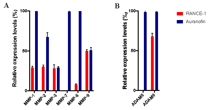
RANCE-1 (1) and Auranofin induced changes in the expression levels of metalloproteases in Caki-1 cells. A. matrix metalloproteases (MMP) family members. B disintegrin and metalloprotease (ADAM) family members. Analysis of 150 ng of protein extracted from cell lysate collected from RANCE-1 or Auranofin (IC20) treated cells. The data shown are representative of triplicates.
Accumulating evidence demonstrates the crucial role of proteolytic enzymes such ADAMs, which are closely related to MMPs facilitate tumor cell invasion and metastasis. In particular, ADAM-9 is reported to be highly expressed in invasive renal cell cancer while ADAM8 is considered a robust hemo— histo— chemical marker for lung cancer and correlated with poor prognosis for patients with pancreatic ductal adenocarcinoma.61,62 From our study, we observed that treatment with RANCE-1 (1) ablated all expression of ADAM-8, and inhibited ADAM-9 expression by 37%, while Auranofin had no effect on the expression levels of those two proteases (Figure 6B). Such inhibition is particularly robust and in combination with the VEGF and IL(s) secretion’s inhibition that RANCE-1 (1) displays, this compound presents a molecular target profile that would potentially make it a very efficient antimetastatic and antiangiogenic agent.
Inhibition of Cathepsin Proteases
Cysteine cathepsin proteases (Cts) are known regulators of cancer progression and therapeutic response. Some members of the cathepsin family are highly expressed in metastatic tumors, including CatsB, CtsL, CtsS and CtsX.63–65 Cathepsin B is of significant importance to cancer therapy as it is involved in various pathologies and oncogenic processes in humans.50,64–66 Therefore, a pancathepsin inhibitor is of great clinical interest. Some ruthenium p-cymene compounds (RAPTA family) have been shown to be inhibitors of cathepsin B and this inhibition has been correlated to their antimetastatic properties.67 We reported on the inhibition of cathepsin B (purified) by a neutral ruthenium-gold compound.27 We observed that RANCE-1 (1) ablated CtsB and CtsD expression (100% suppression), while inhibiting CtsL, CtsS and CtsX/Z by 38%, 35% an 53% respectively, while Auranofin had no inhibitory effect on the expression of the five Cts evaluated.
From our studies, RANCE-1 is emerging as a potent pan-MMP and pan-cathepsin inhibitor with additional notable inhibition of members of the ADAM protease family, which may explain the mechanism by which RANCE-1 inhibits tumor growth, invasion, and angiogenesis (Figure 7).
Figure 7.
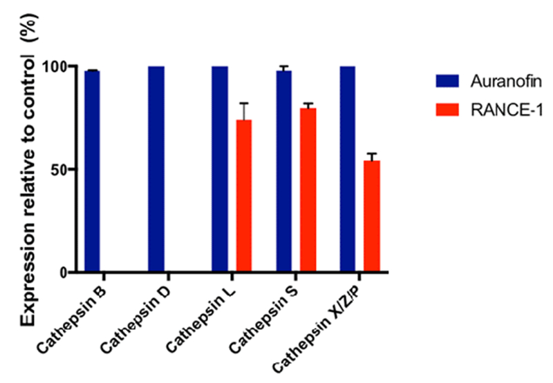
RANCE-1 (1) and Auranofin induced changes in the expression levels of members of the cathepsin proteases family in Caki-1 cells. Analysis of 150 ng of protein extracted from cell lysate collected from RANCE-1 (1) or Auranofin (IC20) treated cells. The data shown are representative of triplicates.
Therefore our studies suggest that bimetallic ruthenium-gold compound RANCE-1 (1) is a potent pan-MMP and pan-cathepsin inhibitor with notable inhibition of members of the ADAM protease family. This inhibition may lay the mechanism by which RANCE-1 (1) inhibits tumor growth, invasion and denovo vasculature.
Conclusions
To summarize, this study demonstrates the synergism of a heterometallic Ru-Au (RANCE-1, 1) compound designed to harness the cytotoxic and apoptotic effects of Au(I) lipophilic cations as well as their TrxR inhibition properties with the potential antimetastatic effects of a Ru p-cymene derivative containing a phosphine. In addition to be cytotoxic and apoptotic, RANCE-1 (1) displays relevant inhibition of migration, invasion and angiogenesis, while also inhibiting molecular pathways associated with these processes. The molecular targets inhibited include different interleukins, metalloproteases and cathepsins, which are involved in tumor metastasis and angiogenesis, and to an even higher degree the angiogenic factor VEGF (also involved in angiogenesis). It is noteworthy that the inhibition observed is in general better than that of the individual monometallic fragments present in the heterometallic compound. In some cases, this inhibition can be correlated with a particular metallic fragment of the bimetallic compound (reinforcing the idea of the positive synergistic effect caused by the two distinct metals).
During this study we evaluated Auranofin and observed it had a very similar effect to RANCE-1 (1) in the renal cancer cell line Caki-1 in terms of its antiproliferative and antimetastatic properties and some of the sets of targets inhibited. RANCE-1 (1) however was a better VEGF inhibitor than Auranofin and a much better pan-MMP and pan-cathepsin inhibitor. Moreover, RANCE-1 (1) block all angiogenic formation while Auranofin merely induced branching disturbances of de novo angiogenesis in an in vitro model.
These results are very relevant in the search for multi targeted therapies that would best avert target specific induced resistance, and thus warrant further evaluation of the efficacy and mechanism of RANCE-1 (1) in renal cancer in vivo. In addition, the relevant results found for Auranofin (an old anti-rheumatic drug being currently ‘repurposed’ as chemotherapeutic for different diseases including cancer) also warrant further exploration of this agent in renal cancer treatment.
Figure 2.
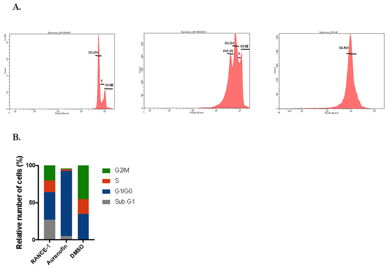
Cell cycle arrest induced by RANCE-1 (1), and Auranofin. Cells were treated with or without IC20 concentration of RANCE-1 (1), and Auranofin for 24 h. A. Flow cytometry histogram of RANCE-1 increased G1/G0 (37%) and Sub G1 (26%) population accumulation while Auranofin increased G1/G0 (82 %) population accumulation. B. bar-graph representation of from flow cytometry histogram.
Chart 1.

Compounds used in this study: bimetallic [Cl2(p-cymene)Ru(μ-dppm)Au(IMes)]ClO4 (RANCE-1, 1),28 monometallic [Ru(p-cymene)Cl2(dppm-κP)] R (2),30 and [AuCl(IMes)] ANCE-1 (3),31 and Auranofin.
ACKNOWLEDGMENT
We thank the National Cancer Institute (NCI) for grant 1SC1CA182844 (M.C.) and Dr. Karen Hubbard for providing infrastructure and advice. We are grateful to Dr. Natalia Curado and Yu-Fung Mui for their assistance in the preparation of the metallic compounds, Mike A. Cornejo for his assistance with some IC50 assays, and Ciara Bagnall for her technical expertise in imaging and immunocytochemistry.
Abbreviations:
- ANCE-1
[AuCl(IMes)]
- ATCC
American Type Culture Collection
- ADAM
a disintegrin and metalloproteinase
- Auranofin
[(2R,3R,4S,5R,6S)-3,4,5-tris(acetyloxy)-6-{[(triethyl-λ5-phosphanylidene)aurio]sulfanyl}oxan-2-yl]methyl acetate
- Caki-1
human clear cell renal cell carcinoma
- Cisplatin
cis-Diamminedichloroplatinum(II)
- dppm
diphenylphosphanylmethyl(diphenyl)phosphane
- DTNB
3,3’-Disulfanediylbis(6-nitrobenzoic acid)
- HCT 116
human colorectal carcinoma
- HUVEC
human umbilical vein endothelial cells
- ILs
Interleukins
- IMes
1,3-bis(2,4,6-trimethylphenyl)imidazol-2-ylidene
- IRM90
human foetal lung fibroblast
- MMPs
matrix metalloproteinase
- NHC
N-heterocyclic carbene
- p-cymene
4-Isopropyltoluene
- R
[Ru(p-cymene)Cl2(dppm-κP)]
- RANCE-1
[Cl2(p-cymene)Ru(μ-dppm)Au(IMes)]ClO4
- TNB2−
5-thio-2-nitrobenzoic acid
- TrRx
thioredoxin reductase
- VEGF
vascular endothelial growth factor
References
- 1.Pelletier F, Comte V, Massard A, Wenzel M, Toulot S, Richard P, Picquet M, Le Gendre P, Zava O, Edafe F, Casini A, Dyson PJ (2010) J Med Chem 53:6923–6933. [DOI] [PubMed] [Google Scholar]
- 2.Liu Y, Chen TF, W Y-S, Mei W-J, Huang X-M, Yang F, Liu J, Zheng W-J. Chem-Biol Interact (2010) 183:349–356. [DOI] [PubMed] [Google Scholar]
- 3.Wenzel M, Bertrand B, Eymin M–J, Comte V, Harvey JA, Richard P, Groessl M, Zava O, Amrouche H, Harvey PD, Le Gendre P, Picquet M, Casini A (2011) Inorg Chem 50:9472–9480. [DOI] [PubMed] [Google Scholar]
- 4.Bjelosevic H, Guzei IA, Spencer LC, Persson T, Kriel FH, Hewer R, Nell MJ, Gut J, van Resburg CEJ, Rosenthal P, Coates J, Darkwa J, Elmroth SKC (2102) J Organomet Chem 720: 52–59. [Google Scholar]
- 5.Kaushik NK, Mishra A, Ali A, Adhikari JS, Verma AK, Gupta R (2012) J Inorg Biol Chem 17: 1217–1230. [DOI] [PubMed] [Google Scholar]
- 6.Nieto D, Gonzalez-Vadillo AM, Bruna S, Pastor CJ, Rios-Luci C, Leon LG, Padron JM, Navarro-Ranninger C, Cuadrado I. Dalton Trans (2012) 41:432–441. [DOI] [PubMed] [Google Scholar]
- 7.Lease N, Vasileviski V, Carreira M, de Almeida A, Sanaú M, Hirva P, Casini A, Contel M (2013) J Med Chem 56:5806–5818. [DOI] [PMC free article] [PubMed] [Google Scholar]
- 8.Garcia-Moreno E, Gascon S, Rodriguez-Yoldi MJ, Cerrada E, Laguna M (2013) Organometallics 32: 3710–3720. [Google Scholar]
- 9.Barreiro E, Casas JS, Couce MD, Sanchez A.; Sordo J, Vazquez-Lopez EM (2104) J Inorg Biochem 131: 68–75. [DOI] [PubMed] [Google Scholar]
- 10.Wenzel M, Bigaeva E, Richard P, Le Gendre P, Picquet M, Casini A, Bodio E (2014) J Inorg Biochem 141: 10–16. [DOI] [PubMed] [Google Scholar]
- 11.Fernandez-Moreira V, Marzo I, Gimeno C (2014) Chem Sci 5: 4434–4446. [Google Scholar]
- 12.Zaidi Y, Arjmand F.; Zaidi N, Usmani JA, Zubair H, Akhatar K Hossain M, Shadab GGHA. Metallomics (2014) 6: 1469–1479. [DOI] [PubMed] [Google Scholar]
- 13.Govender P, Lemmerhirt H, Hutton AT, Therrien B, Bednarski PJ, Smith GS (2014) Organometallics 33: 5535–5545. [Google Scholar]
- 14.Bertrand B, Citta A, Franken IL, Picquet M, Folda A, Scalcon V, Rigobello MP, Le Gendre P, Casini A, Bodio E (2015) J Biol Inorg Chem 20: 1005–1020. [DOI] [PubMed] [Google Scholar]
- 15.Boselli L, Carraz M, Mazeres S, Paloque L, Gonzalez G, Benoit-Vical F, Valentin A, Hemmert C, Gornitzka H (2015) Organometallics 34: 1046–1055. [Google Scholar]
- 16.Nieto D, Bruna S, Gonzalez-Vadillo AM, Perles J, Carrillo-Hermosilla F, Antiniolo A, Padron JM, Plata GB, Cuadrado I (2015) Organometallics 34: 5407–5417. [Google Scholar]
- 17.Nithyakumar A, Alexander V (2015) Dalton Trans 44:17800–17809. [DOI] [PubMed] [Google Scholar]
- 18.Fenton JB, Busse M, Rendina LM (2015) Aus J Chem 68: 576–580. [Google Scholar]
- 19.Ma L, Ma R, Wang Z, Yiu S-M, Zhu G (2016) Chem Commun 52: 10735–10738. [DOI] [PubMed] [Google Scholar]
- 20.Singh N, Jang S, J J-H, Kim DH, Park DW, Kim I, Kim H, Kang SC, Chi K-W (2016) Chemistry-A Eur J 22: 16157–16164. [DOI] [PubMed] [Google Scholar]
- 21.Lopes J, Alves D, Morais TS, Costa PJ, Piedade M, Fatima M, Marques F, Villa de Brito MJ, Garcia MH (2017) J Inorg Biochem 169:68–78. [DOI] [PubMed] [Google Scholar]
- 22.Luengo A, Fernandez-Moreira V, Marzo I, Gimeno MC (2017) Inorg Chem 56: 15159–15170. [DOI] [PubMed] [Google Scholar]
- 23.González-Pantoja JF, Stern M, Jarzecki AA, Royo E, Robles-Escajeda E, Varela-Ramirez A, Aguilera RJ, Contel M (2011) Inorg Chem 50:11099–11110. [DOI] [PMC free article] [PubMed] [Google Scholar]
- 24.Fernández-Gallardo J, Elie BT, Sulzmaier F, Sanaú M, Ramos JW, Contel M (2014) Organometallics 33: 6669–6681. [DOI] [PMC free article] [PubMed] [Google Scholar]
- 25.(a) Fernández-Gallardo J, Elie BT, Sadhukha T, Prabha S, Sanaú M, Rotenberg SA, Ramos JW, Contel M (2015) Chem Sci 6: 5269–5283. [DOI] [PMC free article] [PubMed] [Google Scholar]; (b) Contel M, Fernández-Gallardo J, Elie BT, Ramos JW. US Patent 9,315,531 (April/19/2016).
- 26.Mui YF, Fernández-Gallardo J, Gubran A; Elie BT, Maluenda I, Sanaú M, Navarro O, Contel M (2016) Organometallics 35: 1218–1227. [DOI] [PMC free article] [PubMed] [Google Scholar]
- 27.Massai L, Fernández-Gallardo J, Guerri A, Arcangeli A, Pillozzi S, Contel M, Messori L (2015) Dalton Trans 44: 11067–11076. [DOI] [PMC free article] [PubMed] [Google Scholar]
- 28.Fernández-Gallardo J, Elie BT, Sanaú M, Contel M (2016) Chem. Commun 52: 3155–3158. [DOI] [PMC free article] [PubMed] [Google Scholar]
- 29.Dean TC, Yang M, Liu M, Grayson JM, DeMartino AW, Day CS, Lee J, Furdui CM, Bierbach U (2017) ACS Med Chem Lett 8: 572–576. [DOI] [PMC free article] [PubMed] [Google Scholar]
- 30.Das S, Sinha S, Britto R, Somasundaram K, Samuelson AG (2010) J Inorg Biochem 104: 93–104. [DOI] [PubMed] [Google Scholar]
- 31.Fremont P, Scott NM, Stevens ED, Nolan SP (2005) Organometallics 24: 2411–2418. [Google Scholar]
- 32.Varghese E, Busselberg D (2014) Cancers 6: 2243–2258 and refs therein. [DOI] [PMC free article] [PubMed] [Google Scholar]
- 33.Roder C, Thomson MJ (2015) Drugs R D 15: 13–20. [DOI] [PMC free article] [PubMed] [Google Scholar]
- 34.Adhireksan Z, Davey GE, Campomanes P, Groessl M, Clavel CM, Yu H, Nazarov AA, Yeo CHF, Ang WH, Droge P, Rothlisberger U, Dyson PJ, Davey CA (2014) Nature Comm 5:3462. [DOI] [PMC free article] [PubMed] [Google Scholar]
- 35.Nakayaa A, Sagawa M, Muto A, Uchida H, Ikedaa Y, Kizari M (2011) Leukemia Res 35: 243–249. [DOI] [PubMed] [Google Scholar]
- 36.Liu C, Liu Z, Li M, Li X, Wong Y-S, Ngai S-M, Zheng W, Zhang Y, Chen T (2013) PLOS one 8: e53945. [DOI] [PMC free article] [PubMed] [Google Scholar]
- 37.Fan C, Zheng W, Fu X, Li X, Wong Y-S, Chen T (2014) Cell Death and Disease 5: e191. [DOI] [PMC free article] [PubMed] [Google Scholar]
- 38.Park S-H, Lee JH, Berek JS, Hu MC-T (2014) Int J Oncol 45: 1691–1698. [DOI] [PMC free article] [PubMed] [Google Scholar]
- 39.Denkert C, Furstenberg A, Daniel PT, Koch I, Kobell M, Weichert W, Siegert A, Hauptmann S (2013) Oncogene, 22, 8653–8661. [DOI] [PubMed] [Google Scholar]
- 40.Moutasim KA, Nystrom ML, Thomas GJ (2011). Methods Mol Biol. 731: 333–343. [DOI] [PubMed] [Google Scholar]
- 41.Hulkower KI, Herber RL (2011). Pharmaceutics 3: 107–124. [DOI] [PMC free article] [PubMed] [Google Scholar]
- 42.DeCicco‐Skinner KL, Henry GH, Cataisson C, Tabib T, Gwilliam JC, Watson NJ, Bullwinkle EM, Falkenburg L, O’Neill RC, Morin A, Wiest JS (2014) J Vis Exp 91:e51312. [DOI] [PMC free article] [PubMed] [Google Scholar]
- 43.Cheng X, Holenya P, Can S, Alborzinia H, Rubbiani R, Ott I, Wölfl S (2014) Mol Cancer 13:221. [DOI] [PMC free article] [PubMed] [Google Scholar]
- 44.Farina AR, Tacconelli A, Cappabianca L, Masciulli MP, Holmgren A, Beckett GJ, Gulino A, Mackay AR (2001). Eur J Biochem. 268: 405–413. [DOI] [PubMed] [Google Scholar]
- 45.Lim HV, Hong S, Jin W, Lim S, Kim SJ, Kang HJ, Park EH, Ahn K, Lim CJ (2005). Exp Mol Med 37: 497–506. [DOI] [PubMed] [Google Scholar]
- 46.Marzano C, Gandin V, Folda A, Scutari G, Bindoli A, Rigobello AP (2007). Free Radic Biol Med 42: 872–881. [DOI] [PubMed] [Google Scholar]
- 47.Zou M, Zhang X, Xu C (2016). Cell Oncol 39: 47–57. [DOI] [PubMed] [Google Scholar]
- 48.Wei LH, Kuo ML, Chen CA, Chou CH, Lai KB, Lee CN, Hsieh CY (2003). Oncogene 22: 1517–1527. [DOI] [PubMed] [Google Scholar]
- 49.Suzuki M, Tetsuka T, Yoshida S, Watanabe N, Kobayashi M, Matsui N, Okamoto T (2000). FEBS Lett. 465: 23–27. [DOI] [PubMed] [Google Scholar]
- 50.Quail DF and Joyce JA (2013). Nat. Med. 19:1423–1437. [DOI] [PMC free article] [PubMed] [Google Scholar]
- 51.Kwong W-L, Lam K-Y, Lok C-N, Lai Y-T, Lee P-Y, Che C-M (2016). Angew Chem Int Ed. 55: 13524–13528. [DOI] [PubMed] [Google Scholar]
- 52.Lippitz BE, Harris RA(2016). Oncoimmunology.11(5):e1093722. [DOI] [PMC free article] [PubMed] [Google Scholar]
- 53.Welte T, Zhang XH (2015). Mediators Inflamm 804347. [DOI] [PMC free article] [PubMed] [Google Scholar]
- 54.Kim N-M, Lee M-Y, Park S-J, Choi J-S, Oh M-K, Kim In-S (2007) Immunology 122: 607–614. [DOI] [PMC free article] [PubMed] [Google Scholar]
- 55.Li Q, Chen B, Cai J, Sun Y, Wang G, Li Y, Li R, Feng Y, Han B, Li J, Tian Y, Yi L, Jiang C (2016). PLoS One 11, e0151815. [DOI] [PMC free article] [PubMed] [Google Scholar]
- 56.Overall CM, Kleifeld O (2006). Nat Rev Cancer 6: 227–239. [DOI] [PubMed] [Google Scholar]
- 57.He ZH, Chen N, Wang D, Bin Y, She JX (2014). Eur. J. Pharmacol. 740: 240–247. [DOI] [PubMed] [Google Scholar]
- 58.Woo JH, Lim JH, Kim YH, Suh SI, Min DS, Chang JS, Lee YH, Park JW, Kwon TK (2004). Oncogene 23: 1845–1853. [DOI] [PubMed] [Google Scholar]
- 59.Woo JH, Lim JH, Kim YH, Suh SI, Min DS, Chang JS, Lee YH, Park JW, Kwon TK (2004). Oncogene 23: 1845–1853. [DOI] [PubMed] [Google Scholar]
- 60.Woessner JF and Nagase H (2000) Matrix Metalloproteinases and TIMPs: Protein Profile, Oxford University Press, Oxford. [Google Scholar]
- 61.Rocks N, Paulissen G, El Hour M, Quesada F, Crahay C, Gueders M, Foidart JM, Noel A, Cataldo D (2008). Biochimie 90: 369–79. [DOI] [PubMed] [Google Scholar]
- 62.Schlomann U, Koller G, Conrad C, Ferdous T, Golfi P, Garcia AM, Höfling S, Parsons M, Costa P, Soper R, Bossard M, Hagemann T, Roshani R, Sewald N, Ketchem RR, Moss ML, Rasmussen FH, Miller MA, Lauffenburger DA, Tuveson DA, Nimsky C, Bartsch JW (2015). Nat Commun 6: 6175. [DOI] [PMC free article] [PubMed] [Google Scholar]
- 63.Yousef EM, Tahir MR, St-Pierre Y, Gaboury LA (2014). BMC Cancer 14:609. [DOI] [PMC free article] [PubMed] [Google Scholar]
- 64.Olson OC, Joyce JA (2015) Nat Rev Cancer 15:712–729. [DOI] [PubMed] [Google Scholar]
- 65.Sevenich L and Joyce JA (2014). Genes Dev 28: 2331–2347. [DOI] [PMC free article] [PubMed] [Google Scholar]
- 66.Bakst RL, Xiong H, Chen CH, Deborde S, Lyubchik A, Zhou Y, He S, McNamara W, Lee SY, Olson OC, Leiner IM, Marcadis AR, Keith JW, Al-Ahmadie HA, Katabi N, Gil Z, Vakiani E, Joyce JA, Pamer E, Wong RJ (2017). Cancer Res 22:6400–6414. [DOI] [PMC free article] [PubMed] [Google Scholar]
- 67.Casini A, Gabbiani C, Sorrentino F, Rigobello MP, Bindoli A, Geldbach TJ, Marrone A, Re N, Hartinger CG, Dyson PJ, Messori L (2008). J Med Chem 51: 6773–6781. [DOI] [PubMed] [Google Scholar]


