Abstract
Microbiome sequencing has become the standard procedure in the study of new ecological and human-constructed niches. To our knowledge, this is the first report of a metagenome from the water of a greenhouse drain. We found that the greenhouse is not a diverse niche, mainly dominated by Rhizobiales and Rodobacterales. The analysis of the functions encoded in the metagenome showed enrichment of characteristic features of soil and root-associated bacteria such as ABC-transporters and hydrolase enzymes. Additionally, we found antibiotic resistances genes principally for spectinomycin, tetracycline, and aminoglycosides. This study aimed to identify the bacteria and functional gene composition of a greenhouse water drain sample and also provide a genomic resource to search novel proteins from a previously unexplored niche. All the metagenome proteins and their annotations are available to the scientific community via http://microbiomics.ibt.unam.mx/tools/metagreenhouse/.
Keywords: Shotgun sequencing, Greenhouse, Metagenome, Environmental sample, Water drain
Introduction
All the environments in the world contain millions of microorganisms. However, most of them are uncultivable, difficulting their study under laboratory conditions using traditional culture techniques. In contrast, the rapid development of sequencing technologies and the lower of their associated costs has allowed exploring the microbial composition of almost any ecological niche using metagenomic approaches, ranging from human gut to hot springs [1–3]. In this regard, metagenomic approaches have been used to answer two central questions: (i) which microorganisms are present and (ii) what is their functional contribution [4]. Metagenomic has opened the opportunity to find new microbial phyla [5] and novel protein families in previously unexplored niches [6], due to uncultivable microorganisms from there. Thus, the metagenomic resource provides the capacity of bioprospecting on the discovery of novel enzymes for research or industrial applications [7]. According to this idea, some new challenges in functional metagenomics, phylogenomics, ecology, and biotechnology have emerged. There are numerous applications of metagenomic analysis, ranging from prevention of diseases to solve industrial problems [8]. In recent years, the scientific community has tried to identify the role that microbial communities have in several disciplines such as human health [9, 10], and industry [11–13]. Metagenomics also has been applied to explore the impact of microorganisms in human-constructed niches [11, 14].
A greenhouse is an ecological niche entirely human manipulated, with the continuous exposure to pesticides, fertilizers, antibiotics and different chemicals for research purposes. Thus, subjecting the microbial communities under selective pressures. These effects can be analyzed using the high throughput sequencing methods. This allowed us the possibility to design new strategies for monitoring the microbial evolution of the structure and dynamics in particular human-constructed niches such as a greenhouse, plus comparing it to similar conditions somewhere else and eventually trace back any emerging problem. To our knowledge, this is the first report of a shotgun metagenome from a water sample of a greenhouse drain. Our work aimed to determine the microbial and functional composition of the water from a greenhouse drain. Our results indicated that this environment has low bacterial diversity, mainly dominated by Alphaproteobacteria, which is composed of Rhizobiales and Rhodobacterales orders. Interestingly, we found several antibiotic resistance genes and a functional enrichment for de novo amino acid synthesis in the metagenome.
Site information
The sampling site corresponds to the water of a greenhouse drain. The greenhouse is on the top of a building, located at the Institute of Biotechnology (IBt) of the National Autonomous University of Mexico (UNAM), in Cuernavaca City in México. The greenhouse is used for the cultivation of several plant species for research purposes.
Metagenome sequencing information
Metagenome project history
The collected sample was part of a pilot project to identify the novel bacterial composition of the water in the experimental greenhouse drain at the Institute of Biotechnology (IBt) of the National Autonomous University of Mexico (UNAM). We deposited the sequencing reads in the NCBI under the SRA accession number SRR5689218 and SRR5689219 and the Bioproject PRJNA390663. Additionally, the reads were uploaded to the MG-RAST server under the ids mgm4717011.3, mgm4717032.3, mgm4716707.3, mgm4716832.3, mgm4716680.3, mgm4716681.3, mgm4716833.3, mgm4717034.3. For more details see the study information in Table 1.
Table 1.
Study information
| Label | Greenhouse Drain-IBt |
|---|---|
| MG-RAST ID | mgm4717011.3, mgm4717032.3, mgm4716707.3, mgm4716832.3, mgm4716680.3, mgm4716681.3, mgm4716833.3, mgm4717034.3 |
| SRA ID | SRR5689218 (Drain A) SRR5689219 (Drain B) |
| Study | NA |
| GOLD ID (sequencing project) | NA |
| GOLD ID (analysis project) | NA |
| NCBI BIOPROJECT | PRJNA390663 |
| Relevance | Water drain sample |
Sample information
We collected the sample on 14 September 2015 at 18:00 h (GMT-5) at the IBt (Latitude: 18.918611, Longitude: − 99.234167). In Table 2 the sample information according to the minimal information standards is showed [15].
Table 2.
Sample information
| Label | Greenhouse Drain-IBt |
|---|---|
| GOLD ID (biosample) | NA |
| Biome | Culturing environment |
| Feature | Water of greenhouse drain system |
| Material | Water |
| Latitude and Longitude | 18.918611, −99.234167 |
| Vertical distance | 1510 m over sea level |
| Geographic location | Cuernavaca, Morelos. México |
| Collection date and time | 14/09/15, 18:00 h (GMT-5) |
Sample preparation, DNA extraction, library generation, and sequencing technology
Sample preparation (collection, transport, and storage)
A sample of 170 ml of water was directly collected from the greenhouse drain and immediately transported to the laboratory, located in the same building. Microbes were obtained by filtering this water through a sterilized PTFE 0.45 μm filter (Cat. 728–2045, Nalgene, NY, USA) using a vacuum pump. After filtration, we extracted the total DNA from the membranes.
DNA extraction (kits used, protocols used)
Total DNA was recovered from the filter membrane by shaking the filter for 5 min in a tube containing lysis solution and beads from ZR Soil Microbe DNA MicroPrep Kit (Cat. D6003 Zymo Research, Irvine, CA, USA). The following steps for DNA isolation were carried out following the manufacturer’s instructions for the ZR Soil Microbe DNA kit. After extraction, we assessed the DNA quality by agarose gel electrophoresis and quantity determined by the Thermo Fisher Qubit High-sensitivity fluorometric assay (Cat. Q32851, Life Technologies, Carlsbad, CA, USA).
Library generation (kits used, protocols used)
We constructed two DNA libraries containing different insert sizes: Drain-A and Drain-B with an insert size of 400 and 2000 bp, respectively (Table 3). Furthermore, different amounts of input DNA were used to construct the libraries: 1 ng for Drain-A and 25 ng for Drain-B. Both libraries were created following the manufacturer’s instructions for the Nextera XT DNA Library Preparation kit (Cat. FC-131-1024, Illumina, CA, USA). First, DNA was fragmented (tagmented) using the Nextera transposase. Second, the tagmented DNA was amplified using 12 PCR cycles to add the Index 1 (i7), Index 2 (i5), and full adapter sequences. The program on the thermal cycler was as follows: 72 °C for 3 min, 95 °C for 30 s; 12 cycles (95 °C for 10 s, 55 °C for 30 s and 72 °C for 30 s) and 72 °C for 5 min. After PCR amplification, both libraries were carefully size selected using Agencourt Ampure XP beads (Cat. A63882, Beckman Coulter, CA, USA) and the size was verified using a DNA Agilent Bioanalyzer 2100 (Cat. 5067–1504, Agilent Technologies, CA, USA).
Table 3.
Library information
| Label | Drain-A | Drain-B |
|---|---|---|
| Sample Label(s) | Drain-A | Drain-B |
| Sample prep method | ZR Soil Microbe DNA (Zymo) | ZR Soil Microbe DNA (Zymo) |
| Library prep method(s) | Nextera XT | Nextera XT |
| Sequencing platform(s) | Illumina NextSeq 500 | Illumina NextSeq 500 |
| Sequencing chemistry | V2 SBS Kit | V2 SBS Kit |
| Sequence size (GBp) | 10.4GBp | 0.60GBp |
| Number of reads | 6,976,736 | 401,466 |
| Single-read or paired-end sequencing? | Paired-end | Paired-end |
| Sequencing library insert size | 500 bp | 2000 bp |
| Average read length | 150 bp | 150 bp |
Sequencing technology
The Illumina NextSeq 500 Mid Output cell was used for sequencing in a 2 × 150 bp paired-end format, resulting in a total of 7,378,202 of reads for a sum of 11 Gbp of DNA data. Each sample yielded 6,976,736 and 401,466 of reads for Drain-A and Drain-B libraries, respectively (Table 4).
Table 4.
Sequence processing
| Label | Greenhouse Drain-IBt (merged library name) |
|---|---|
| Tool(s) used for quality control | Fast QC, Dynamic Trimm |
| Number of sequences removed by quality control procedures | 169,936 |
| Number of sequences that passed quality control procedures | 7,208,266 |
| Number of artificial duplicate reads | 664,856 |
Sequence processing, annotation, and data analysis
Sequence processing
Pair-end raw reads were quality filtered using DynamicTrimm [16]. To this end, we eliminated the barcodes and primers, removed the reads containing ambiguous bases and trimmed the sequences with quality >Q20 (6 bp sliding window). We mapped the raw reads against Homo sapiens genome (GRCh38) using BWA with default parameters [17] to remove human DNA for downstream analysis.
Metagenome processing
All the quality-filtered reads of the two libraries were used to construct two de novo metagenomic assemblies, one using IDBA-UD [18] with 20–125 of k-mer length range and other using MetaSpades [19] with a k-mer range of 21–121 with steps of 10 (Table 5). After that, we used Mummer (nucmer) with a cluster match of 80 nucleotides (contig coverage) and 99% of identity to merge all the contigs of both assemblies [20]. The merged metagenome was selected because it contained a minor number of contigs and the best N50 and L50 among the others. The contigs were validated mapping back the reads using BWA [17] with default parameters, resulting 79% of the reads mapped back to the final assembly with coverage of 26X [21]. This percentage and coverage are adequate for a metagenome assembly [22]. All the contigs larger than 1 Kb were selected for gene prediction and functional annotation, resulting in 7003 contigs with an N50 and N75 of 4246 and 1807 bp, respectively.
Table 5.
Metagenome statistics
| Label | Metagenome Label | Comment |
|---|---|---|
| Libraries used | Drain-A and Drain-B | We performed the assembly using all the reads of the two libraries that passed quality filters. |
| Assembly tool(s) used | IDBA-UD and MetaSpades and merged with nucmer | 20–125 of k-mer length (IDBA-UD) 21–121 (MetaSpades) |
| Number of contigs after assembly | 7003 | These numbers correspond to the best assembly merged using nucmer. |
| Number of singletons after assembly | N/A | MetaSpades and IDBA-UD were used in pre-correction mode to discard singletons k-mers. |
| Total bases assembled | 859,091,400 | Total base pairs in the assembly. |
| Contig n50 | 4246 | |
| % of Sequences assembled | 97% | The fraction of the input data in the assembly. |
| Measure for % assembled | 79% | The method used for calculating % assembled was determinate by read mapping using BWA (default parameters) against final assembly and considering the total reads (7,208,266 reads) |
Metagenome annotation
All classified reads (at different taxonomic levels) for each library were merged into a single library (Greenhouse Drain-IBt) to determine their relative abundances. After that, all quality-filtered reads were functionally and taxonomically classified using the MG-RAST server [23]. The annotations are available under the accession numbers mgm4717011.3, mgm4717032.3, mgm4716707.3, mgm4716832.3, mgm4716680.3, mgm4716681.3, mgm4716833.3, mgm4717034.3. The taxonomic and functional classification was performed with MG-RAST server using the RefSeq and SEED subsystem databases with default parameters, respectively (Table 6). Normalized raw count was used to determine the relative abundances of reads for each taxonomic level, using an in-house developed Perl script. Additionally, the reads were also taxonomically classified by Kraken [24] using the RefSeq bacterial database from NCBI (ftp://ftp.ncbi.nlm.nih.gov/genomes/refseq/bacteria/). Taxonomic abundances were calculated using an in-house developed Perl script based on the number of reads for each taxonomic group. Furthermore, the reads were also functionally annotated by HUMAnN2 using the UniRef90 database. The taxonomic association in HUMAnN2 [25] was performed with Meta PhlAn2 using ChocoPhlAn database.
Table 6.
Annotation parameters
| Label | Metagenome Label | Comment |
|---|---|---|
| Annotation system | Drain-IBt | The functional annotation using the reads was obtained using MG-RAST, Kraken, and HUMAnN2, while the functional protein annotation of the assembly was obtained from GO, InterPro, and KEGG using Blast2GO. |
| Gene calling program | Frag Gene Scan | FragGeneScan was training with Illumina reads. |
| Annotation algorithm | ||
| Database(s) used | RefSeq, SEED, ChocoPhlAn, UniRef90, Interpro (data bases) Blast NR data base | |
Post-processing
Final contigs of the metagenome were used to predict 25,735 proteins by FragGeneScan (Table 7) [26]. Out of the total of predicted proteins, 21,700 were functionally annotated by Blast2GO PRO version 2.8 [27], using BLASTp against NR, Gene Ontology (GOs) and InterProScan version 5.25 [28]. Antibiotic resistance genes (ARGs) were determined using the antibiotic resistance genes database (ARDB).
Table 7.
Metagenome properties
| Label | Metagenome label | Comment |
|---|---|---|
| Number of contigs | 7003 | |
| GBp | 11.0 GBp | |
| Number of features identified | 25,735 | Total number of predicted protein features from the assembly |
| CDS | 21,700 | Total number of proteins annotated by Blast2GO and Interpro. |
| rRNA | 18,612 | Total number of reads determined as ribosomal genes using RiboPicker version 0.4.3. |
| CDSs with GO | 14,328 | Number of proteins with GO terms. Number of reads mapped to a protein. |
| CDSs with UniRef90 | 1,619,062 | |
| CDS with SEED subsystem | 786,622 | |
| Alpha diversity | 2.04 and 1.99 | Alpha diversity was determinate at order level comparing MG-RAST and Kraken results. Shannon index was measured using Phyloseq. |
Metagenome properties
Shotgun sequence data generated a total of 7,378,202 of reads that were quality processed (see Sequence processing section) and got 7,206,754 of quality reads. Next, we assigned the taxonomy classification to 1,966,261 reads using MG-RAST and to 877,380 reads using Kraken, both of them using the LCA method [29]. Additionally, high-quality reads were used to obtain a de novo assembly consisting of 7003 contigs with an N50 and N75 of 4246 and 1807 bp, respectively. The largest contig of the assembly had 221,208 bp length (Table 5).
Taxonomic diversity
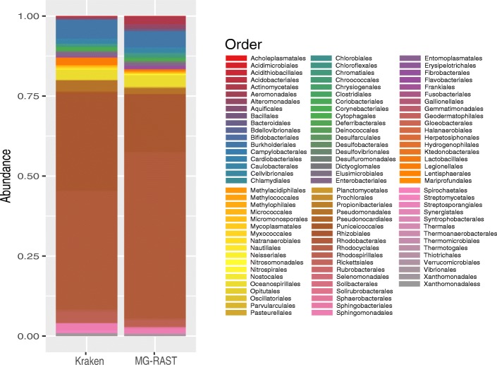
After using MG-RAST, we found that Proteobacteria was the most abundant phylum with 91% of the reads, followed by Actinobacteria (2.6%) and Bacteroidetes (2%) (Table 8). Additionally, at the class level, Alphaproteobacteria (74%) was highly present, and Gammaproteobacteria (10%), Betaproteobacteria (6%), and Actinobacteria (3%) showed the lowest abundances. The Rhizobiales and Rhodobacterales orders (Alphaproteobacteria) were the most abundant in the metagenome (Fig. 1). Next, we used Kraken to compare the taxonomy classification obtained by MG-RAST. Although Kraken assigned a lower number of reads (877,380) than MG-RAST (1,966,261), we found similar results in taxonomy abundance. Proteobacteria was the most abundant phylum (94%), followed by Actinobacteria (3%) and Bacteroidetes (0.4%) (Table 8). Also, similar to MG-RAST classification, Rhodobacterales and Rhizobiales orders were highly abundant. The most critical difference in taxonomic classification between the two algorithms was the number of orders identified, MG-RAST identified 92 and Kraken 51 orders (Fig. 1). To evaluate if this difference could impact on the microbial alpha diversity metrics, we used the relative abundance tables from MG-RAST and Kraken to measure the richness (Simpson) and evenness (Shannon) using Phyloseq [30]. This analysis showed a Shannon index of 2.04 and 1.99 for Kraken and MG-RAST, respectively (Table 7). In contrast, the Simpson index was 0.75 and 0.69 for MG-RAST and Kraken, respectively. However, these different values between MG-RAST and Kraken were not significant, suggesting that there is no difference between both algorithms for alpha diversity classification. The observed diversity metrics indicate that the greenhouse water drain is not a diverse niche.
Table 8.
Taxonomic composition
| Phylum | Greenhouse MG-RAST | Greenhouse Kraken |
|---|---|---|
| Acidobacteria | 0.0015405 | 0.0012883 |
| Actinobacteria | 0.0267721 | 0.0316892 |
| Aquificae | 0.0003382 | 0.0000208 |
| Armatimonadetes | NA | 0.0000139 |
| Bacteroidetes | 0.0208696 | 0.0049244 |
| Chlamydiae | 0.0002807 | 0.0000231 |
| Chlorobi | 0.0012536 | 0.0005910 |
| Chloroflexi | 0.0030123 | 0.0010620 |
| Chrysiogenetes | 0.0003128 | 0.0001454 |
| Crenarchaeota | NA | 0.0001524 |
| Cyanobacteria | 0.0047115 | 0.0015376 |
| Deferribacteres | 0.0002573 | 0.0000185 |
| Deinococcus-Thermus | 0.0024569 | 0.0021171 |
| Dictyoglomi | 0.0000570 | NA |
| Elusimicrobia | 0.0000478 | NA |
| Euryarchaeota | NA | 0.0005772 |
| Fibrobacteres | 0.0000351 | 0.0000277 |
| Firmicutes | 0.0107219 | 0.0047444 |
| Fusobacteria | 0.0002528 | 0.0000300 |
| Gemmatimonadetes | 0.0003763 | 0.0001316 |
| Ignavibacteriae | NA | 0.0000162 |
| Lentisphaerae | 0.0001322 | NA |
| Nitrospirae | 0.0001816 | 0.0001247 |
| Planctomycetes | 0.0019784 | 0.0006049 |
| Proteobacteria | 0.9125950 | 0.9467869 |
| Spirochaetes | 0.0007344 | 0.0008519 |
| Synergistetes | 0.0005554 | 0.0000139 |
| Tenericutes | 0.0000585 | 0.0001893 |
| Thermodesulfobacteria | NA | 0.0000231 |
| Thermotogae | 0.0004699 | 0.0001732 |
| Verrucomicrobia | 0.0099982 | 0.0021217 |
Relative abundances at phylum level using MG-RAST and Kraken, Relative abundances were determinate using the normalized number of reads of each order divided into the total number of reads
Fig. 1.
Bacterial abundance at order level. Relative abundance of bacterial orders classified by Kraken and MG-RAST
Functional diversity
Next, to know the encoded functions directly from the reads we classified them using MG-RAST and HUMAnN2. According to MG-RAST, we observed that most abundant functions were related to vitamins, protein biosynthesis and central carbohydrate metabolism (Table 9). In contrast, using HUMAnN2 the de Novo nucleotide biosynthesis, vitamins, and prosthetic groups were the most abundant functions (Table 10).
Table 9.
Functional diversity
| Level 2 category | Relative Abundance |
|---|---|
| Vitamins | 0.071243175 |
| Protein biosynthesis | 0.055205552 |
| Central carbohydrate metabolism | 0.052705254 |
| ABC transporters | 0.046491929 |
| Lipids | 0.043826646 |
| Disease and Defense | 0.037793615 |
| Prophages | 0.037587808 |
| Branched-chain amino acids | 0.033340704 |
| Arginine | 0.032626333 |
| Lysine | 0.031525862 |
Top ten of the most abundant functions annotated by MG-RAST against SEED database. Relative abundances were determined normalizing by the total number of reads
Table 10.
Functional diversity (UniRef90)
| Pathway | Relative abundance |
|---|---|
| Gondoate biosynthesis | 0.071311068 |
| L-isoleucine biosynthesis I | 0.058340078 |
| Adenosine ribonucleotides de novo biosynthesis | 0.05276 |
| Superpathway of guanosine nucleotides de novo biosynthesis I | 0.039785498 |
| L-valine biosynthesis | 0.037358283 |
| Guanosine ribonucleotides de novo biosynthesis | 0.029089431 |
| Superpathway of pyrimidine deoxyribonucleotides de novo biosynthesis | 0.027701595 |
| 5-aminoimidazole ribonucleotide biosynthesis II | 0.026628851 |
| Superpathway of L-threonine biosynthesis | 0.026026654 |
| Mycolate biosynthesis | 0.02416827 |
Top ten of most abundant function annotated by HUMAnN2 against UniRef90 database. Relative abundances were determined normalizing by the total number of reads
Additional results
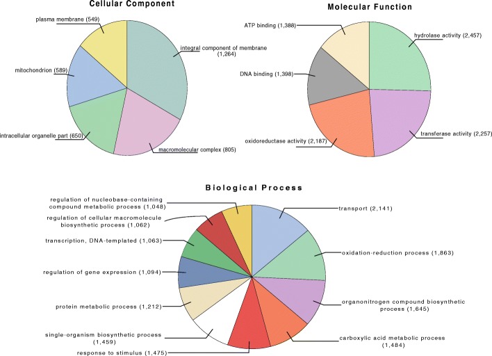
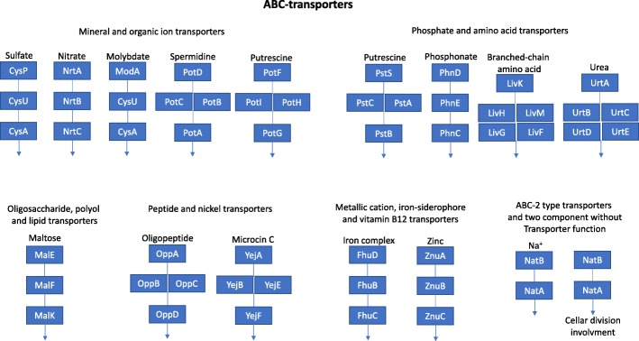
We assembled the metagenome to get more insight into the protein functional composition of the water sample. In this regard, we used the contigs to predict 25,735 proteins from which only 35.8% had significant blastp match (E-value 10− 5) against the NR RefSeq proteins database. The 64.2% of unknown proteins could represent novel proteins. A total of 14,328 (55.6%) proteins were classified using Gene Ontology (GOs) using Blast2GO and Interpro. We found that the term “transport” in the Biological process category was the most abundant function encoded in the greenhouse metagenome (Fig. 2). To get insights on this observation, we found that of the 2141 transporters annotated by Interpro, the 17.23% (369 proteins) are ABC-type transporters. Interestingly, has been reported that Rhizobiales has enriched the ABC transporter genes in their genomes (Fig. 2) [31, 32]. Furthermore, we use KAAS-KEGG [33] to identify the pathways containing ABC-transporters. After that, we only found six complete pathways in the greenhouse metagenome such as the vitamin B12 transporter (Fig. 3). Interestingly, the ABC-transporters were also present when we used only the reads for functional analysis (Table 9 and Table 10). Additionally, the hydrolase, transferase, and oxidoreductase were the most abundant GO molecular functions (Fig. 2). This result was in agreement with reports in which rhizobial bacteria associated with the nodules and seed of plants has many genes for these molecular functions in their genomes [34].
Fig. 2.
Gene Ontology (GO) terms distribution. The pie graphs show the number of genes annotated for cellular component, biological process, and molecular function categories
Fig. 3.
KEGG pathways associated with ABC-transporters. KEGG pathways of the ABC-transporters with all genes present in the metagenome
Finally, we searched for Antibiotic Resistance Genes (ARGs) in the metagenome, using the ARDB database [35]. We found a total of 31 ARGs and the most abundant genes were for resistance to spectinomycin (16%), tetracycline (12%) and aminoglycosides (9%) (Table 11). These findings are consistent with previous studies in chicken and vegetable greenhouse soil samples [36]. Although ARG genes are common in most ecological niches more investigation is needed to explore the role that these genes could play in microbial dynamics of human-constructed niches such as the greenhouse. Furthermore, these ARGs genes could be used for further analysis in phylogenomics to aim their evolutionary history and trace the adquision of these genes [37].
Table 11.
Antibiotics resistance genes
| Type | Number of genes | Resistance |
|---|---|---|
| vatb, aad9ib, aph6ic | 5 | Spectinomycin |
| tetpb, tetm, tetx | 4 | Tetracycline |
| emre | 3 | Aminoglycoside |
| aac3ia | 3 | Astromicin, Gentamicin, Sisomicin |
| baca | 3 | Bacitracin |
| ceob, catb1 | 2 | Chloramphenicol |
| cara, tlrc | 2 | Lincosamide, Macrolide, Streptogramin_b |
| acrb | 1 | Acriflavin, Aminoglycoside, beta_lactam, Glycylcycline |
| bl2d_oxa2 | 1 | Cloxacillin, Penicillin |
| mexd | 1 | Erythromycin, Fluoroquinolone, Glycylcycline, Roxithromycin |
| fosa | 1 | Fosfomycin |
| ksga | 1 | Kasugamycin |
| macb | 1 | Macrolide |
| pbp1b | 1 | Penicillin |
| arna | 1 | Polymyxin |
| dfra26 | 1 | Trimethoprim |
Conclusions
The use of metagenomic approaches to characterize new environments such as a research-greenhouse has the potential to unveil novel bacterial dynamics, enzyme functions, and metabolic pathways. To the best of our knowledge, this is the first report of the bacterial and functional contribution of the water from a greenhouse drain. We consider it exemplifies how the utilization of a metagenomic approach provides a more comprehensive view regarding the structure and functional composition of a bacterial community. Our results indicated that soil Rhizobiales bacteria and their genome functions mainly dominate the greenhouse water drain. This study aimed to identify the bacteria and functional gene composition of a greenhouse water drain sample and also represent a genomic resource to search novel proteins from a previously unexplored niche. Interestingly, we found over 400 proteins containing unintegrated signatures, which are highly conserved domains with unknown function according to Interpro, representing potential novel enzymes. All the metagenome proteins and their annotations are available to the scientific community via http://microbiomics.ibt.unam.mx/tools/metagreenhouse/.
Acknowledgments
Funding
This work was supported by Consejo Nacional de Ciencia y Tecnología (CONACyT) from México with grant SALUD-2014-C01–234188 and by Universidad Nacional Autónoma de México (UNAM) with grant DGAPA PAPPIT UNAM (IA203118). GLL acknowledges the support of CONACyT as a postdoctoral fellow.
Abbreviations
- ARDB
Antibiotic resistance genes database
- ARGs
Antibiotic resistance genes
- GO
Gene Ontology
- IBt
Institute of Biotechnology
- NA
Not available
Authors’ contributions
AOL formulated the project and provided the materials. FCG collected the sample, performed the DNA isolation and constructed the libraries. AVM carry out the sample sequencing. GLL performed the taxonomic and functional classification. GLL conducted the richness and evenness analysis and carried out the de Novo assembly. JMH designed the web server site for the availability of the metagenome sequences. AOL, FCG, and GLL wrote the manuscript. All the authors reviewed and approved the final document.
Competing interests
The authors declare that the research was conducted in the absence of any commercial or financial relationships that could be construed as a potential conflict of interest.
Publisher’s Note
Springer Nature remains neutral with regard to jurisdictional claims in published maps and institutional affiliations.
References
- 1.Bikel S, Valdez-Lara A, Cornejo-Granados F, Rico K, Canizales-Quinteros S, Soberon X, Del Pozo-Yauner L, Ochoa-Leyva A. Combining metagenomics, metatranscriptomics and viromics to explore novel microbial interactions: towards a systems-level understanding of human microbiome. Comput Struct Biotechnol J. 2015;13:390–401. doi: 10.1016/j.csbj.2015.06.001. [DOI] [PMC free article] [PubMed] [Google Scholar]
- 2.Lopez-Lopez O, Cerdan ME, Gonzalez-Siso MI. Hot spring metagenomics. Life (Basel) 2013;3:308–320. doi: 10.3390/life3020308. [DOI] [PMC free article] [PubMed] [Google Scholar]
- 3.Wang WL, Xu SY, Ren ZG, Tao L, Jiang JW, Zheng SS. Application of metagenomics in the human gut microbiome. World J Gastroenterol. 2015;21:803–814. doi: 10.3748/wjg.v21.i3.803. [DOI] [PMC free article] [PubMed] [Google Scholar]
- 4.Oulas A, Pavloudi C, Polymenakou P, Pavlopoulos GA, Papanikolaou N, Kotoulas G, Arvanitidis C, Iliopoulos I. Metagenomics: tools and insights for analyzing next-generation sequencing data derived from biodiversity studies. Bioinform Biol Insights. 2015;9:75–88. doi: 10.4137/BBI.S12462. [DOI] [PMC free article] [PubMed] [Google Scholar]
- 5.Eloe-Fadrosh EA, Paez-Espino D, Jarett J, Dunfield PF, Hedlund BP, Dekas AE, Grasby SE, Brady AL, Dong H, Briggs BR, et al. Global metagenomic survey reveals a new bacterial candidate phylum in geothermal springs. Nat Commun. 2016;7:10476. doi: 10.1038/ncomms10476. [DOI] [PMC free article] [PubMed] [Google Scholar]
- 6.Ufarte L, Potocki-Veronese G, Laville E. Discovery of new protein families and functions: new challenges in functional metagenomics for biotechnologies and microbial ecology. Front Microbiol. 2015;6:563. doi: 10.3389/fmicb.2015.00563. [DOI] [PMC free article] [PubMed] [Google Scholar]
- 7.Segata N, Izard J, Waldron L, Gevers D, Miropolsky L, Garrett WS, Huttenhower C. Metagenomic biomarker discovery and explanation. Genome Biol. 2011;12:R60. doi: 10.1186/gb-2011-12-6-r60. [DOI] [PMC free article] [PubMed] [Google Scholar]
- 8.Coughlan LM, Cotter PD, Hill C, Alvarez-Ordonez A. Biotechnological applications of functional metagenomics in the food and pharmaceutical industries. Front Microbiol. 2015;6:672. doi: 10.3389/fmicb.2015.00672. [DOI] [PMC free article] [PubMed] [Google Scholar]
- 9.Critchfield JW, van Hemert S, Ash M, Mulder L, Ashwood P. The potential role of probiotics in the management of childhood autism spectrum disorders. Gastroenterol Res Pract. 2011;2011:161358. doi: 10.1155/2011/161358. [DOI] [PMC free article] [PubMed] [Google Scholar]
- 10.Major G, Spiller R. Irritable bowel syndrome, inflammatory bowel disease and the microbiome. Curr Opin Endocrinol Diabetes Obes. 2014;21:15–21. doi: 10.1097/MED.0000000000000032. [DOI] [PMC free article] [PubMed] [Google Scholar]
- 11.Bonifay V, Wawrik B, Sunner J, Snodgrass EC, Aydin E, Duncan KE, Callaghan AV, Oldham A, Liengen T, Beech I. Metabolomic and metagenomic analysis of two crude oil production pipelines experiencing differential rates of corrosion. Front Microbiol. 2017;8:99. doi: 10.3389/fmicb.2017.00099. [DOI] [PMC free article] [PubMed] [Google Scholar]
- 12.Buie Timothy, Campbell Daniel B., Fuchs George J., Furuta Glenn T., Levy Joseph, VandeWater Judy, Whitaker Agnes H., Atkins Dan, Bauman Margaret L., Beaudet Arthur L., Carr Edward G., Gershon Michael D., Hyman Susan L., Jirapinyo Pipop, Jyonouchi Harumi, Kooros Koorosh, Kushak Rafail, Levitt Pat, Levy Susan E., Lewis Jeffery D., Murray Katherine F., Natowicz Marvin R., Sabra Aderbal, Wershil Barry K., Weston Sharon C., Zeltzer Lonnie, Winter Harland. Evaluation, Diagnosis, and Treatment of Gastrointestinal Disorders in Individuals With ASDs: A Consensus Report. Pediatrics. 2010;125(Supplement 1):S1–S18. doi: 10.1542/peds.2009-1878C. [DOI] [PubMed] [Google Scholar]
- 13.Turnbaugh PJ, Ley RE, Mahowald MA, Magrini V, Mardis ER, Gordon JI. An obesity-associated gut microbiome with increased capacity for energy harvest. Nature. 2006;444:1027–1031. doi: 10.1038/nature05414. [DOI] [PubMed] [Google Scholar]
- 14.Gomez-Alvarez V, Revetta RP, Santo Domingo JW. Metagenome analyses of corroded concrete wastewater pipe biofilms reveal a complex microbial system. BMC Microbiol. 2012;12:122. doi: 10.1186/1471-2180-12-122. [DOI] [PMC free article] [PubMed] [Google Scholar]
- 15.Yilmaz P, Kottmann R, Field D, Knight R, Cole JR, Amaral-Zettler L, Gilbert JA, Karsch-Mizrachi I, Johnston A, Cochrane G, et al. Minimum information about a marker gene sequence (MIMARKS) and minimum information about any (x) sequence (MIxS) specifications. Nat Biotech. 2011;29:415–420. doi: 10.1038/nbt.1823. [DOI] [PMC free article] [PubMed] [Google Scholar]
- 16.Cox MP, Peterson DA, Biggs PJ. SolexaQA: at-a-glance quality assessment of Illumina second-generation sequencing data. BMC Bioinformatics. 2010;11:485. doi: 10.1186/1471-2105-11-485. [DOI] [PMC free article] [PubMed] [Google Scholar]
- 17.Li H, Durbin R. Fast and accurate short read alignment with burrows-wheeler transform. Bioinformatics. 2009;25:1754–1760. doi: 10.1093/bioinformatics/btp324. [DOI] [PMC free article] [PubMed] [Google Scholar]
- 18.Peng Y, Leung HC, Yiu SM, Chin FY. IDBA-UD: a de novo assembler for single-cell and metagenomic sequencing data with highly uneven depth. Bioinformatics. 2012;28:1420–1428. doi: 10.1093/bioinformatics/bts174. [DOI] [PubMed] [Google Scholar]
- 19.Nurk S, Meleshko D, Korobeynikov A, Pevzner PA. metaSPAdes: a new versatile metagenomic assembler. Genome Res. 2017;27:824–834. doi: 10.1101/gr.213959.116. [DOI] [PMC free article] [PubMed] [Google Scholar]
- 20.Delcher AL, Phillippy A, Carlton J, Salzberg SL. Fast algorithms for large-scale genome alignment and comparison. Nucleic Acids Res. 2002;30:2478–2483. doi: 10.1093/nar/30.11.2478. [DOI] [PMC free article] [PubMed] [Google Scholar]
- 21.Hunt M, Newbold C, Berriman M, Otto TD. A comprehensive evaluation of assembly scaffolding tools. Genome Biol. 2014;15:R42. doi: 10.1186/gb-2014-15-3-r42. [DOI] [PMC free article] [PubMed] [Google Scholar]
- 22.Van Belleghem SM, Roelofs D, Van Houdt J, Hendrickx F. De novo transcriptome assembly and SNP discovery in the wing polymorphic salt marsh beetle Pogonus chalceus (Coleoptera, Carabidae) PLoS One. 2012;7:e42605. doi: 10.1371/journal.pone.0042605. [DOI] [PMC free article] [PubMed] [Google Scholar]
- 23.Keegan KP, Glass EM, Meyer F. MG-RAST, a metagenomics Service for Analysis of microbial community structure and function. Methods Mol Biol. 2016;1399:207–233. doi: 10.1007/978-1-4939-3369-3_13. [DOI] [PubMed] [Google Scholar]
- 24.Wood DE, Salzberg SL. Kraken: ultrafast metagenomic sequence classification using exact alignments. Genome Biol. 2014;15:R46. doi: 10.1186/gb-2014-15-3-r46. [DOI] [PMC free article] [PubMed] [Google Scholar]
- 25.Abubucker S, Segata N, Goll J, Schubert AM, Izard J, Cantarel BL, Rodriguez-Mueller B, Zucker J, Thiagarajan M, Henrissat B, et al. Metabolic reconstruction for metagenomic data and its application to the human microbiome. PLoS Comput Biol. 2012;8:e1002358. doi: 10.1371/journal.pcbi.1002358. [DOI] [PMC free article] [PubMed] [Google Scholar]
- 26.Rho M, Tang H, Ye Y. FragGeneScan: predicting genes in short and error-prone reads. Nucleic Acids Res. 2010;38:e191. doi: 10.1093/nar/gkq747. [DOI] [PMC free article] [PubMed] [Google Scholar]
- 27.Conesa A, Gotz S. Blast2GO: a comprehensive suite for functional analysis in plant genomics. Int J Plant Genomics. 2008;2008:619832. doi: 10.1155/2008/619832. [DOI] [PMC free article] [PubMed] [Google Scholar]
- 28.Quevillon E, Silventoinen V, Pillai S, Harte N, Mulder N, Apweiler R, Lopez R. InterProScan: protein domains identifier. Nucleic Acids Res. 2005;33:W116–W120. doi: 10.1093/nar/gki442. [DOI] [PMC free article] [PubMed] [Google Scholar]
- 29.Huson DH, Auch AF, Qi J, Schuster SC. MEGAN analysis of metagenomic data. Genome Res. 2007;17:377–386. doi: 10.1101/gr.5969107. [DOI] [PMC free article] [PubMed] [Google Scholar]
- 30.McMurdie PJ, Holmes S. Phyloseq: an R package for reproducible interactive analysis and graphics of microbiome census data. PLoS One. 2013;8:e61217. doi: 10.1371/journal.pone.0061217. [DOI] [PMC free article] [PubMed] [Google Scholar]
- 31.Konstantinidis KT, Tiedje JM. Trends between gene content and genome size in prokaryotic species with larger genomes. Proc Natl Acad Sci U S A. 2004;101:3160–3165. doi: 10.1073/pnas.0308653100. [DOI] [PMC free article] [PubMed] [Google Scholar]
- 32.Lopez-Leal G, Tabche ML, Castillo-Ramirez S, Mendoza-Vargas A, Ramirez-Romero MA, Davila G. RNA-Seq analysis of the multipartite genome of rhizobium etli CE3 shows different replicon contributions under heat and saline shock. BMC Genomics. 2014;15:770. doi: 10.1186/1471-2164-15-770. [DOI] [PMC free article] [PubMed] [Google Scholar]
- 33.Moriya Y, Itoh M, Okuda S, Yoshizawa AC, Kanehisa M. KAAS: an automatic genome annotation and pathway reconstruction server. Nucleic Acids Res. 2007;35:W182–W185. doi: 10.1093/nar/gkm321. [DOI] [PMC free article] [PubMed] [Google Scholar]
- 34.Peralta H, Aguilar A, Diaz R, Mora Y, Martinez-Batallar G, Salazar E, Vargas-Lagunas C, Martinez E, Encarnacion S, Girard L, Mora J. Genomic studies of nitrogen-fixing rhizobial strains from Phaseolus vulgaris seeds and nodules. BMC Genomics. 2016;17:711. doi: 10.1186/s12864-016-3053-z. [DOI] [PMC free article] [PubMed] [Google Scholar]
- 35.Liu B, Pop M. ARDB--antibiotic resistance genes database. Nucleic Acids Res. 2009;37:D443–D447. doi: 10.1093/nar/gkn656. [DOI] [PMC free article] [PubMed] [Google Scholar]
- 36.Fang H, Wang H, Cai L, Yu Y. Prevalence of antibiotic resistance genes and bacterial pathogens in long-term manured greenhouse soils as revealed by metagenomic survey. Environ Sci Technol. 2015;49:1095–1104. doi: 10.1021/es504157v. [DOI] [PubMed] [Google Scholar]
- 37.Davies J, Davies D. Origins and evolution of antibiotic resistance. Microbiol Mol Biol Rev. 2010;74:417–433. doi: 10.1128/MMBR.00016-10. [DOI] [PMC free article] [PubMed] [Google Scholar]


