Abstract
Introduction
The mechanism underlying beneficial outcomes of bariatric surgery still remains unclear. Especially little is known about hormonal and metabolic changes induced by the novel bariatric procedure mini gastric bypass (MGB).
Aim
To evaluate pre- and post-prandial changes in both ghrelin isoforms in obese patients without diabetes and cardiovascular complications treated with MGB, sleeve gastrectomy (SG) or Roux-en-Y gastric bypass (RYGB) surgery.
Material and methods
From 45 patients initially enrolled in the study, 23 persons completed a one-year follow-up period. Venous blood for acyl and desacyl ghrelin (AG and DAG) as well as other metabolic assays was collected 3 months before and 6 and 12 months after bariatric surgery (MGB, RYGB, SG) – in the fasting state and 2 h after the consumption of a standard 300 kcal-mixed meal (Nutridrink standard, Nutricia).
Results
AG and DAG levels (both fasting and prandial) as well as AG/DAG ratio did not change after 6 and 12 months in MGB and RYGB groups. In the SG group we observed a significant decrease in fasting and postprandial DAG levels and consecutively an increase in the fasting AG/DAG ratio after 6 and 12 months. Six months after surgery we observed some differences between carbohydrate metabolism measures in the MGB group (lower HbA1c, HOMA-IR and fasting insulinaemia) in comparison to the rest of the participants, but 12 months after each type of surgery body mass index and indices of carbohydrate and lipid metabolism did not differ.
Conclusions
The results of our study demonstrate that all studied bariatric procedures can successfully reduce overall body weight and suggest also that the mechanisms of weight loss and improvement in carbohydrate and lipid metabolism after all three types of surgery are independent of ghrelin and the acyl/desacyl ghrelin ratio.
Keywords: acyl ghrelin, desacyl ghrelin, acyl/desacyl ratio, post-prandial, obesity, bariatric, mini gastric bypass
Introduction
The mechanism underlying the beneficial effect of bariatric surgery still remains unclear. Changes in incretin hormone levels are proposed to be important consequence of that treatment. Ghrelin is a gastroenteric hormone with the strongest orexigenic signal [1], whose levels are affected by bariatric treatment [2]. It is known that ghrelin exists in two different forms: acylated (acyl ghrelin – AG) and desacylated (desacyl ghrelin – DAG). Desacyl ghrelin was long considered to be an inactive degradation product of the acylated form, but emerging evidence suggests that the desacylated form of ghrelin may induce metabolic effects independently of AG. The interactions between both isoforms may be important in the energy balance [3, 4]. It is also important to assess the fasting as well as postprandial level of both ghrelin isoforms, because they are obviously affected by food intake and their proportions can be changed in obesity [5, 6]. There are suggestions that obese people with metabolic syndrome have a higher acyl/desacyl ratio (AG/DAG) [7] and that this ratio may be modulated by some form of medical intervention [8]. Probably ghrelin isoform changes may be an important element of the metabolic response to bariatric treatment.
Recently, there has been dynamic development of bariatric surgery and a rapid increase in the number of procedures performed.
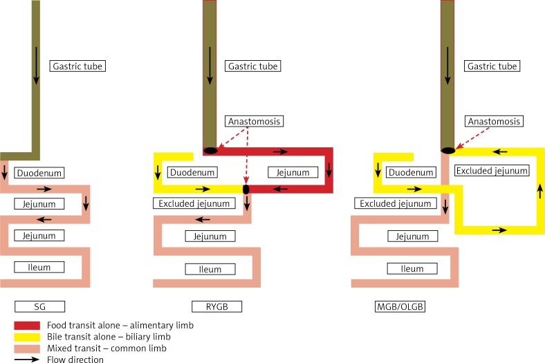
There are different kinds of operations – not only the commonly accepted Roux-en-Y gastric bypass (RYGB) and sleeve gastrectomy (SG), but also the novel mini gastric bypass (MGB) [9, 10]. The aforementioned procedures are presented in Figure 1. Their mechanisms of action may vary depending on the different changes in the anatomy of the digestive tract (Figure 1) – from SG where no biliary exclusion is performed to MGB with significantly increased length of the biliary limb [11, 12]. As for hormonal changes, especially ghrelin, most of the data are for RYGB and some for SG [13, 14], but there is a lack of data on hormonal effects of MGB [15].
Figure 1.
The schematic presentation of sleeve gastrectomy (SG), Roux-en-Y gastric bypass (RYGB) and Mini Gastric Bypass (MGB) from [16]
Aim
The aim of this study was to evaluate pre- and post-prandial changes in both ghrelin isoforms in obese patients without diabetes and cardiovascular complications treated with MGB, SG or RYGB surgery.
Material and methods
Forty-five patients (34 female/11 male) with simple obesity, without cardiovascular events in anamnesis, not taking any diabetic medicaments, were accepted initially for surgery and enrolled in the prospectively designed study. They were selected from patients who reported to the Outpatient Clinic of the Department of General, Endocrine and Transplant Surgery, Medical University of Gdansk, Poland, in 2013–2014 with the purpose of surgical obesity treatment. All patients met IFSO criteria [16].
The inclusion criteria for the study were the maximum body mass index (BMI) > 35 kg/m2 and informed consent to participate in the study.
The exclusion criteria included use of anti-diabetic medications, history of cardiovascular events, chronic liver or kidney disease and pregnancy. The study was approved by the university ethics committee.
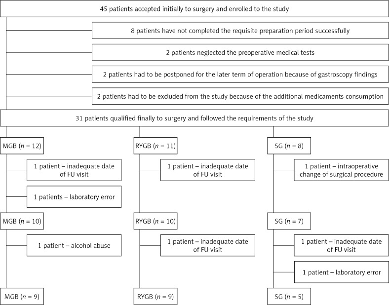
Only 31 patients followed the requirements of the study and qualified finally for surgery at that time. They were operated on after a standard 3-month preparation period. The RYGB procedure was offered to 11 subjects with lower oesophageal sphincter dysfunction. Twelve others were qualified for the MGB and 8 persons for the SG. For both types of gastric bypass the restriction was provided by a long, narrow (calibrated for 36 F) 50 ml gastric pouch. The length of the alimentary limb (AL) in RYGB patients was maintained for 150 cm and respectively 50 cm for the excluded biliary limb (BL) while in MGB the biliary transit was designed for 200 cm. The common limb (CL) was proportionally left the same in both operations. In contrast to the bypass procedures there is no jejunal exclusion in SG, but 80–90% of the stomach capacity is resected. The size of the pouch in the SG procedure has also been determined by the 36 F calibration tube, the same as it has been used for both subtypes of bypass. The first stapler was launched 4 cm proximally to the pylorus. The volume of the stomach reservoir was measured for 60 ml. A detailed description of the surgical technique has been included in a previous publication from our institution [16]. The intra- and postoperative course was uneventful in all patients. From 31 subjects, 8 (2 from RYGB, 3 from MGB and 3 from SG group) had to be excluded from the study. The reasons for exclusion and the study flow chart are presented in Figure 2.
Figure 2.
The draft of the procedure
Anthropometric examination and blood sampling were performed at the beginning of the study (3 months before the surgery) and then 6 and 12 months after surgery. In all patients in the fasting state the waist circumference, weight and body composition were measured. The percentage of fat tissue (FAT %), fat mass (FM), and fat-free mass (FFM) were assessed using the bioelectrical impedance method (TANITA SC 330). The BMI (BMI = body weight (kilograms)/height (meters)2) and fat-free mass index (FFMI: fat-free mass (kilograms/height (meters)2), were then calculated.
The venous blood was collected after a 12-hour period of fasting, then patients received a standard mixed meal containing 300 kcal, 16% protein, 49% carbohydrate and 35% fat (Nutridrink standard 200 ml, Nutricia). Venous blood sampling was repeated 2 h after the start of the meal (MMTT, mixed-meal tolerance test) [17].
Plasma desacylated and acylated ghrelin levels were measured with a commercial enzyme immunoassay (Human Acylated Ghrelin EIA Kit, Human Unacylated Ghrelin EIA Kit, Biovendor, Czech Republic) in accordance with the supplier’s specifications. Blood samples were immediately centrifuged at 3500 × g for 10 min at +4°C and serum was kept at –80°C until further analyses were performed. Glucose was measured by the hexokinase method (Abbott Laboratories, USA). Serum lipid concentrations were measured by the oxidase method (Abbott Laboratories, USA) and the Friedewald formula was used to calculate the LDL-CH concentration. Serum insulin levels were determined by EIA (Abbott Laboratories, USA). Serum HbA1c levels were assessed by a Tosoh G8 HPLC Analyser (TOSOH, Japan).
Insulin resistance was then estimated using the homeostasis model assessment (HOMA-IR), which was calculated in line with the following formula: fasting insulin (μU/ml) × fasting glucose (mg/dl)/405 [18].
Statistical analysis
The data are expressed as the mean ± SD or median (range). The Kolmogorov-Smirnov test was used to verify whether the variable distribution was normal. Differences between means were evaluated by the independent Student’s t test and the Mann-Whitney U test was used when the distribution of the variable was not normal. Fasting and postprandial values were compared using the paired Student’s t test and Wilcoxon’s signed-rank test was used when the distribution of the variable was not normal. Spearman’s rank correlation coefficient (r) was used to evaluate the relationships between the variables. The Friedman ANOVA was used for comparison of continuous data with repeated measures, and to perform the comparison between multiple groups the Kruskal-Wallis test was used. Statistical analysis was performed using Statistica version 12 (StatSoft, Poland). P-values < 0.05 were considered statistically significant.
Results
At the beginning of the study, the mean BMI among 45 participants was 45.7 ±6.7 kg/m2 (range: 32.5–56.6 kg/m2), waist circumference: 118.1 ±14.5 cm (range: 94–148 cm) and HOMA-IR: 3.1 ±1.7 (range: 1.2–11.3). The characteristics of the whole study group at the beginning are shown in Table I. The MGB, RYGB and SG groups did not differ in age and metabolic parameters at the baseline.
Table I.
Characteristics of the whole studied group (n = 45)
| Parameters | Value |
|---|---|
| Age [years] | 37.5 ±10.4 |
| Male/female | 11/34 |
| Smoking | 11/45 |
| BMI [kg/m2] | 45.7 ±6.7 |
| Waist circumference (WC) [cm] | 118.1 ±14.5 |
| SBP [mm Hg] | 124.4 ±14.0 |
| DBP [mm Hg] | 81.4 ±8.0 |
| Fat% | 45.7 ±6.7 |
| Fat mass [kg] | 56.5 ±14.7 |
| Fat-free mass [kg] | 66.5 ±13.3 |
| Fat-free mass index (FFMI) [kg/m2] | 23.0 ±3.7 |
| Fasting glucose [mg/dl] | 91.4 ±9.1 |
| Fasting insulin [μU/ml] | 13.4 ±6.9 |
| HOMA-IR | 3.1 ±1.7 |
| Glucose 2 h MMTT [mg/dl] | 94.8 ±19.9 |
| Insulin 2 h MMTT [μU/ml] | 27.5 ±18.4 |
| HbA1c [%] | 5.7 ±0.5 |
| HDL-CH [mg/dl] | 44.8 ±10.2 |
| LDL-CH [mg/dl] | 122.0 ±30.4 |
| TG [mg/dl] | 156.8 ±56.9 |
| TSH [μU/ml] | 1.8 ±0.8 |
MMTT – mixed-meal tolerance test.
Metabolic and hormonal changes in all patients after bariatric treatment
A year after the bariatric treatment the mean decreases in weight and BMI in all patients were 40.6 kg (123.0 vs. 82.4 kg; p < 0.05) and 14.6 kg/m2 (43.5 vs. 28.9 kg/m2; p < 0.005), respectively. The mean reduction in FM was 31.8 kg (56.5 kg vs. 24.7 kg; p < 0.005) and in FFM 8.9 kg (66.5 kg vs. 57.6 kg; p < 0.005). HOMA-IR decreased after surgery by 1.1 (2.6 vs. 1.6; p < 0.05).
Fasting DAG level decreased from 268.5 ±163.0 pg/ml to 197.7 ±140.7 pg/ml after 6 months and to 158.8 ±141.8 pg/ml 12 months after bariatric surgery (p < 0.05). However, the postprandial DAG level and AG levels (both fasting and postprandial) did not change after the surgery in the whole study group. Similarly, we did not find statistical changes in the AG/DAG ratio after bariatric treatment. All metabolic and hormonal parameters before and after bariatric treatment are presented in Table II.
Table II.
Anthropometric and biochemical data before and after bariatric treatment (n = 23)
| Parameter | 3 months before | After 6 months | After 12 months | P-value |
|---|---|---|---|---|
| BMI [kg/m2] | 43.0 ±6.2 | 31.2 ±4.9 | 28.9 ±4.6 | 0.0003 |
| FFMI [kg/m2] | 23.0 ±3.7 | 20.3 ±2.9 | 20.3 ±2.7 | < 0.0001 |
| WC [cm] | 118.1 ±14.7 | 95.4 ±11.6 | 90.5 ±12.8 | < 0.0001 |
| HOMA-IR | 2.5 ±1.1 | 1.6 ±0.9 | 1.6 ±0.8 | 0.001 |
| HbA1c (%) | 5.9 ±0.6 | 5.0 ±0.4 | 4.9 ±0.3 | 0.0005 |
| Glucose [mg/dl]: | ||||
| Fasting | 89.8 ±9.5 | 87.5 ±7.3 | 85.9 ±7.8 | 0.12 |
| 2 h MMTT | 92.4 ±19.7 | 69.1 ±10.3 | 67.6 ±13.4 | 0.00002 |
| Insulin [μU/ml]: | ||||
| Fasting | 11.3 ±4.1 | 7.3 ±4.1 | 7.4 ±3.5 | 0.01 |
| 2 h MMTT | 23.6 ±16.6 | 9.8 ±11.6 | 8.1 ±7.8 | 0.0001 |
| DAG [pg/ml]: | ||||
| Fasting | 268.5 ±163.0 | 197.7 ±140.7 | 158.8 ±141.8 | 0.026 |
| 2 h MMTT | 169.0 ±124.8 | 130.6 ±96.1 | 137.9 ±116.8 | 0.38 |
| AG [pg/ml]: | ||||
| Fasting | 38.5 ±22.7 | 39.1 ±50.2 | 31.6 ±17.7 | 0.38 |
| 2 h MMTT | 34.5 ±22.4 | 27.6 ±18.9 | 28.0 ±15.0 | 0.48 |
| AG/DAG*: | ||||
| Fasting | 0.14 (0.05–0.96) | 0.17 (0.06–1.13) | 0.24 (0.08–2.06) | 0.28 |
| 2 h MMTT | 0.25 (0.03–1.18) | 0.21 (0.10–0.78) | 0.20 (0.10–1.07) | 0.95 |
MMTT – mixed-meal tolerance test. Results presented as mean ± SD or *median (range).
Comparison of metabolic changes between different types of surgery
In all groups (MGB, RYGB and SG) we observed beneficial metabolic outcomes (data included in Table III). The grade of BMI reduction and changes in body composition at 6 and 12 months after the surgery did not differ significantly between the groups. There were some differences between carbohydrate metabolism indices after MGB in comparison to the rest of participants. At 6 months after the surgery the HbA1c level was lower in the MGB group than the RYGB group (4.9 ±0.3% vs. 5.2 ±0.3%, p < 0.02). Also HOMA-IR and fasting insulinaemia after 6 months were lower in the MGB group than the SG group: 1.3 ±0.5 vs. 1.8 ±0.4 (p = 0.029) and 5.8 ±2.3 µU/ml vs. 8.7 ±1.9 µU/ml (p = 0.015) respectively. However, these differences were not found 12 months after the surgery.
Table III.
Comparison between different types of bariatric treatment in anthropometric and biochemical parameters
| Parameter | MGB (n = 9) | RYGB (n = 9) | SG (n = 5) | ||||||
|---|---|---|---|---|---|---|---|---|---|
| 3 months before | After 6 months | After 12 months | 3 months before | After 6 months | After 12 months | 3 months before | After 6 months | After 12 months | |
| Glucose [mg/dl]: | |||||||||
| Fasting | 90.2 ±6.9 | 86.1 ±6.2 | 88.0 ±8.5 | 87.7 ±12.1 | 90.0 ±9.0 | 85.1 ±6.6 | 93.7 ±8.6 | 85.3 ±4.4 | 83.3 ±9.5* |
| 2 h MMTT | 90.8 ±11.4 | 73.1 ±9.2 | 71.7 ±13.1* | 92.7 ±22.5 | 67.4 ±11.4 | 66.0 ±13.3* | 96.7 ±36.1 | 62.3 ±6.5 | 60.0 ±15.5 |
| Insulin [μU/ml]: | |||||||||
| Fasting | 10.9 ±4.9 | 5.4 ±1.9 | 6.4 ±3.4* | 12.1 ±4.1 | 9.1 ±5.7 | 8.5 ±3.7 | 10.5 ±2.9 | 7.5 ±1.1 | 7.3 ±3.3 |
| 2 h MMTT | 25.4 ±19.5 | 5.6 ±3.3 | 5.4 ±3.8* | 21.6 ±16.2 | 14.2 ±17.0 | 10.2 ±10.5 | 22.8 ±23.2 | 16.3 ±12.4 | 9.3 ±2.6 |
| BMI [kg/m2] | 42.3 ±5.5 | 29.7 ±4.5 | 27.5 ±3.5* | 44.5 ±7.0 | 32.3 ±5.7 | 29.7 ±5.4* | 41.5 ±7.5 | 31.9 ±3.5 | 30.0 ±4.9* |
| FFMI [kg/m2] | 24.6 ±5.7 | 20.8 ±3.7 | 20.5 ±3.0* | 22.6 ±2.5 | 19.7 ±2.0 | 19.8 ±2.4* | 21.8 ±2.7 | 20.4 ±3.1 | 20.8 ±3.0 |
| WC [cm] | 115.9 ±14.0 | 92.2 ±11.2 | 86.7 ±8.5* | 119.6 ±15.5 | 97.0 ±12.6 | 92.6 ±14.9* | 115.9 ±14.4 | 96.4 ±8.8 | 93.7 ±14.7* |
| HOMA-IR | 2.5 ±1.2 | 1.5 ±0.4 | 1.4 ±0.8* | 2.7 ±1.6 | 2.1 ±1.3 | 1.8 ±0.9 | 2.4 ±0.7 | 1.6 ±0.2 | 1.5 ±0.8* |
| HbA1c (%) | 5.6 ±0.4 | 4.7 ±0.3 | 4.9 ±0.4* | 6.0 ±0.7 | 5.2 ±0.2 | 5.0 ±0.1* | 5.6 ±0.5 | 5.1 ±0.4 | 5.1 ±0.2 |
| LDL [mg/dl] | 121.0 ±36.3 | 104.9 ±19.3 | 96.9 ±18.6* | 124.5 ±26.1 | 90.4 ±16.5 | 95.6 ±24.7* | 116.8 ±20.0 | 114.8 ±30.7 | 120.0 ±21.5 |
| HDL [mg/dl] | 47.0 ±10.6 | 46.0 ±9.3 | 50.8 ±12.1 | 43.9 ±5.8 | 41.0 ±10.4 | 48.2 ±11.9 | 49.1 ±16.4 | 56.7 ±18.6 | 49.2 ±8.7 |
| TG [mg/dl] | 127.5 ±10.6 | 94.7 ±45.8 | 89.3 ±32.9* | 144.3 ±44.5 | 88.6 ±26.2 | 84.8 ±35.3* | 179.5 ±60.0 | 104.8 ±29.6 | 96.0 ±10.2 |
MMTT – mixed-meal tolerance test.
P for each type of surgery < 0.05.
The only significant metabolic changes after 6 and 12 month between the studied groups were found for total cholesterol (RYGB vs. SG: 149.1 ±23.2 mg/dl vs. 195.8 ±37.2 mg/dl; p = 0.009 and 159.4 ±32.0 mg/dl vs. 193 ±13.5 mg/dl; p = 0.043 respectively) and LDL cholesterol levels (RYGB vs. SG: 90.4 ±16.5 mg/dl vs. 114.8 ±30.7 mg/dl; p = 0.07 and 95.6 ±24.7 mg/dl vs. 120.0 ±21.5 mg/dl; p = 0.08 respectively).
Changes of ghrelin isoforms after different types of surgery
AG and DAG levels (both fasting and prandial) as well as AG/DAG ratio did not change after 6 and 12 months in MGB and RYGB groups. In the SG group we observed a significant decrease in fasting and postprandial DAG levels and consecutively an increase in the fasting AG/DAG ratio after 6 and 12 months (data included in Table IV). There were significant differences in fasting DAG level and postprandial DAG level between SG and RYGB groups after 6 months: 85.9 ±14.3 pg/ml vs. 263.1 ±155.0 pg/ml (p = 0.009) and 52.2 ±22.3 pg/ml vs. 159.6 ±99.6 pg/ml (p = 0.014) respectively. After 12 months we also found lower fasting and postprandial DAG levels in the SG group than the RYGB group (data in Table IV; p = 0.02). The postprandial AG/DAG ratio was also significantly higher in the SG than the RYGB group after 6 months: 0.34 (0.17–0.66) vs. 0.15 (0.12–0.66) (p = 0.002) and 12 months after the surgery (data in Table IV). We also found in the SG group in comparison to the MGB group lower fasting DAG (85.9 ±14.3 pg/ml vs. 178.3 ±112.2 pg/ml; p = 0.04) and fasting AG levels (16.6 ±2.5 pg/ml vs. 31.9 ±14.0 pg/ml; p = 0.012) after 6 months, but there were no significant differences between SG and MGB groups in both isoforms of ghrelin levels after 12 months.
Table IV.
Fasting and postprandial (2h MMTT) AG and DAG before and after MGB, RYGB, SG
| Parameter | 3 months before | After 6 months | After 12 months | P-value |
|---|---|---|---|---|
| MGB (n = 9) | ||||
| DAG [pg/ml]: | ||||
| Fasting | 193.1 ±145.0 | 178.3 ±112.2 | 156.1 ±123.9 | 0.46 |
| 2 h MMTT | 117.2 ±75.7 | 122.8 ±93.4 | 143.2 ±124.9 | 0.46 |
| AG [pg/ml]: | ||||
| Fasting | 41.4 ±31.8 | 31.9 ±14.0 | 28.6 ±12.9 | 0.72 |
| 2 h MMTT | 30.5 ±18.7 | 24.6 ±11.2 | 28.0 ±12.2 | 0.64 |
| AG/DAG*: | ||||
| Fasting | 0.22 (0.08–0.96) | 0.16 (0.11–0.41) | 0.26 (0.08–0.48) | 0.89 |
| 2 h MMTT | 0.31 (0.13–0.88) | 0.22 (0.10–0.78) | 0.20 ( 0.10–1.05) | 0.24 |
| RYGB (n = 9) | ||||
| DAG [pg/ml]: | ||||
| Fasting | 334.9 ±157.5 | 263.1 ±155.0 | 211.9 ±176.0 | 0.37 |
| 2 h MMTT | 225.4 ±144.6 | 159.6 ±99.6 | 185.4 ±116.4 | 0.26 |
| AG [pg/ml]: | ||||
| Fasting | 38.7 ±17.3 | 55.1 ±71.4 | 40.7 ±22.3 | 0.72 |
| 2 h MMTT | 35.0 ±26.1 | 32.5 ±25.1 | 31.5 ±20.2 | 0.64 |
| AG/DAG*: | ||||
| Fasting | 0.12 (0.05–0.51) | 0.25 (0.06–1.13) | 0.18 (0.11–2.06) | 0.72 |
| 2 h MMTT | 0.11 (0.03–1.18) | 0.15 (0.12–0.66) | 0.17 (0.11–0.33) | 0.46 |
| SG (n = 5) | ||||
| DAG [pg/ml]: | ||||
| Fasting | 281.5 ±196.7 | 85.9 ±14.3 | 68.2 ±36.3 | 0.038 |
| 2 h MMTT | 138.2 ±69.7 | 58.0 ±15.8 | 42.9 ±18.4 | 0.038 |
| AG [pg/ml]: | ||||
| Fasting | 26.3 ±6.2 | 16.6 ±2.5 | 20.8 ±5.9 | 0.11 |
| 2 h MMTT | 29.8 ±14.9 | 19.6 ±5.8 | 21.7 ±7.5 | 0.31 |
| AG/DAG*: | ||||
| Fasting | 0.12 (0.08–0.76) | 0.17 (0.16–0.20) | 0.34 (0.17–0.75) | 0.02 |
| 2 h MMTT | 0.28 (0.07–0.65) | 0.34 (0.17–0.66) | 0.51 (0.32–1.07) | 0.17 |
Mean ± SD or *median (range).
Discussion
Bariatric surgery is nowadays the only effective treatment for severe obesity and such co-morbidities as diabetes mellitus, but the exact mechanisms underlying the beneficial outcomes still remain unclear. It is proposed that hormonal changes induced by the surgery might be involved, especially ghrelin, the strongest peripheral orexigenic hormone, produced primarily in the stomach. It is not obvious how the RYGB procedure influences ghrelin levels – the data are conflicting [13, 14, 19, 20]. We know even less about the effects of MGB, a novel bariatric procedure, on ghrelin levels. Hence the aim of the study was to evaluate fasting and postprandial levels of both ghrelin isoforms after MGB, RYGB and SG, which is also commonly used in bariatric treatment.
To our knowledge, there are only two reports about the effects of MGB on ghrelin levels [21, 22]. They cover a total of 6 pediatric patients with a genetically conditioned morbid obesity in the course of Prader-Willi syndrome (PWS). The MGB procedure appeared to reduce fasting acyl ghrelin levels and provided effective weight reduction in those patients. Prader-Willi syndrome is known to be characterized by the presence of hyperghrelinemia, and simple obesity is associated with reduced ghrelin levels [2, 6]. According to available data we demonstrated for the first time that levels of both ghrelin isoforms (in the fasting state as well as prandial) did not change in a year of observation after MGB. We also observed that ghrelin levels did not change after RYGB. We found, however, that SG leads to a decrease in desacyl ghrelin levels, which is not a surprise as the resection of the stomach with oxyntic cells is an essential part of the SG procedure. However, we did not observe any changes in the degree of weight loss after SG and the other procedures.
It was reported by Barazzoni that obese people with metabolic syndrome have a higher acyl/desacyl ratio [7]. However, in our earlier study we found the opposite – the fasting AG/DAG ratio was significantly higher in non-obese controls than in obese non-diabetic patients [6]. In spite of those discrepancies, it is not explained how bariatric procedures modulate the AG/DAG ratio [23]. In our 1-year observation study the AG/DAG ratio did not change after RYGB or after MGB and increased significantly after SG in the fasting but not in the postprandial state. The grade of BMI reduction and changes in body composition did not differ significantly between the groups. We can conclude that probably ghrelin isoform changes are not so important in beneficial metabolic outcomes induced by bariatric surgery. Some of our results may suggest, however, that the MGB procedure might be the most beneficial for patients with insulin resistance, as we observed at 6 months after the surgery lower fasting insulinaemia, HbA1c and HOMA-IR in the MGB group. However, these differences were not significant 12 months after the surgery. We also reported previously decreased level of postprandial insulinaemia in the MGB group after 6 months from the surgery [15]. These observations comply with the concept of bile acids as a novel “hormone” [14], as the MGB procedure can be associated with increased circulating bile acid concentrations because of the significantly increased length of the biliary limb [11, 12], and probably ghrelin does not mediate in that mechanism.
The important limitation of our study is the small number of participants. Because of that some differences could not reach statistical significance.
Conclusions
The results of this study demonstrate that all studied bariatric procedures can reduce overall body weight. The data also suggest that the mechanisms of weight loss and improvement in carbohydrate and lipid metabolism after all three types of surgery are independent of ghrelin. Prospective randomized studies based on a larger number of patients and a longer follow-up period are needed to establish the role of ghrelin after bariatric surgery.
Conflict of interest
The authors declare no conflict of interest.
References
- 1.Wren AM, Seal LJ, Cohen MA, et al. Ghrelin enhances appetite and increases food intake in humans. J Clin Endocrinol Metab. 2001;86:5992. doi: 10.1210/jcem.86.12.8111. [DOI] [PubMed] [Google Scholar]
- 2.Cummings DE, Weigle DS, Frayo RS, et al. Plasma ghrelin levels after diet-induced weight loss or gastric bypass surgery. N Engl J Med. 2002;346:1623–30. doi: 10.1056/NEJMoa012908. [DOI] [PubMed] [Google Scholar]
- 3.Delhanty PJ, van der Lely AJ. Ghrelin and glucose homeostasis. Peptides. 2011;32:2309–18. doi: 10.1016/j.peptides.2011.03.001. [DOI] [PubMed] [Google Scholar]
- 4.Delhanty PJ, Neggers SJ, van der Lely AJ. Mechanisms in endocrinology. Ghrelin: the differences between acyl- and des-acyl ghrelin. Eur J Endocrinol. 2012;167:601–8. doi: 10.1530/EJE-12-0456. [DOI] [PubMed] [Google Scholar]
- 5.Erdmann J, Töpsch R, Lippl F, et al. Postprandial response of plasma ghrelin levels to various test meals in relation to food intake, plasma insulin, and glucose. J Clin Endocrinol Metab. 2004;89:3048–54. doi: 10.1210/jc.2003-031610. [DOI] [PubMed] [Google Scholar]
- 6.Dardzińska JA, Małgorzewicz S, Kaska Ł, et al. Fasting and postprandial acyl and desacyl ghrelin levels in obese and non-obese subjects. Endokrynol Pol. 2014;65:377–81. doi: 10.5603/EP.2014.0052. [DOI] [PubMed] [Google Scholar]
- 7.Barazzoni R, Zanetti M, Ferreira C, et al. Relationships between desacylated and acylated ghrelin and insulin sensitivity in the metabolic syndrome. J Clin Endocrinol Metab. 2007;92:3935–40. doi: 10.1210/jc.2006-2527. [DOI] [PubMed] [Google Scholar]
- 8.Kuppens RJ, Delhanty PJ, Huisman TM, et al. Acylated and unacylated ghrelin during OGTT in Prader-Willi syndrome: support for normal response to food intake. Clin Endocrinol (Oxf) 2016;85:488–94. doi: 10.1111/cen.13036. [DOI] [PubMed] [Google Scholar]
- 9.Victorzon M. Single-anastomosis gastric bypass: better, faster, and safer? Scand J Surg. 2015;104:48–53. doi: 10.1177/1457496914564106. [DOI] [PubMed] [Google Scholar]
- 10.Janik MR, Stanowski E, Paśnik K. Present status of bariatric surgery in Poland. Videosurgery Mininv. 2016;11:22–5. doi: 10.5114/wiitm.2016.58742. [DOI] [PMC free article] [PubMed] [Google Scholar]
- 11.Lee WJ, Lin YH. Single-anastomosis gastric bypass (SAGB): appraisal of clinical evidence. Obes Surg. 2014;24:1749–56. doi: 10.1007/s11695-014-1369-9. [DOI] [PubMed] [Google Scholar]
- 12.Kaska L, Sledzinski T, Chomiczewska A, et al. Improved glucose metabolism following bariatric surgery is associated with increased circulating bile acid concentrations and remodeling of the gut microbiome. World J Gastroenterol. 2016;22:8698–19. doi: 10.3748/wjg.v22.i39.8698. [DOI] [PMC free article] [PubMed] [Google Scholar]
- 13.Yousseif A, Emmanuel J, Karra E, et al. Differential effects of laparoscopic sleeve gastrectomy and laparoscopic gastric bypass on appetite, circulating acyl-ghrelin, peptide YY3-36 and active GLP-1 levels in non-diabetic humans. Obes Surg. 2014;24:241–52. doi: 10.1007/s11695-013-1066-0. [DOI] [PMC free article] [PubMed] [Google Scholar]
- 14.Malin SK, Kashyap SR. Differences in weight loss and gut hormones: Rouen-Y gastric bypass and sleeve gastrectomy surgery. Curr Obes Rep. 2015;4:279–86. doi: 10.1007/s13679-015-0151-1. [DOI] [PubMed] [Google Scholar]
- 15.Dardzińska JA, Kaska Ł, Wiśniewski P, et al. Fasting and post-prandial peptide YY levels in obese patients before and after mini versus Roux-en-Y gastric bypass. Minerva Chir. 2017;72:24–30. doi: 10.23736/S0026-4733.16.07212-6. [DOI] [PubMed] [Google Scholar]
- 16.Fried M, Yumuk V, Oppert JM, et al. Interdisciplinary European guidelines on metabolic and bariatric surgery. International Federation for Surgery of Obesity and Metabolic Disorders-European Chapter (IFSO-EC); European Association for the Study of Obesity (EASO); European Association for the Study of Obesity Obesity Management Task Force (EASO OMTF) Obes Surg. 2014;24:42–55. doi: 10.1007/s11695-013-1079-8. [DOI] [PubMed] [Google Scholar]
- 17.Kaska Ł, Proczko M, Wiśniewski P, et al. A prospective evaluation of the influence of three bariatric procedures on insulin resistance improvement. Should the extent of undiluted bile transit be considered a key postoperative factor altering glucose metabolism? Videosurgery Miniinv. 2015;10:213–28. doi: 10.5114/wiitm.2015.52062. [DOI] [PMC free article] [PubMed] [Google Scholar]
- 18.Matthews DR, Hosker JP, Rudenski AS, et al. Homeostasis model assessment: insulin resistance and beta-cell function from fasting plasma glucose and insulin concentrations in man. Diabetologia. 1985;28:412–9. doi: 10.1007/BF00280883. [DOI] [PubMed] [Google Scholar]
- 19.Tymitz K, Engel A, McDonough S, et al. Changes in ghrelin levels following bariatric surgery: review of the literature. Obes Surg. 2011;21:125–30. doi: 10.1007/s11695-010-0311-z. [DOI] [PubMed] [Google Scholar]
- 20.Kalinowski P, Paluszkiewicz R, Wróblewski T, et al. Ghrelin, leptin, and glycemic control after sleeve gastrectomy versus Roux-en-Y gastric bypass-results of a randomized clinical trial. Surg Obes Relat Dis. 2017;13:181–8. doi: 10.1016/j.soard.2016.08.025. [DOI] [PubMed] [Google Scholar]
- 21.Fong AK, Wong SK, Lam CC, Ng EK. Ghrelin level and weight loss after laparoscopic sleeve gastrectomy and gastric mini-bypass for Prader-Willi syndrome in Chinese. Obes Surg. 2012;22:1742–5. doi: 10.1007/s11695-012-0725-x. [DOI] [PubMed] [Google Scholar]
- 22.Musella M, Milone M, Leongito M, et al. The mini-gastric bypass in the management of morbid obesity in Prader-Willi syndrome: a viable option? J Invest Surg. 2014;27:102–5. doi: 10.3109/08941939.2013.832824. [DOI] [PubMed] [Google Scholar]
- 23.Barazzoni R, Zanetti M, Nagliati C, et al. Gastric bypass does not normalize obesity-related changes in ghrelin profile and leads to higher acylated ghrelin fraction. Obesity (Silver Spring) 2013;21:718–22. doi: 10.1002/oby.20272. [DOI] [PubMed] [Google Scholar]


