Abstract
Roots are one of the three fundamental organ systems of vascular plants1, where they play roles in anchorage, symbiosis, nutrient and water uptake2–4. However, the fragmentary nature of the fossil record obscures their origins and makes it difficult to identify the sole defining characteristic of extant roots – the presence of self-renewing structures called root meristems covered by a root cap at their apex1–9. Here we report the discovery of the oldest meristems of rooting axes preserved in the 407 million year old Rhynie chert, the earliest preserved terrestrial ecosystem10. These meristems, of the lycopsid Asteroxylon mackiei11–14, lacked root caps and instead developed a continuous epidermis over the surface of the meristem. A. mackiei rooting axes and meristems are therefore unique among vascular plants. These data support the hypothesis that roots, as defined in extant vascular plants by the presence of a root cap7, were a late innovation in the vascular lineage. Roots therefore acquired traits in a stepwise fashion. The relatively late origin of roots with caps in lycophytes is consistent with the hypothesis that roots evolved multiple times2, rather than having a single origin1, and the extensive similarities between lycophyte and euphyllophyte roots15–18 therefore represent examples of convergent evolution. The key phylogenetic position of A. mackiei, with its transitional rooting organ, between early diverging land plants that lacked roots and the derived plants that developed roots, demonstrates how roots were “assembled” during the course of plant evolution.
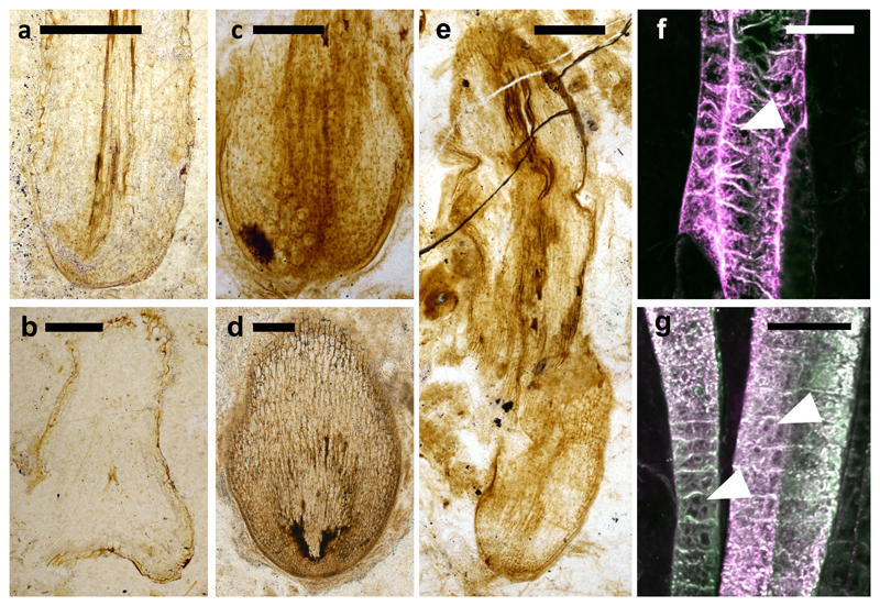
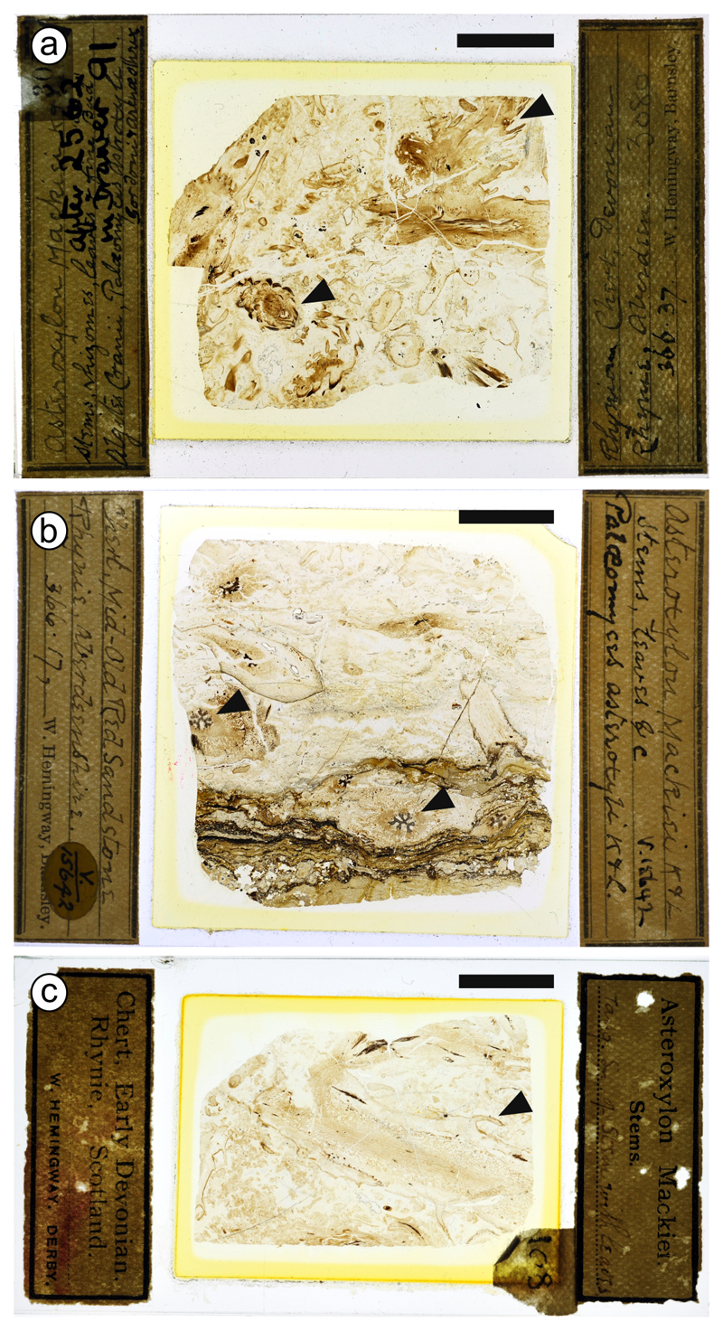
The body plan of A. mackiei comprised three axes types; leafy shoot axes, rooting axes and the transition region between these axes11–13,19,20. To identify meristems at the apices of Asteroxylon mackiei rooting axes we visually inspected 641 thin sections prepared from the Rhynie chert. We discovered the apices of five rooting axes among three thin sections (Fig. 1a–e). These five apices were assigned to A. mackiei based on two pieces of evidence. First, each of the five apices were discovered on thin sections in which A. mackiei was the only plant species present (Extended Data Fig. 1). Second, the morphology of the G-type tracheids in two of the rooting axes is diagnostic of A. mackiei21,22 (highlighted with white arrowheads in Fig. 1f,g). These data indicate that the five apices are the tips of axes of A. mackiei.
Figure 1. Five Asteroxylon mackiei rooting axes apices lacking root caps.
a–e The tips of five A. mackiei rooting axes terminating in apices. All apices are covered by a continuous layer of tissue and lack tapered root caps characteristic of the roots of extant vascular plants. f, g Confocal image of the tracheids in the axes of (a) and (e), respectively. The sheet perforations between thickening (arrowheads) are a characteristic of G-type tracheids diagnostic of A. mackiei. (a, b, f) NHMUK V.15642, (c, e, g) GLAHM Kid 3080, (d) OXF 108. Scale bars (a, b, e) 500 µm, (c, d) 250 µm, (f, g) 20 µm.
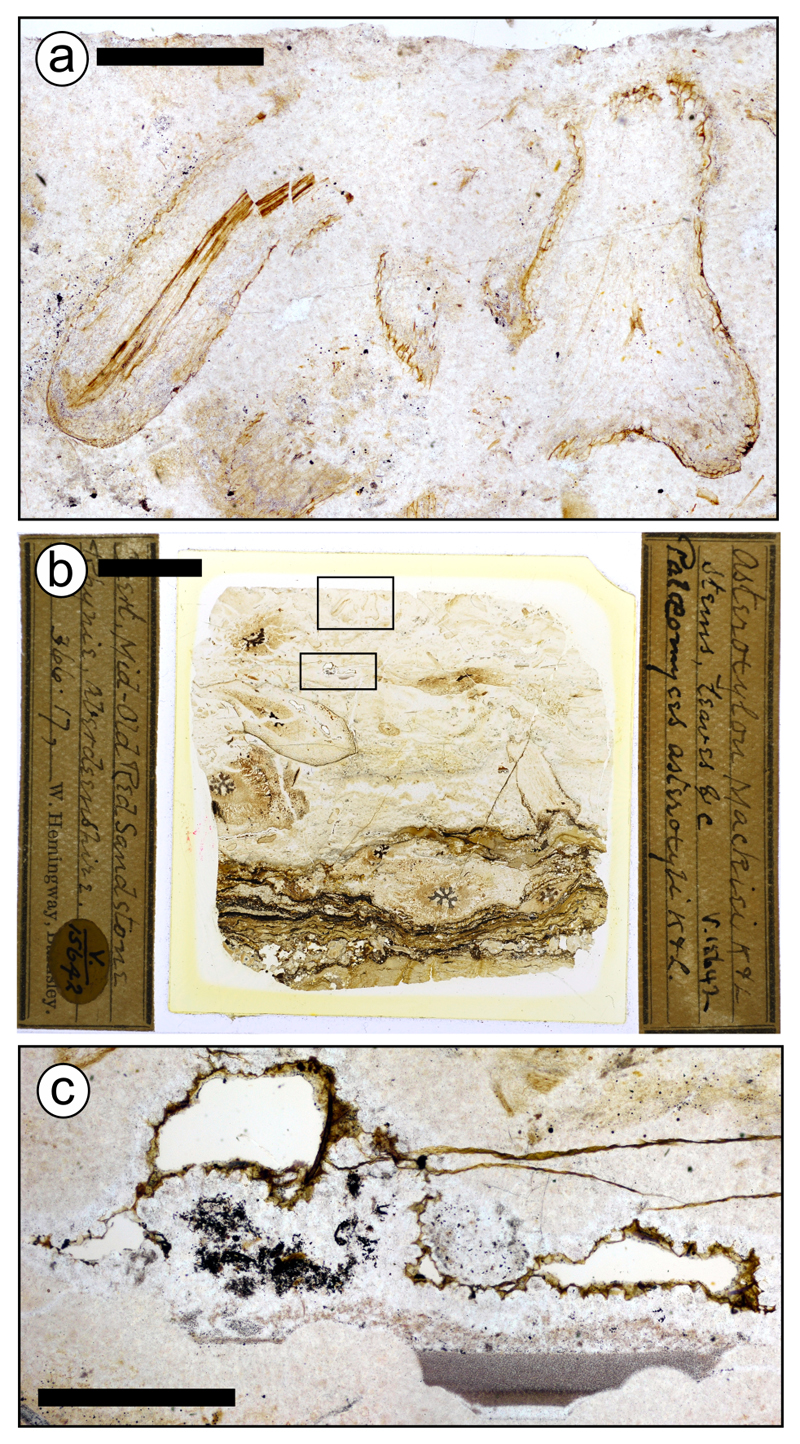
Three characteristics allowed us to identify the five apices as A. mackiei rooting axes. First, A. mackiei rooting axes lacked leaves and stomata (Fig. 1a) 11–13,19. Neither stomata, leaf primordia nor leaves were present on either apical or subapical surfaces on any of the apices (Fig. 1a–e). Second, A. mackiei rooting axes grew in the direction of the gravity vector11–13,19. The positively gravitropic growth of two apices (Fig. 1a,b) was inferred from their orientation relative to sediment layers in both the growth substrate and a geopetally infilled void preserved in the thin section (Extended Data Fig. 2). The two axes that terminated in apices (Fig. 1a,b; Extended Data Fig. 2) were orientated perpendicular to the sediment layers, this indicated that the axes grew gravitropically into the sediment. Third, A. mackiei rooting axes were isotomously branching systems11–13,19. The organisation of the pair of apices on the rooting axis in Fig. 1b, indicates that they had recently branched isotomously. Taken together the lack of leaves and stomata, the positively gravitropic growth and isotomous branching indicate that we identified five apices of A. mackiei rooting axes.
Having verified the identity of the A. mackiei apices, we next characterised the organisation of tissues in these meristems and compared their organisation with those of the roots of extant vascular plants. Three fundamental tissues were present in the apices of A. mackiei rooting axes (Fig. 1): vascular tissue developed at the centre of the axis, surrounded by layers of ground tissue covered by a distinct epidermis that comprised cells that divided anticlinally (Fig. 1; Extended Data Fig. 3). None of the epidermal cells developed hair cells resembling rhizoids or root hairs. The region where these tissues converged on the tip constituted the promeristem23. The promeristem was multicellular, like the root meristems of many extant lycopsids17,24 and the shoot meristem of A. mackiei25. In all five cases the promeristem constituted the apical-most cells of the axis; there was no tissue beyond the apical region of the promeristem where a root cap would be located in roots of extant vascular plants17,23,24,26. The roots of extant vascular plants comprise four tissues – vascular, ground, epidermis and root cap – that are derived from the promeristem17,23,24,26. The vascular tissue, ground tissues and epidermis differentiate basally as in A. mackiei. However, a root cap also develops apically and laterally in extant members17,23,24,26. The discovery that five rooting axes apices of A. mackiei lacked root caps suggests that rooting axes of early-diverging lycopsids did not develop root caps.
Given that there are no examples of extant plant species that do not develop root caps at their root apex17,23,24,26 we sought explanations that might account for the absence of the root cap in these fossils. We first considered that taphonomic processes could have led to the selective preservation of vascular, ground and dermal tissues without the preservation of root caps. Three of the apices (Fig. 1c–e), were preserved growing through a thin layer of degraded organic material called mulm27, which coats the apex and flanks. It is formally possible that mulm around the apex of the rooting structure could represent a decayed root cap. However, mulm also coats the external surfaces of many plants preserved in the Rhynie chert27 including leafy shoots of A. mackiei preserved on the same thin sections as the three apices (Fig. 1c–e; Extended Data Fig. 4). The preservation of mulm around leafy shoots indicates that the presence of this substance at the apices of rooting axes does not represent the remains of root caps. Furthermore, it is unlikely that root caps were not preserved when all other tissues in the apices were well preserved (Fig. 1). Finally, root caps were readily preserved in permineralised plant deposits in the Carboniferous and Permian Periods demonstrating that root caps can be fossilised28,29. We therefore rule out the possibility that root caps formed but were not preserved.
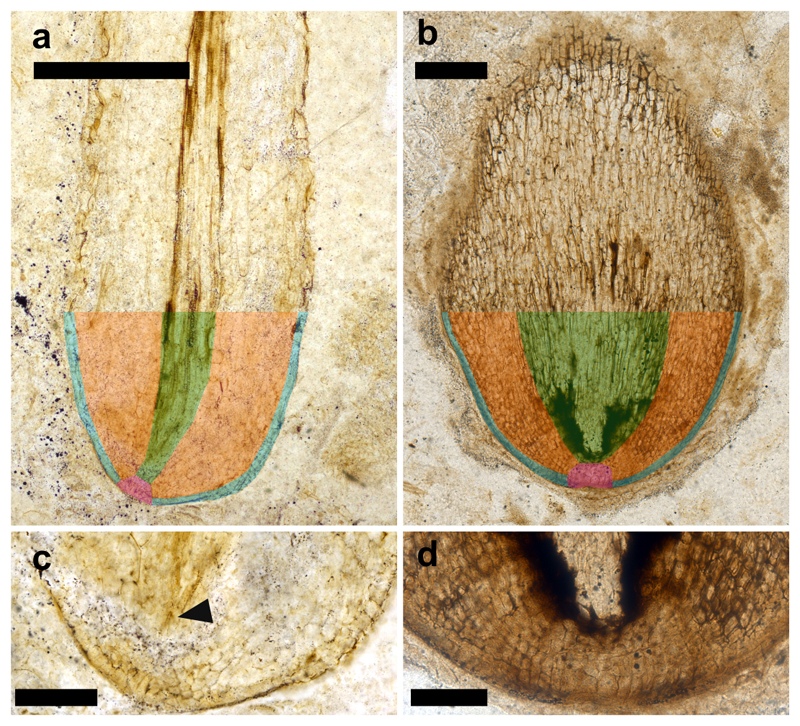
An alternative explanation for the lack of root caps in these apices is that the root cap may not have been present on apices that had stopped growing. We therefore established whether the apices were fossilised during active growth or after growth had ceased. During active growth, root meristems of extant plants comprise large numbers of relatively small, dividing cells, and cells increase in size with distance from the apex as they elongate and differentiate23,28,30. In three of the five A. mackiei apices, large numbers of small cells were located at the apex and cell size gradually increased with distance from the tip (Fig. 1c–e; Extended Data Fig. 3). This cellular organisation indicated that the apices without root caps were actively growing when fossilised. In the two remaining apices, the differentiation of vascular tissue near the tip of the apex28,30 indicated that neither apex was active at the time of preservation (Fig. 1a,b; Extended Data Fig. 3). The absence of root caps from the three actively growing fossilised meristems is consistent with the hypothesis that root caps did not develop on A. mackiei rooting axes.
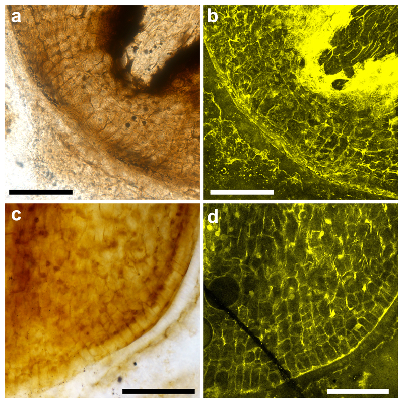
If root caps developed in A. mackiei there would be evidence of cell divisions from which the root caps developed in the preserved promeristems. We characterised cellular organisation of the two well preserved promeristems that were active when fossilised (Fig. 1d,e). We determined the orientation of cell walls at the apex to test if these were consistent with the development of a root cap23. If a root cap was formed, periclinal divisions (where the new wall is parallel to the surface) would occur near the apex of the promeristem23. Figure 2a,b is an oblique section through the meristem preserving the apical promeristem. There are no periclinal divisions in the outer layer of the apical promeristem (Fig. 2a,b). Instead, cells in the outermost layer of the apical region divided anticlinally (new cell walls oriented perpendicular to the surface). This indicates that the cell division pattern of the apical region of the meristem was inconsistent with the development of a root cap but consistent with the development of a continuous epidermis over the surface of the meristem. If a lateral root cap formed it would develop from a combination of both anticlinal and periclinal cell divisions, producing layers of cells that taper with distance from the apex as cells are sloughed off23. Figure 2c, d is a longitudinal section through the meristem in which the outer layer of the apex is preserved. Only anticlinal cell divisions occur in the outer layer and there is no evidence of periclinal divisions. The cell division pattern of these meristems is inconsistent with the development of a lateral root cap but consistent with the development of a continuous epidermal surface. Together the cellular organisation of two promeristems allowed us to rule out the development of a root cap in A. mackiei, instead we predict meristems were covered by a continuous epidermis.
Figure 2. Cell division patterns in Asteroxylon mackiei rooting axes meristems are inconsistent with the formation of root caps.
a, c, Transmitted light and b, d, confocal laser microscopy images showing cellular organisation of two A. mackiei rooting axes meristems. (a, b) The absence of periclinal cell divisions in the apical promeristem is inconsistent with the formation of a apical root cap. (c, d) The absence of periclinal cell divisions on the flanks of the meristem is inconsistent with the formation of a lateral root cap. Anticlinal cell divisions in both the promeristem (a, b) and lateral flanks (c, d) are consistent with the meristem being covered by a continuous epidermis. (a, b) OXF 108 (Fig. 1d), (c, d) GLAHM Kid 3080 (Fig. 1c). Scale bars 100 µm.
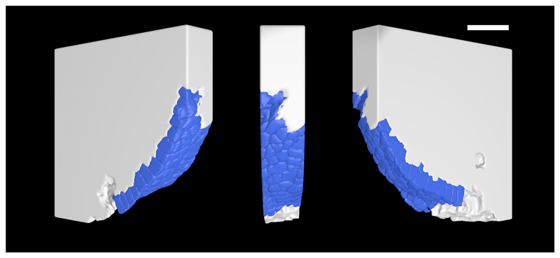
To test the hypothesis that a continuous epidermal surface covered the rooting axes meristems of A. mackiei we reconstructed a three-dimensional (3-D) model of the surface of the meristem from a z-stack of images captured on a confocal microscope (Fig. 3; see Supplementary Videos 1, 2). A continuous and smooth layer of epidermis covered the meristem and there was no evidence of tapering or cells sloughing off. We conclude that rooting axes meristems of A. mackiei developed a continuous epidermis that covered the apex.
Figure 3. Asteroxylon mackiei rooting axis meristems were covered by a continuous layer of epidermis and lacked a root cap.
Three-dimensional model from a z-stack of images captured on a confocal microscope of the meristem of an A. mackiei rooting axis with the epidermal layer highlighted in blue. The cells in the epidermis form a smooth continuous surface over the meristem. GLAHM Kid 3080 (Figs 1c, 2c,d). Scale bar 500 µm.
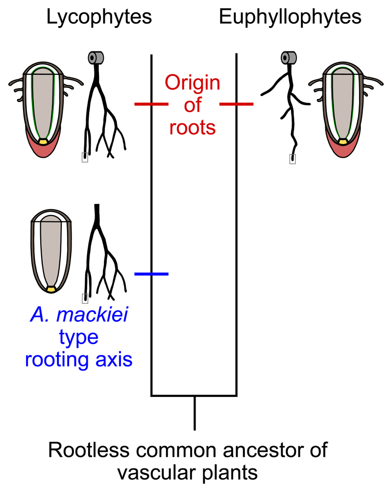
Taken together the cellular organisation of the five apices demonstrates that A. mackiei rooting axes developed from a novel meristem type that lacked both root caps and root hairs. We conclude that the evolution of rooting axes in lycopsids occurred in a stepwise manner. There was a stage, represented by A. mackiei, that was characterised by the presence of radially symmetric, positively gravitropic, isotomously branching axes with vascular tissue organisation that is distinct from that in the leafy shoot and a lack of leaves and stomata11–13,19 (Fig. 4). Subsequently, a root cap, root hairs, endogenous development, and an endodermis evolved16–18,24 (Fig. 4). This sequence of events is consistent with the hypothesis that the common ancestor of all extant vascular plants was rootless, and that roots with caps had at least two separate origins among lycophytes and euphyllophytes, respectively2 (Fig. 4). The similarities in root anatomy (the presence of a root cap, root hairs, an endodermis and quiescent centre16–18,24), development (endogenous origin)18 and gene expression15 shared by extant lycophytes and euphyllophytes, therefore represent examples of convergent evolution. The simultaneous presence of rooting axes and leaves on shoots6,9,14,16 in A. mackiei suggests that the co-evolution of roots and leaf-bearing shoots may have contributed to the evolution of an efficient transpiration stream among early lycopsids.
Figure 4. Stepwise and independent origins of roots among vascular plants.
The roots of extant lycophytes evolved in a stepwise manner. There was a stage, represented by Asteroxylon mackiei, characterised by the presence of radially symmetric, positively gravitropic, isotomously branching axes. Subsequently, a root cap, root hairs, endogenous development, and an endodermis evolved. This sequence of events is consistent with the hypothesis that the common ancestor of all extant vascular plants was rootless, and roots with caps had at least two independent origins among lycophytes and euphyllophytes. The extensive similarities between roots of extant lycophytes and euphyllophytes therefore represent examples of convergent evolution.
Methods
Identifying Asteroxylon mackiei rooting axes apices
To identify meristems at the apices of A. mackiei rooting axes we visually inspected 641 thin sections prepared from the Rhynie chert from the following collections: Eleven from the collections of the School of Biology, University of St Andrews, UK; Seven from the Oxford University Herbaria, UK; 33 from the collections of the Manchester Museum, The University of Manchester, UK; 299 from the London Natural History Museum, UK; 291 from The Hunterian, University of Glasgow, UK.
Imaging of rooting apices
Photographs of thin sections were taken using a Nikon D80 with a 60-mm macro lens set up on a copystand. Thin sections were lit from below with a lightbox and lit from above with aerial lights (Extended Data Fig. 1). High resolution images of the apices were taken using a Nikon Eclipse LV100ND (Figs 1a–c,e, 2c; Extended Data Figs 2, 3, 4a,b) and Olympus BX50 (Figs 1d, 2a; Extended Data Fig. 4c) compound microscopes and a Leica M165 FC (Extended Data Fig. 4c) stereo microscope. To create high definition images multiple overlapping photographs were taken and combined to make a single image using AutoStitch31.
Confocal laser scanning microscopy was also used to image A. mackiei rooting axes meristems. Confocal images were acquired with a Nikon A1-Si laser-scanning confocal microscope (London Natural History Museum, UK) (Figs 1f,g, 2d) and a Leica SP5 confocal microscope (Department of Plant Sciences, University of Oxford, UK) (Fig. 2c). The Nikon A1-Si laser-scanning confocal microscope was used with a ×40 oil-immersion objective with a 1.3 numerical aperture and 29.37 μm pinhole. Autofluorescence of the sample was excited with a 561 nm and 640 nm laser and emission was collected with windows of 570–620 nm and 675–725 nm for each laser, respectively. The Leica SP5 confocal microscope was used with a ×20 oil-immersion objective with a 0.7 numerical aperture and a 60.7 μm pinhole. Autofluorescence of the sample was excited with a 633 nm laser and emission was collected with a window of 645–800 nm.
Segmentation and visualisation of A. mackiei meristem
Images were processed using FIJI32. Segmentation of the epidermal surface of the meristem was carried out in MorphoGraphX33 (Supplementary Video 1). The three-dimensional model was visualised in Blender™ (Fig. 3, Supplementary Video 2)34.
Extended Data
Extended Data Figure 1. Five root apices were found on three thin sections in which Asteroxylon mackiei was the only plant species present.
Diagnostic features of A. mackiei include the apices of leafy shoots (black arrowhead in a), star shaped xylem (black arrowhead in b) and leaves (black arrowhead in c). (a) GLAHM Kid 3080, (b) NHMUK V.15642, (c) OXF 108. Scale bars 1 cm.
Extended Data Figure 2. Asteroxylon mackiei rooting axes grew in the direction of the gravity vector.
Positively gravitropic growth of two apices (a) was inferred from their orientation relative to sediment layers in both the growth substrate (dark brown and black bands at base of sample b) and a geopetally infilled void (c) preserved in the thin section. The position of both the apices and geopetally infilled void are highlighted with black boxes within the thin section (b). (a–c) NHMUK V.15642. Scale bars (a, c) 1 mm, (b) 1 cm.
Extended Data Figure 3. Fundamental tissues present in a rooting axis apex preserved after growth had finished (a) and a rooting axis meristem preserved during active growth (b).
a, b, Root apices with fundamental tissue types colour coded (blue, epidermis; pink, promeristem; orange, cortex; and green, procambium). c, d, Magnified images of the apical regions of (a) and (b), respectively. The presence of differentiated vascular tissue (arrowhead c) close to the tip of the apex indicates this apex was not active at the time of preservation. By contrast, in (d) there is no differentiated vascular tissue. Instead the apex is characterised by large numbers of cells and cell size gradually increases with distance from the tip indicating the apex was active when fossilised. (a, c) NHMUK V.15642 – same specimen as illustrated in (Fig. 1a), (b, c) OXF 108 – same specimen as illustrated in (Fig. 1d; 2a, b). Scale bars (a) 500 µm, (b) 250 µm, (c) 150 µm, (d) 100 µm.
Extended Data Figure 4. Mulm27 coats the rooting axes and leafy shoots of Asteroxylon mackiei.
A thin layer of degraded organic material called mulm27 (highlighted with arrowheads a–d) coats both rooting axes (a, c) and leafy shoots of A. mackiei. (a, b) GLAHM Kid 3080, (c, d) OXF 108. Scale bars (a, c, d) 500 µm, (b) 1 mm.
Supplementary Material
Acknowledgments
AJH was funded by the George Grosvenor Freeman Fellowship by Examination in Sciences, Magdalen College, Oxford, UK. LD was funded by a European Research Council Advanced Grant (EVO500, contract 25028) and a European Commission Framework 7 Initial Training Network (PLANTORIGINS, project identifier 238640). We are grateful to the Oxford University Herbaria, the University of Manchester, Manchester Museum, The Hunterian, University of Glasgow and the London Natural History Museum for access to fossil thin sections, and to the curators of these collections; Dr Stephen Harris, Kate Sherburn, Dr Neil Clark and Dr Peta Hayes for their assistance. We thank Dr Tomasz Goral, Dr Christine Strullu-Derrien, Dr Charlotte Kirchhelle, Prof. Ian Moore and Dr Imran Rahmen for assistance and advice for confocal imaging, segmentation and 3D reconstructions and Mr John Baker for photographic advice. We are grateful to Prof. A M Hetherington, Dr N J Hetherington and members of the Dolan lab for helpful comments and discussions.
Footnotes
Data availability statement
The three thin sections analysed in this study are available in publically availably collections: GLAHM Kid 3080; OXF 108 and NHMUK V.15642. The confocal laser scanning microscopy data sets generated are available from the corresponding author on reasonable request. All other data supporting the findings of this study are included in this published article (and its Extended Data Figures).
Author Contributions
AJH and LD designed the project. AJH carried out the analyses. AJH and LD wrote the paper.
Competing interests
The authors declare no competing financial interests.
References
- 1.Schneider H, Pryer KM, Cranfill R, Smith AR, Wolf PG. Developmental genetics and plant evolution. Taylor & Francis; 2002. Evolution of vascular plant body plans: a phylogentic perspective; pp. 330–364. [Google Scholar]
- 2.Raven JA, Edwards D. Roots: evolutionary origins and biogeochemical significance. J Exp Bot. 2001;52:381–401. doi: 10.1093/jexbot/52.suppl_1.381. [DOI] [PubMed] [Google Scholar]
- 3.Kenrick P, Strullu-Derrien C. The origin and early evolution of roots. Plant Physiol. 2014;166:570–580. doi: 10.1104/pp.114.244517. [DOI] [PMC free article] [PubMed] [Google Scholar]
- 4.Kenrick P. The Origin of Roots. In: Eshel A, Beeckamn T, editors. Plant roots: the hidden half. Taylor & Francis; 2013. pp. 1–14. [Google Scholar]
- 5.Gensel PG, Berry CM. Early lycophyte evolution. Am Fern J. 2001;91:74–98. [Google Scholar]
- 6.Matsunaga KKS, Tomescu AMF. Root evolution at the base of the lycophyte clade: insights from an Early Devonian lycophyte. Ann Bot. 2016;117:585–598. doi: 10.1093/aob/mcw006. [DOI] [PMC free article] [PubMed] [Google Scholar]
- 7.Sachs J. In: Text-book of botany, morphological and physiological. Bennett AW, Thiselton Dyer WT, translators. Clarendon Press; Oxford (UK): 1875. [Google Scholar]
- 8.Hao S, Xue J, Guo D, Wang D. Earliest rooting system and root : shoot ratio from a new Zosterophyllum plant. New Phytol. 2010;185:217–225. doi: 10.1111/j.1469-8137.2009.03056.x. [DOI] [PubMed] [Google Scholar]
- 9.Matsunaga KKS, Tomescu AMF. An organismal concept for Sengelia radicans gen. et sp. nov. – morphology and natural history of an Early Devonian lycophyte. Ann Bot. 2017;119:1097–1113. doi: 10.1093/aob/mcw277. [DOI] [PMC free article] [PubMed] [Google Scholar]
- 10.Edwards D, Kenrick P, Dolan L. History and contemporary significance of the Rhynie cherts—our earliest preserved terrestrial ecosystem. Philos Trans R Soc B Biol Sci. 2018;373 doi: 10.1098/rstb.2016.0489. 20160489. [DOI] [PMC free article] [PubMed] [Google Scholar]
- 11.Edwards D. Embryophytic sporophytes in the Rhynie and Windyfield cherts. Trans R Soc Edinburgh Earth Sci. 2004;94:397–410. [Google Scholar]
- 12.Kidston R, Lang WH. On Old Red Sandstone plants showing structure, from the Rhynie Chert Bed, Aberdeenshire. Part III. Asteroxylon mackiei, Kidston and Lang. Trans R Soc Edinb Earth Sci. 1920;52:643–680. [Google Scholar]
- 13.Kidston R, Lang WH. On Old Red Sandstone plants showing structure, from the Rhynie Chert Bed, Aberdeenshire. Part IV. Restorations of the vascular cryptogams, and discussion of their bearing on the general morphology of the Pteridophyta and the origin of the organisation of land plants. Trans R Soc Edinb Earth Sci. 1921;52:831–854. [Google Scholar]
- 14.Kenrick P, Crane PR. The origin and early diversification of land plants: a cladistic study. Smithsonian Series in Comparative Evolutionary Biology. Washington, DC, USA: Smithsonian Institute Press; 1997. [Google Scholar]
- 15.Huang L, Schiefelbein J. Conserved gene expression programs in developing roots from diverse plants. Plant Cell. 2015;27:2119–2132. doi: 10.1105/tpc.15.00328. [DOI] [PMC free article] [PubMed] [Google Scholar]
- 16.Hetherington AJ, Dolan L. The evolution of lycopsid rooting structures: conservatism and disparity. New Phytol. 2017;215:538–544. doi: 10.1111/nph.14324. [DOI] [PubMed] [Google Scholar]
- 17.Fujinami R, et al. Root apical meristem diversity in extant lycophytes and implications for root origins. New Phytol. 2017;215:1210–1220. doi: 10.1111/nph.14630. [DOI] [PubMed] [Google Scholar]
- 18.Foster AS, Gifford EM. Comparative morphology of vascular plants. W. H. Freeman and Company; 1959. [Google Scholar]
- 19.Bhutta AA. Studies on the flora of the Rhynie Chert. PhD thesis. University of Wales; Cardiff (UK): 1969. [Google Scholar]
- 20.Kerp H. Organs and tissues of Rhynie chert plants. Philos Trans R Soc B Biol. Sci. 2018;373 doi: 10.1098/rstb.2016.0495. 20160495. [DOI] [PMC free article] [PubMed] [Google Scholar]
- 21.Kenrick P, Crane PR. Water-conducting cells in early fossil land plants: implications for the early evolution of tracheophytes. Bot Gaz. 1991;152:335–356. [Google Scholar]
- 22.Strullu-Derrien C, Wawrzyniak Z, Goral T, Kenrick P. Fungal colonization of the rooting system of the early land plant Asteroxylon mackiei from the 407-Myr-old Rhynie Chert (Scotland, UK) Bot J Linn Soc. 2015;179:201–213. [Google Scholar]
- 23.Clowes FAL. Apical meristems. Blackwell; 1961. [Google Scholar]
- 24.Guttenberg HV. Histogenese der Pteridophyten. Handbuch der Pflanzenanatomie. VII. 2 Gebrüder Borntraeger; 1966. [Google Scholar]
- 25.Hueber FM. Thoughts on the early Lycopsids and Zosterophylls. Ann Missouri Bot Gard. 1992;79:474–499. [Google Scholar]
- 26.Heimsch C, Seago JL. Organization of the root apical meristem in angiosperms. Am J Bot. 2008;95:1–21. doi: 10.3732/ajb.95.1.1. [DOI] [PubMed] [Google Scholar]
- 27.Kelman R, Feist M, Trewin NH, Hass H. Charophyte algae from the Rhynie chert. Trans R Soc Edinb Earth Sci. 2004;94:445–455. [Google Scholar]
- 28.Hetherington AJ, Dubrovsky JG, Dolan L. Unique cellular organization in the oldest root meristem. Curr Biol. 2016;26:1629–1633. doi: 10.1016/j.cub.2016.04.072. [DOI] [PMC free article] [PubMed] [Google Scholar]
- 29.Strullu-Derrien C, McLoughlin S, Philippe M, Mørk A, Strullu DG. Arthropod interactions with bennettitalean roots in a Triassic permineralized peat from Hopen, Svalbard Archipelago (Arctic) Palaeogeogr Palaeoclimatol Palaeoecol. 2012;348–349:45–58. [Google Scholar]
- 30.Shishkova S, Rost TL, Dubrovsky JG. Determinate root growth and meristem maintenance in angiosperms. Ann Bot. 2008;101:319–40. doi: 10.1093/aob/mcm251. [DOI] [PMC free article] [PubMed] [Google Scholar]
- 31.Brown M, Lowe DG. Automatic panoramic image stitching using invariant features. Int J Comput Vis. 2007;74:59–73. [Google Scholar]
- 32.Schindelin J, et al. Fiji: an open-source platform for biological-image analysis. Nat Methods. 2012;9:676–682. doi: 10.1038/nmeth.2019. [DOI] [PMC free article] [PubMed] [Google Scholar]
- 33.Barbier de Reuille P, et al. MorphoGraphX: A platform for quantifying morphogenesis in 4D. Elife. 2015;4:e05864. doi: 10.7554/eLife.05864. [DOI] [PMC free article] [PubMed] [Google Scholar]
- 34.Garwood R, Dunlop J. The walking dead: Blender as a tool for paleontologists with a case study on extinct arachnids. J Paleontol. 2014;88:735–746. [Google Scholar]
Associated Data
This section collects any data citations, data availability statements, or supplementary materials included in this article.


