Summary
Background
Foals can follow the herd within hours of birth, but it has been shown that kinetic gait parameters and static balance still have to mature. However, development of dynamic balance has not been investigated.
Objectives
To objectively quantify landing and pressure pattern dynamics under the hoof during the first half year of life.
Study design
Prospective, cohort study performed at a single stud farm.
Methods
Pressure plate measurements at walk and trot from ten Dutch warmblood foals during the first 24 weeks of life were used to quantify toe‐heel and medial‐lateral hoof balance asymmetry indexes and to determine preferred landing strategy. Concurrently, radiographs of the tarsocrural and femoropatellar joints were taken at 4–6 weeks and after 6 months to check for osteochondrosis. A linear mixed model was used to determine the effects of time point, limb pair (front/hind), side (left/right) and osteochondrosis status of every foal.
Results
At 25% of stance duration at walk, front limbs were more loaded in the heel region in weeks 6–20 (P≤0.04), the medial‐lateral balance was more to the lateral side from week 6 onwards at both walk and trot (P≤0.04). Landing preference gradually changed in the same directions. Variability in pressure distribution decreased over time. (Subclinical) osteochondrosis did not influence any of the measured parameters.
Main limitations
This study is limited by the relatively small sample size only containing one breed from a single stud farm.
Conclusions
Dynamic hoof balance in new‐born foals is more variable and less oriented towards the lateral side of the hoof and to the heel than in mature horses. This pattern changes gradually during the first weeks of life. Knowledge of this process is essential for the clinician when considering interventions in this area in early life.
Keywords: horse, foal, gait development, kinetics, hoof balance
Introduction
As precocious fright‐and‐flight animals, foals stand and walk within hours after birth. While being able to keep up with their dam immediately after birth, their gait has to mature 1, 2. This also applies to their static balance, as stabilographic measurements showed that both amplitude and velocity of the body's centre of pressure (COP) movements decrease rapidly after birth 3. Simultaneously, the variability of stance duration at walk and trot is reduced, suggesting maturation of the musculoskeletal system and gait control 2. It is not unlikely that dynamic balance, i.e. the way of landing and the pressure pattern under the hoof during stance phase, also goes through rapid development during this period; this requires further investigation.
Pressure plate systems can objectively quantify pressure distribution underneath the hoof during the stance phase 4. Previous studies have successfully used this technique to quantify toe‐heel and medial‐lateral hoof balance using asymmetry indices (ASI) in the forefeet of adult warmblood horses and ponies 5, 6 and by determination of COP patterns in adult horses 7. These results suggested that adult horses present a distinct loading pattern at the walk, generally loading the lateral side of the hooves more 5. Although van Heel et al. 4 reported lateral landing as preferred way in both front and hindlimbs at trot, Oosterlinck et al. 5 reported contralateral differences in medial‐lateral loading pattern between front hooves at impact, whereas at the end of stance, lateral parts of both front feet were loaded more. In ponies, these contralateral differences were not observed 6. Using high‐speed video recordings, Wilson et al. 8 reported a substantial variation, both within horses and between them, in front hoof placement. Nevertheless, the authors reported that for the studied population of adult horses (age range 4–23 years), the most common hoof placement pattern at walk was the lateral heel and at trot the lateral hoof wall.
For this study, we hypothesised that foals initially show a relatively high degree of variation in their toe‐heel and medial‐lateral loading patterns during the stance phase at walk and trot due to immature coordination. This would be translated as relatively high variability in medial‐lateral and toe‐heel ASIs, which would then gradually decrease, stabilising in the patterns observed in adult horses. For hoof placement at landing, we similarly hypothesised that young foals would show more variation in their preferred landing than older foals.
Materials and methods
Foals
The data for this study were collected during the experimental sessions that have also yielded data for another study on the longitudinal development of kinetic gait parameters and the possible influence of osteochondrosis (OC) in warmblood foals 2. In brief, 11 privately owned Royal Dutch Sport Horse foals (5 fillies and 6 colts) bred for showjumping, were used. All foals were born and housed at the same stud farm and kept together with their dams in a stable bedded with straw and daily access to pasture. After weaning, between weeks 20 and 24, the foals were housed in a large, half‐open group stable with straw bedding. During the study period, no hoof trimming was performed.
One colt was excluded from this study due to asymmetrical front feet. Before each measurement session, foals were observed by an experienced equine veterinarian (B.M.C.G.) at walk and trot on a straight line and hard surface to ensure that they were clinically sound. Radiographic evaluation assessed the OC status of the femoropatellar, femorotibial and tarsocrural joints at the age of 4–6 weeks and at the age of at least 6 months (age range 6–9 months). Three radiographs (dorso‐plantar, latero‐medial and dorsomedial‐planterolateral oblique) were taken of the tarsocrural joints, and two (latero‐medial and caudolateral‐craniomedial oblique) from the femoropatellar and femorotibial joints. Two board‐certified veterinary radiologists evaluated the radiographs.
Data collection
Data were collected using a pressure plate with a measuring surface of 1.95 × 0.32 m, (Footscan 3D, 2 m system)a, at a sampling rate of 125 Hz, connected to a laptop computer with the dedicated software (Gait Scientific, version 7.99–27.05.2014)a. The pressure plate was embedded in a wooden frame, with a small ramp in front of and behind the plate to avoid stumbling. The runway, including the pressure plate, was covered with a 10 m long, 1.5 m wide and 5 mm thick rubber matb (natural rubber/styrene‐butadiene rubber, shore hardness 65 ± 5) to protect the plate. Before each measuring session, the pressure plate was calibrated according to the manufacturer's instructions and offset was manually adjusted to avoid sensor saturation.
All measurements were performed at the stud farm in an empty stable building. After a short warm‐up period, foals were consistently led over the pressure plate at their preferred speed by an experienced handler. Before weaning the foals followed their dams, led by another person next to the pressure plate; after weaning, foals were handled alone. A trial was considered valid if the foal looked ahead, moved over the pressure plate in a consistent way and the hooves made full ground contact within the measuring area. When the foals were small, all limbs could be measured during one run, but with increasing size, left and right‐side data needed to be recorded in separate trials. At each measuring session, at least five valid measurements were collected from each hoof at walk and trot. For this, in general between 10–25 trails were needed.
During the first 2 weeks after birth, data were recorded solely at the walk. From week 2 until week 12, foals were measured every fortnight at both walk and trot and subsequently at 16, 20 and 24 weeks. All trials were recorded with a small digital video camera (Philips full HD 1080p camcorder)c for retrospective visual control.
Data processing
Collected footprints were manually assigned to left fore (LF), right fore (RF), left hind (LH) or right hind (RH) based on the video images. With the help of the RsScan softwarea, hoof prints were manually divided in a toe and heel region by a line through the maximal hoof width and in a medial and lateral zone by a line through the middle of the hoof, as described by Oosterlinck et al. 5. Any footprints that were not aligned with the pressure plate coordinate system were excluded from the analysis. After exporting the raw data from the pressure plate system, data were processed using custom‐written matlabd scripts. Toe‐heel and medial‐lateral hoof balance ASI of each data frame was calculated using the vertical forces obtained for the four hoof zones by the following formulas 5:
For the toe‐heel ASI, a positive value indicates higher loading towards the toe zone and a negative value higher loading towards the heel zone. For the medial‐lateral ASI, a positive value indicates higher loading towards the medial zone and a negative value higher loading towards the lateral zone.
All ASI data were normalised to the percentage of stance time to compare the data obtained from the different measurements. This was performed by interpolating each measurement to 200 samples using a shape‐preserving, piecewise cubic interpolation method. For each hoof‐impact, the preferred hoof placement zone was determined based on the force distribution over the four zones at impact with the plate (first recorded data frame). For this, we have analysed the four zones in two pairs, medial‐lateral and toe‐heel. Based on the zone pair which had the highest force value, the measurement was later classified as toe or heel first and as lateral or medial first.
Data analysis
Open software (R version 3.3.1)e was used for statistical analysis using the package ‘nlme’ (version 3.1–121) for linear mixed effects model, package ‘lme4’ (version 1.1–12) for logistic regression analysis. To evaluate the preferred landing site, data from all hoof impacts was used. For each trial, relative frequencies of lateral/medial and toe/heel landing were calculated and used in a logistic regression analysis model. Odds ratios for preferred landing sites were determined by calculating the exponential function of the model estimates 9.
Both for walk and trot, a linear mixed effect model, with foal ID used as a random effect and time point, limb pair (front/hind), side (left/right) and OC status as fixed effects, were used for statistical evaluation of the data. Correction for multiple comparisons was done for both models with the False Discovery Rate method of Benjamini–Hochberg 10.
For all models, the first measurement moment (i.e. week 0.5 for walk and week 2 for trot) was used as a reference for the comparison between time points.
Results
Foals
All foals were considered clinically sound at visual inspection before each measurement session, except for two foals at 24 weeks. As these animals were judged to be sound again 1 week later, pressure plate measurements were taken then and included in the dataset as being representative for week 24. At the first radiographic screening, five foals were negative for OC, whereas in the other five at least one lesion was found. At the second screening moment, two out of the five foals were still positive for OC.
Figure 1 presents a front, hind and lateral view of a representative foal used in the study, illustrating the changes in conformation observed between week 1 and 12. At birth, this foal showed a subtle valgus deviation and relatively wide base of support. At the next measuring moment limbs were straight with a relatively smaller base of support.
Figure 1.
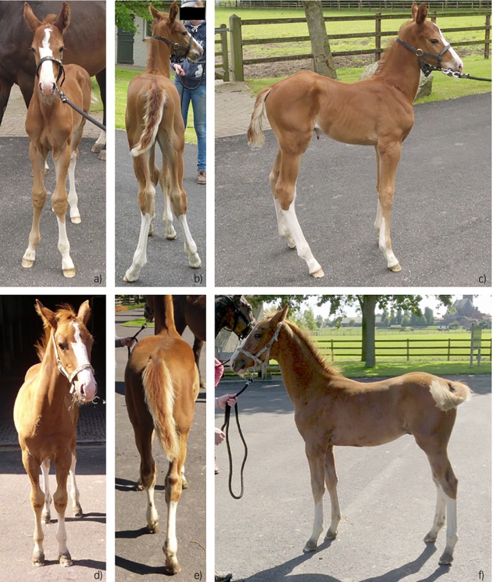
Front a) & d), hind b) & e) and lateral c) & f) views of the same foal at the age of 1 week a), b) and c) and 12 weeks d), e) and f) illustrating conformational changes over time. Note the subtle valgus deviation at week 1 in all 4 limbs.
Hoof balance – walk ASI
Details of model estimates and significances for the toe‐heel/medial‐lateral ASIs can be found in the Supplementary Item 1.
Median toe‐heel ASI values during the whole stance phase are presented in Fig 2. At one percent of the stance phase, only the front limbs showed significantly more loading of the heel region at week 12 in comparison with the first measurement at week 0.5 (P‐value 0.04). At 25% of the stance duration, front limbs showed significantly more loading of the heel region in weeks 6 until 20 (P≤0.04), whereas in the hindlimbs the difference in this phase of stance with the first measurement was only significant at week 24 (P‐value 0.001), which was the only significant difference found in the hindlimbs. In the front limbs, there were also differences at 50% from week 6 until week 24 (P≤0.01) and at 75% from week 16 until week 24 (P≤0.04). Statistical analysis at the end of stance (i.e. 99%), was not possible due to skewness of the data, even after transformation. No significant effects of side (left/right) or OC status were found on the toe‐heel balance.
Figure 2.
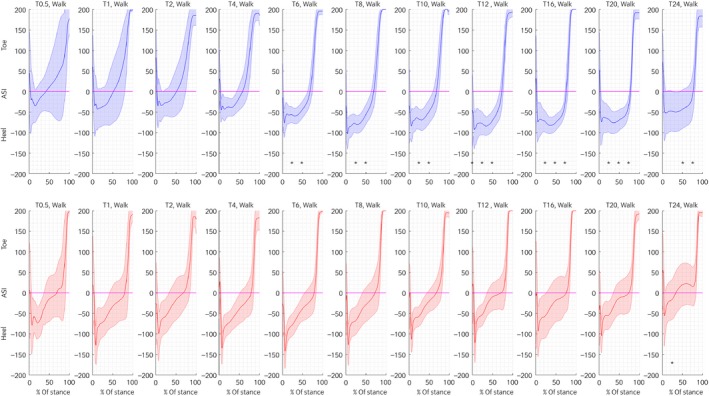
Front (upper row, blue) and hind (lower row, red) Toe‐Heel ASIs calculated for the complete stance duration at walk for the different ages. The line represents the median ASI of all foals and the shaded area the median absolute deviation of all foals. Statistically significant differences at the different time points of stance (1%, 25%, 50% and 75% of stance) compared to the first measurement (age 0.5 weeks at walk and 2 weeks at trot) are indicated with an *.
Medial – lateral ASI at the walk is presented in Fig 3. At one percent of the stance duration, both the front (P≤0.02) and hindlimbs (P≤0.04) showed significantly more loading of the lateral side of the hooves from week 6 until 24. At 25% of stance, the same trend was visible with significantly more loading of the lateral side of the front hoofs at weeks 8–12 (P≤0.04) and from week 6 until 24 (P≤0.04) for the hindlimbs.
Figure 3.
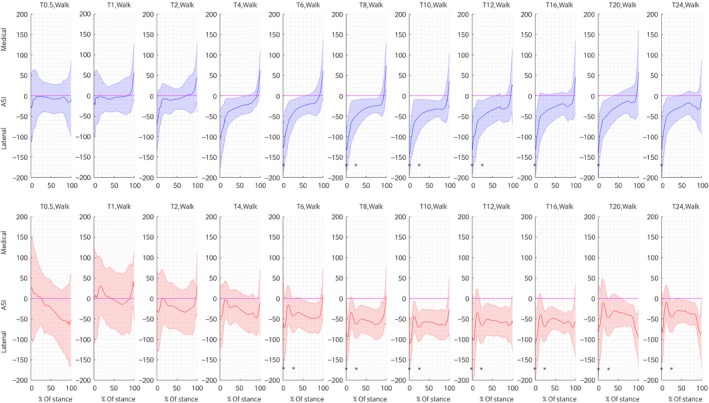
Average front (upper row, blue) and hind (lower row, red) Medial‐Lateral ASIs calculated for the complete stance duration at walk for the different ages. The line represents the mean ASI of all foals and the shaded area the median absolute deviation of all foals. Statistically significant differences at the different time points of stance (1, 25, 50 and 75% of stance) compared to the first measurement (age 0.5 weeks at walk and 2 weeks at trot) are indicated with an *.
At half and 75% of stance, no significant changes in the medial – lateral loading distribution were found. A significant difference between the left and right limb, with more lateral loading of the right side was observed at 1 (P‐value 0.0002), 25 (P‐value 0.0001) and 50% (P‐value 0.002) of the stance duration, but not at 75%. No significant effect of OC status was found for the medial‐lateral balance.
Hoof balance – trot ASI
For the toe‐heel ASI at trot, only at week 16 (P‐value 0.02) for the front and week 10 (P‐value 0.02) in the hindlimbs, statistically significant differences, indicative of more loading of the heel region, were found at the beginning of the stance phase (1%) (Fig 4). At 25% of stance, the front limbs showed significantly more loading of the heel at weeks 12–20 (P≤0.05). During mid‐stance, significantly more loading towards the heel was observed at weeks 4 and 6 (P≤0.012) in the hindlimbs. At 75% of stance, significantly more loading of the heel region in the front limbs was observed in weeks 16 and 20 (P≤0.03), for the hindlimbs this was only the case at week 4 (P‐value 0.04). No significant effects of side (left/right) or OC status were found on the toe‐heel balance.
Figure 4.
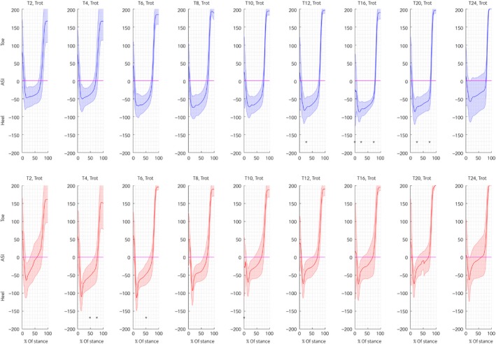
Average front (upper row, blue) and hind (lower row, red) Toe‐Heel ASIs calculated for the complete stance duration at trot for the different ages. The line represents the mean ASI of all foals and the shaded area the median absolute deviation of all foals. Statistically significant differences at the different time points of stance (1, 25, 50 and 75% of stance) compared to the first measurement (age 0.5 weeks at walk and 2 weeks at trot) are indicated with an *.
Medial‐lateral hoof balance at trot is presented in Fig 5. At one percent of stance, significantly more loading of the lateral part of the front hooves was observed at weeks 8 until 12 (P≤0.02). For the hindlimbs, this was only the case at weeks 10 and 24 (P≤0.04).
Figure 5.
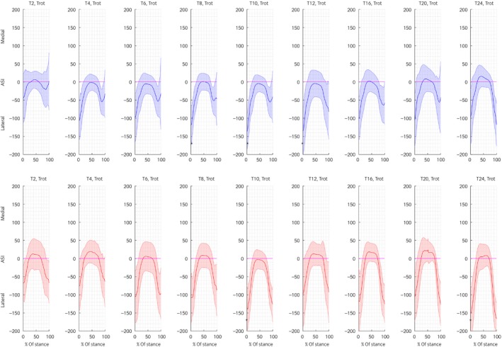
Average front (upper row, blue) and hind (lower row, red) Medial‐Lateral ASIs calculated for the complete stance duration at trot for the different ages. The line represents the mean ASI of all foals and the shaded area the median absolute deviation of all foals. Statistically significant differences at the different time points of stance (1, 25, 50 and 75% of stance) compared to the first measurement (age 0.5 weeks at walk and 2 weeks at trot) are indicated with an *.
At all points during stance, the right hooves showed significantly more loading of the lateral side (P≤0.01). There were no effects of OC status on the medial‐lateral hoof loading pattern.
Variation in hoof balance
A general trend of decreasing intra‐trial variability over time was observed, which was more prominent in the hindlimbs (Table 1). At walk, front limb intra‐trial variability of the toe‐heel ASI only changed significantly at 25% (week 16, P‐value 0.02) and 50% (weeks 8, 16 and 20, P≤0.03) of stance. In all cases, variability was lower than during the first measurement. Hindlimb variability of the toe‐heel balance showed, besides significantly lower variability at weeks 10 and 16 (P≤0.03) (25% of stance) and in week 16 (P‐value 0.03) (50% of stance), also significantly lower variability at 75% of stance at weeks 6, 8, 16 and 24 (P≤0.03). At trot, only one difference was significant, in the hindlimbs at week 10 at 75% percent of stance (P‐value 0.05).
Table 1.
Average intra‐trial variability (median absolute variation) of the Toe–heel ASI and Medial–lateral ASI of the front and hind limbs at walk and trot at 1, 25, 50 and 75% of stance duration
| Time (weeks) | Limb | Gait | Toe‐heel | Medial–lateral | ||||||
|---|---|---|---|---|---|---|---|---|---|---|
| 1% | 25% | 50% | 75% | 1% | 25% | 50% | 75% | |||
| 0.5 | Front | Walk | 49.18 | 23.86 | 23.41 | 18.04 | 54.14 | 20.41 | 18.19 | 20.25 |
| 1 | 48.28 | 18.30 | 18.11 | 19.31 | 64.85 | 24.47 | 26.14 | 37.28* | ||
| 2 | 33.16 | 30.57 | 22.69 | 21.56 | 51.19 | 23.97 | 22.96 | 23.49 | ||
| 4 | 49.97 | 19.17 | 16.92 | 23.85 | 36.11 | 27.68 | 21.59 | 22.04 | ||
| 6 | 61.99 | 14.66 | 15.47 | 17.96 | 32.69 | 20.97 | 21.70 | 23.36 | ||
| 8 | 49.82 | 15.01 | 13.25* | 23.41 | 36.17 | 16.11 | 20.23 | 21.25 | ||
| 10 | 33.63 | 14.22 | 14.70 | 19.66 | 22.09 | 18.72 | 15.57 | 16.97 | ||
| 12 | 41.13 | 18.23 | 16.56 | 22.47 | 29.11 | 26.24 | 19.85 | 19.15 | ||
| 16 | 34.02 | 12.85* | 13.07* | 17.43 | 27.87 | 18.68 | 17.63 | 18.91 | ||
| 20 | 50.81 | 18.76 | 13.89* | 15.35 | 18.00* | 15.91 | 15.93 | 16.50 | ||
| 24 | 48.67 | 15.07 | 14.75 | 15.15 | 33.51 | 26.37 | 17.65 | 20.06 | ||
| 0.5 | Hind | Walk | 54.47 | 24.81 | 24.15 | 27.05 | 44.39 | 24.15 | 31.13 | 33.76 |
| 1 | 43.80 | 26.60 | 27.37 | 25.16 | 41.51 | 32.41 | 32.05 | 32.09 | ||
| 2 | 34.70 | 17.33 | 18.25 | 21.31 | 50.43 | 25.65 | 20.86* | 23.45* | ||
| 4 | 60.59 | 19.55 | 23.05 | 24.59 | 46.24 | 16.52 | 26.65 | 26.36* | ||
| 6 | 37.51 | 19.70 | 19.47 | 16.91* | 35.27 | 21.34 | 14.94* | 16.92* | ||
| 8 | 53.61 | 18.40 | 17.34 | 15.85* | 41.24 | 17.84 | 15.32* | 17.21* | ||
| 10 | 45.51 | 16.06* | 20.47 | 21.64 | 27.68 | 19.62 | 17.29* | 14.91* | ||
| 12 | 52.32 | 20.39 | 23.02 | 17.88 | 33.80 | 24.12 | 23.42 | 15.78* | ||
| 16 | 21.55 | 14.06* | 14.25* | 16.06* | 23.62 | 15.01 | 13.37* | 17.70* | ||
| 20 | 52.98 | 16.20 | 21.32 | 16.50 | 38.72 | 14.48 | 16.56* | 16.66* | ||
| 24 | 55.40 | 18.92 | 19.04 | 15.11* | 42.20 | 25.76 | 20.99* | 21.62* | ||
| 2 | Front | Trot | 39.23 | 23.60 | 20.58 | 22.96 | 49.32 | 19.54 | 18.07 | 19.89 |
| 4 | 46.32 | 17.30 | 17.77 | 19.80 | 32.75 | 25.84 | 19.91 | 20.85 | ||
| 6 | 63.08 | 14.84 | 14.99 | 18.38 | 55.45 | 20.20 | 13.53 | 15.65 | ||
| 8 | 68.24 | 18.20 | 17.15 | 22.69 | 25.78 | 22.93 | 18.23 | 18.45 | ||
| 10 | 56.27 | 14.05 | 11.61 | 17.16 | 23.43 | 18.24 | 15.09 | 16.54 | ||
| 12 | 43.34 | 18.10 | 15.91 | 14.72 | 24.52 | 23.34 | 18.56 | 22.27 | ||
| 16 | 46.27 | 15.64 | 10.73 | 16.27 | 28.52 | 17.15 | 13.18 | 18.68 | ||
| 20 | 45.61 | 14.28 | 12.82 | 13.95 | 42.52 | 17.21 | 11.74 | 14.84 | ||
| 24 | 64.54 | 16.71 | 15.13 | 16.77 | 29.45 | 17.08 | 12.96 | 12.88 | ||
| 2 | Hind | Trot | 54.23 | 24.98 | 21.08 | 34.85 | 51.64 | 32.21 | 27.67 | 41.86 |
| 4 | 48.00 | 16.37 | 22.58 | 22.84 | 37.65 | 19.42 | 17.56 | 20.16* | ||
| 6 | 40.65 | 17.56 | 16.36 | 22.83 | 40.72 | 17.06* | 14.67* | 18.26* | ||
| 8 | 55.86 | 15.49 | 14.05 | 22.36 | 33.52 | 18.31* | 16.28 | 18.00* | ||
| 10 | 60.22 | 14.68 | 19.18 | 18.81* | 28.07 | 18.13* | 14.78* | 16.52* | ||
| 12 | 42.12 | 17.17 | 17.68 | 29.99 | 29.75 | 19.75* | 17.58 | 23.53* | ||
| 16 | 32.76 | 19.44 | 20.13 | 35.11 | 23.68* | 20.62 | 16.55 | 19.63* | ||
| 20 | 27.27 | 19.65 | 16.10 | 25.96 | 33.68 | 23.20 | 15.75 | 15.33* | ||
| 24 | 38.28 | 14.40 | 15.60 | 24.08 | 27.56 | 21.89 | 16.83* | 18.41* | ||
An asterisk indicates a significant difference when compared with the first measurement (0.5 week for walk and 2 weeks for trot).
Front limb medial‐lateral balance did not show much change over time with only week 20 (P‐value 0.04) at walk and 1% of stance duration having significantly lower variability. Also at 75% of stance, a significant difference was found for the front limbs at week 1 (P‐value 0.01), however, in this case, variability was more compared to the first measurement a few days earlier. In contrast, hindlimb variability showed many significant differences for the medial‐lateral ASI, all decreasing over time when compared to baseline. At trot, there was no change in the front limbs, but many changes over time, again all to values less than baseline, in the hindlimbs (Table 1).
Hoof placement at landing
During the first measurements at walk, more toe landing was observed (OR 1.5), which changed to more heel landing from week six onwards, although no significant changes compared to the first measurement were found (Table 2). At trot, the same trend was observed with young foals showing relatively more toe landing compared to later in life. This difference was significant from week eight on. No effects of OC or differences between front and hindlimbs were observed. However, there was at trot a significant left/right difference with more pronounced heel landing on the right side.
Table 2.
Odds ratios (OR) with 95% confidence intervals (95% CI) for toe or heel (T–H) and medial or lateral (M–L) landing at walk and trot at the different time points
| Time (weeks) | Limb/side | Gait | Toe–heel | Medial–lateral | ||||
|---|---|---|---|---|---|---|---|---|
| Odds ratio | 95% CI | P value | Odds ratio | 95% CI | P value | |||
| 0.5 | Front limbs | Walk | 1.5 | 0.8/2.6 | 1.1 | 0.7/0.2 | ||
| 1 | 1.1 | 0.6/2.0 | 0.8 | 0.7 | 0.4/1.2 | 0.3 | ||
| 2 | 1.1 | 0.6/2.1 | 0.8 | 0.4* | 0.2/0.8 | 0.03 | ||
| 4 | 1.3 | 0.7/2.2 | 0.6 | 0.3* | 0.2/0.5 | <0.001 | ||
| 6 | 0.7 | 0.4/1.3 | 0.5 | 0.2* | 0.1/0.3 | <0.001 | ||
| 8 | 0.6 | 0.4/1.1 | 0.3 | 0.2* | 0.1/0.3 | <0.001 | ||
| 10 | 0.6 | 0.4/1.1 | 0.2 | 0.1* | 0.1/0.3 | <0.001 | ||
| 12 | 0.6 | 0.4/1.1 | 0.3 | 0.2* | 0.1/0.3 | <0.001 | ||
| 16 | 1.1 | 0.6/1.8 | 0.9 | 0.2* | 0.1/0.3 | <0.001 | ||
| 20 | 0.9 | 0.5/1.5 | 0.8 | 0.2* | 0.1/0.3 | <0.001 | ||
| 24 | 1.6 | 0.9/2.9 | 0.2 | 0.2* | 0.1/0.4 | <0.001 | ||
| Hind limbs | 0.9 | 0.7/1.1 | 0.5 | 2.2* | 1.7/2.9 | <0.001 | ||
| Right side | 0.9 | 0.7/1.1 | 0.6 | 0.5* | 0.4/0.7 | <0.001 | ||
| 2 | Front limbs | Trot | 4.1 | 2.3/ 7.1 | 0.7 | 0.4/1.2 | ||
| 4 | 0.8 | 0.4/1.4 | 0.6 | 0.5 | 0.3/0.9 | 0.1 | ||
| 6 | 0.7 | 0.4/1.2 | 0.4 | 0.5 | 0.3/0.9 | 0.1 | ||
| 8 | 0.5 | 0.3/1.0 | 0.1 | 0.4* | 0.2/0.8 | 0.03 | ||
| 10 | 0.3* | 0.2/0.6 | <0.001 | 0.3* | 0.1/0.5 | <0.001 | ||
| 12 | 0.5 | 0.3/0.9 | 0.1 | 0.4* | 0.2/0.7 | 0.02 | ||
| 16 | 0.3* | 0.2/0.5 | <0.001 | 0.5 | 0.3/0.9 | 0.1 | ||
| 20 | 0.5 | 0.3/0.9 | 0.1 | 0.6 | 0.3/1.1 | 0.2 | ||
| 24 | 0.4* | 0.2/1.4 | 0.02 | 0.6 | 0.3/1.0 | 0.2 | ||
| Hind limbs | 1.1 | 0.9/1.4 | 0.7 | 1.0 | 0.7/1.3 | 0.9 | ||
| Right side | 0.7* | 0.5/0.9 | 0.01 | 0.4* | 0.3/0.5f | <0.001 | ||
A value >1 is indicative of preferential toe or medial landing, <1 is indicative of preferential heel or lateral landing. An asterisk indicates a significant difference when compared with the first measurement. (T = 0.5 for walk and T = 2 for trot). Significance between left and right and front and hind is also indicated with an asterisk.
After 2 weeks, foals landed significantly more on the medial side of their hooves when walking. Also, a significant difference between front and hind and left and right was found, whereas OC status did not have a significant effect. Hindlimbs showed relatively more medial landing, and the right side showed more lateral landing. At trot, there was from the beginning more lateral landing compared to medial, which difference increased over time and became significantly different from baseline in weeks 8–12 to get back to the original level afterwards. At trot, no significant differences were found when comparing front and hind and again, significantly more lateral loading was observed at the right side.
Discussion
This study confirmed our initial hypothesis that foals would show a relatively high variation in foot placement patterns that decreases over time. We also saw a systemic shift in dynamic hoof balance in this period. In young foals, there is relatively more loading towards the toe and towards the medial hoof side than in mature horses. The dynamic balance changes to be more like the mature horse within the first 24 weeks of life.
In general, intra‐trial variability decreases over time as expected, although this was not the case for all parameters investigated. The most prominent reduction in intra‐trial variability was observed for the medial‐lateral hoof balance of the hind limbs.
The hoof balance in mature horses is influenced by conformation, their locomotor pattern, the quality and frequency of trimming and the equestrian discipline the horse is bred for or competing in 5, 11, 12. In the foal, subtle limb deviations are a common finding with carpal and fetlock valgus deviations being most prevalent 13. New‐born foals also tend to splay their limbs, hereby increasing the width of their base of support 14, 15. This is thought to be a compensation for poor balance and muscle tone early in life 3. In the current study population, mild deviations in conformation were also observed (Fig 1) during the first few weeks of life. Where these were mild and resolved spontaneously without treatment or farrier intervention, they might help to explain the relatively higher loading of the medial part of the hooves observed during the first weeks of life. Their gradual resolution can be related to the decrease in percentage and odds ratios for medial landing over time, as observed primarily in the hindlimbs at walk. Retrospective evaluation of photographs taken before each measurement showed a gradual increase in hock straightness after birth, and it is more likely that the change in loading pattern is related to this phenomenon. The toe‐heel balance shows less evolution over time and better resembles the situation in adult warmblood horses from the start 5. During the first weeks, hindlimbs are relatively more loaded at the heel than front limbs. This could be related to some degree of flexor tendon laxity and consequent digital hyperextension, as frequently seen in new‐born foals 16, but clear clinical cases were not seen in our population.
Especially at trot, there is a marked preference to start loading at the lateral side of the hoof; after that, loading becomes more even with the hoof balance centring around zero and at the end of the stance phase, the lateral side is again loaded more. This resembles the loading patterns described previously in adult warmblood horses 4, 5. We observed significant side influences, with right limbs being loaded more laterally than left limbs. This observation is in line with the findings of Oosterlinck et al. 5 in adult Warmbloods, but the phenomenon was not observed in the work of van Heel et al. 4, or in adult ponies 6. Possible explanations are the effect of the handler 5, laterality 17, 18, 19, functional differences between the limbs as also encountered in humans 5, 20, and conformational differences between warmblood horses and ponies. In our study, the pressure plate was positioned next to the wall (at the right side of the foal). Most likely, due to the effects of the handler, who has been shown to play a role in very young and untrained foals 21, and of the mare, the foals leant slightly to the right side, probably responsible for the significantly higher loading of the lateral side of their right hooves.
A substantial decrease in the variability of the hoof loading pattern over time was observed confirming our initial hypothesis. The hypothesis was based on the observation that static balance development in young foals, as studied using stabilography, is characterised by relatively large swaying amplitudes and velocities immediately after birth that decreases later in life 3. Comparable suboptimal balance during locomotion could lead to inconsistent pressure patterns in the different zones of the hooves during subsequent strides and hence to increased variation when calculating the intra‐trial variability of an individual foal. Most prominent changes were observed in the medial‐lateral hoof balance of the hindlimbs in the second part of the stance phase. Already during the first weeks of life, a significant reduction in variability was observed. This is in line with the rapid initial improvement of static balance 3 and the significant reduction in variability of the stance duration 2 observed in young foals and is likely due to better control thanks to the maturation of the neuromuscular system. In a stabilographic study cranio‐caudal sway amplitudes that were larger than those in medial‐lateral direction shortly after birth 3. In our study, the reverse was the case. The difference might be explained by the momentum along the line of progression that is created by motion, which has a stabilising action along that line but naturally not perpendicular to it. Similar observations were made in horses suffering from spinal ataxia, a situation that is to some extent comparable with an immature neurological system. These horses also showed increased forces in the medial‐lateral, but not in the sagittal plane 22.
The fact that the age‐related decrease in variability was much more apparent in the hindlimbs than in the front limbs may have several explanations. The stabilising effect of the handler that mainly acts on the forequarter of the horse may be a factor here.
Another difference is that the placement of the front limbs can benefit from visual input, whereas hindlimbs solely have to rely on proprioception 22. Post‐natal development of the proprioceptive system may, therefore, be responsible for the gradual decrease in variability observed in the hindlimbs. The functional difference between the front limbs that carry more weight and the hindlimbs that are more involved in propulsion may play a role here too.
Some between‐foal variability was seen at weeks 20 and 24, as can be deduced from the larger shaded area at these time points in Figs 2, 3, 4, 5. This may have been related to the fact that at week 20 about half of the foals and week 24, all foals were weaned and consequently handled without their dam. Whereas handling of the foals without the presence of the mare was uneventful and all animals were accustomed to the location and procedure of the pressure plate data collection, the absence of the dam may have led to more variability in the trials. In the earlier study, an increase in variability of kinetic gait parameters was also observed in the stressful period directly after weaning 2.
Osteochondrosis was not a significant factor in our models, while there was a small, yet significant effect of OC on kinetic parameters in our previous study 2. Hence, the minor changes observed in kinetic parameters due to subclinical lameness (resulting in a reduction of nPVF in the affected limb) are not reflected in the hoof‐balance ASI. This might be explained by the higher sensitivity of the force plate, which directly measures absolute ground reaction forces, for detecting lameness 23 compared to the pressure plate measures the relative distribution of load.
Through the study period, hoof development and conformation was normal, and no hoof trimming was performed, which is considered standard practice in horses with acceptable limb conformation 24. This was a deliberate choice. Whereas uneven hoof growth could have induced some variability in this study due to changes in hoof‐balance 4, hoof trimming would most likely have induced more variation unrelated to development, ultimately inducing a bias in the data.
There were several limitations to this study. The number of animals included in the study was limited, and all animals were bred for showjumping, making it impossible to draw firm conclusions regarding the whole warmblood population. The hoof regions were visually determined, which is more prone to error than the recently published approach that uses a hoof‐bound coordinate system 7. However, that approach was used for a static situation concerning hoof size, not for repeated measurements of growing foals with changing hoof shapes. Furthermore, only the hoof prints that were aligned with the pressure plate coordinate system could be used, which made it necessary to collect more hoof prints. The number of trials needed to collect sufficient hoof prints was variable between foals and measurement sessions; this can potentially influence the results due to possible differences in intra‐trial variability.
For the understanding of the development of hoof asymmetries as often observed in adult horses and their relation to lameness, further studies monitoring hoof pressure patterns for more extended periods than the first weeks of life are needed.
Conclusions
In the foal, dynamic hoof balance is changing substantially in the first few weeks of life. These alterations seem to follow the conformational changes in this same period. The toe‐heel balance gradually shifts to the heel and the medial‐lateral balance to lateral. This is accompanied by a gradual shift of landing preference in the same directions. Variability in pressure distribution, especially of the medial‐lateral balance in the hindlimbs, decreased, which concurs with the decrease in the variability of some kinematic parameters in the same phase of life. Knowledge of this phenomenon of gradual stabilisation of hoof balance and preferred landing is helpful for the clinician when considering interventions in hoof balance of foals in early life.
Authors’ declaration of interests
No competing interests have been declared.
Ethical animal research
This study was reviewed and approved by the ethical committee of Utrecht University (DEC no.2014.III.04.038). Informed consent was obtained from the owner of the animals.
Sources of funding
None.
Authorship
B. Gorissen contributed to the study design, data collection and data analysis. F. Serra Bragança contributed to data collection and analysis. C. Wolschrijn, W. Back and P.R. van Weeren contributed to study design. All authors contributed to writing the manuscript.
Supporting information
Supplementary Item 1: Model estimates and significances.
Acknowledgements
The authors would like to sincerely thank the owner and staff of the stud farm for permission to use the foals and M. Oosterlinck and S. Nauwelaerts for their valuable input, discussing the methods and results. Furthermore, A.L Wiertz, S.P. Kooij and M. den Heijer are acknowledged for their assistance with the data analysis and J.C.M. Vernooij for his advice on the statistical analysis.
Manufacturers' addresses
aRsScan International N.V., Paal, Belgium.
bDe Mulder Rubber and Plastics, Gent, Belgium.
cKoninklijke Philips N.V., Eindhoven, the Netherlands.
dMathWorks, Natick, Massachusetts, USA.
eR‐Studio, Boston, Massachusetts, USA.
References
- 1. Denham, S.F. , Staniar, W.B. , Dascanio, J.J. , Phillips, A.B. and Splan, R.K. (2012) Linear and temporal kinematics of the walk in Warmblood foals. J. Equine. Vet. Sci. 32, 112‐115. [Google Scholar]
- 2. Gorissen, B.M.C. , Wolschrijn, C.F. , Serra Bragança, F.M. , Geerts, A.A.J. , Leenders, W.O.J.L. , Back, W. and van Weeren, P.R. (2017) The development of locomotor kinetics in the foal and the effect of osteochondrosis. Equine Vet. J. 49, 467‐474. [DOI] [PMC free article] [PubMed] [Google Scholar]
- 3. Nauwelaerts, S. , Malone, S.R. and Clayton, H.M. (2013) Development of postural balance in foals. Vet. J. 198, 70‐74. [DOI] [PubMed] [Google Scholar]
- 4. Van Heel, M.C. , Barneveld, A. , van Weeren, P.R. and Back, W. (2004) Dynamic pressure measurements for the detailed study of hoof balance: the effect of trimming. Equine Vet. J. 36, 778‐782. [DOI] [PubMed] [Google Scholar]
- 5. Oosterlinck, M. , Hardeman, L.C. , van der Meij, B.R. , Veraa, S. , van der Kolk, J.H. , Wijnberg, I.D. , Pille, F. and Back, W. (2013) Pressure plate analysis of toe‐heel and medial‐lateral hoof balance at the walk and trot in sound sport horses. Vet. J. 198, Suppl 1, e9‐e13. [DOI] [PubMed] [Google Scholar]
- 6. Oosterlinck, M. , Royaux, E. , Back, W. and Pille, F. (2014) A preliminary study on pressure‐plate evaluation of forelimb toe‐heel and mediolateral hoof balance on a hard vs. a soft surface in sound ponies at the walk and trot. Equine Vet. J. 46, 751‐755. [DOI] [PubMed] [Google Scholar]
- 7. Nauwelaerts, S. , Hobbs, S.J. and Back, W. (2017) A horse's locomotor signature: COP path determined by the individual limb. PLoS ONE 12, e0167477. [DOI] [PMC free article] [PubMed] [Google Scholar]
- 8. Wilson, A. , Agass, R. , Vaux, S. , Sherlock, E. , Day, P. , Pfau, T. and Weller, R. (2016) Foot placement of the equine forelimb: relationship between foot conformation, foot placement and movement asymmetry. Equine Vet. J. 48, 90‐96. [DOI] [PubMed] [Google Scholar]
- 9. Hogan, J.W. and Blazar, A.S. (2000) Hierarchical logistic regression models for clustered binary outcomes in studies of IVF‐ET. Fertil. Steril. 73, 575‐581. [DOI] [PubMed] [Google Scholar]
- 10. Benjamini, Y. and Hochberg, Y. (1995) Controlling the false discovery rate: a practical and powerful approach to multiple testing. J. R. Stat. Soc. Series B Stat. Methodol. 57, 289‐300. [Google Scholar]
- 11. Johnston, C. and Back, W. (2006) Hoof ground interaction: when biomechanical stimuli challenge the tissues of the distal limb. Equine Vet. J. 38, 634‐641. [DOI] [PubMed] [Google Scholar]
- 12. Trotter, G.W. (2004) Hoof balance in equine lameness. J. Equine. Vet. Sci. 24, 494‐495. [Google Scholar]
- 13. Robert, C. , Valette, J.P. and Denoix, J.M. (2013) Longitudinal development of equine forelimb conformation from birth to weaning in three different horse breeds. Vet. J. 198, Suppl 1, e75‐e80. [DOI] [PubMed] [Google Scholar]
- 14. Adams, R. and Mayhew, I.G. (1984) Neurological examination of newborn foals. Equine Vet. J. 16, 306‐312. [DOI] [PubMed] [Google Scholar]
- 15. Acworth, N.R.J. (2003) The healthy neonatal foal: routine examinations and preventative medicine. Equine Vet. Educ. 6, 45‐49. [Google Scholar]
- 16. Korosue, K. , Endo, Y. , Murase, H. , Ishimaru, M. , Nambo, Y. and Sato, F. (2015) The cross‐sectional area changes in digital flexor tendons and suspensory ligament in foals by ultrasonographic examination. Equine Vet. J. 47, 548‐552. [DOI] [PubMed] [Google Scholar]
- 17. McGreevy, D. and Rogers, L.J. (2005) Differences in motor laterality between breeds of performance horse. Appl. Anim. Behav. Sci. 99, 183‐190. [Google Scholar]
- 18. Van Heel, M. , Kroekenstoel, A.M. , Van Dierendonck, M.C. , Van Weeren, P.R. and Back, W. (2006) Uneven feet may develop as a consequence of lateral grazing behaviour induced by the conformation of a foal. Equine Vet. J. 38, 646‐651. [DOI] [PubMed] [Google Scholar]
- 19. Kroekenstoel, A.M. , Van Heel, M.C.V. , Van Weeren, P.R. and Back, W. (2006) Developmental aspects of distal limb conformation in the horse: potential consequences of uneven feet in foals. Equine Vet. J. 38, 652‐656. [DOI] [PubMed] [Google Scholar]
- 20. Sadeghi, H. , Allard, P. , Prince, F. and Labelle, H. (2000) Symmetry and limb dominance in able‐bodied gait: a review. Gait Posture. 12, 34‐45. [DOI] [PubMed] [Google Scholar]
- 21. Lucidi, P. , Bacco, G. , Sticco, M. , Mazzoleni, G. , Benvenuti, M. , Bernabò, N. and Trentini, R. (2012) Assessment of motor laterality in foals and young horses (Equus caballus) through an analysis of derailment at trot. Physiol. Behav. 109, 8‐13. [DOI] [PubMed] [Google Scholar]
- 22. Ishihara, A. , Reed, S.M. , Rajala‐Schultz, P.J. , Robertson, J.T. and Bertone, A.L. (2009) Use of kinetic gait analysis for detection, quantification, and differentiation of hind limb lameness and spinal ataxia in horses. J. Am. Vet. Med. Assoc. 234, 644‐651. [DOI] [PubMed] [Google Scholar]
- 23. Weishaupt, M.A. , Wiestner, T. , Hogg, H.P. , Jordan, P. and Auer, J.A. (2004) Compensatory load redistribution of horses with induced weightbearing hindlimb lameness trotting on a treadmill. Equine Vet. J. 36, 727‐733. [DOI] [PubMed] [Google Scholar]
- 24. O'Grady, S.E. (2017) Routine trimming and therapeutic farriery in foals. Vet. Clin. N. Am.: Equine Pract. 33, 267‐288. [DOI] [PubMed] [Google Scholar]
Associated Data
This section collects any data citations, data availability statements, or supplementary materials included in this article.
Supplementary Materials
Supplementary Item 1: Model estimates and significances.


