Abstract
Attention deficit hyperactivity disorder (ADHD) is now among the most commonly diagnosed chronic psychological dysfunctions of childhood. By varying estimates, it has increased by 30% in the past 20 years. Environmental factors that might explain this increase have been explored. One such factor may be audiovisual media exposure during early childhood. Observational studies in humans have linked exposure to fast-paced television in the first 3 years of life with subsequent attentional deficits in later childhood. Although longitudinal and well controlled, the observational nature of these studies precludes definitive conclusions regarding a causal relationship. As experimental studies in humans are neither ethical nor practical, mouse models of excessive sensory stimulation (ESS) during childhood, akin to the enrichment studies that have previously shown benefits of stimulation in rodents, have been developed. Experimental studies using this model have corroborated that ESS leads to cognitive and behavioral deficits, some of which may be potentially detrimental. Given the ubiquity of media during childhood, these findings in humansand rodents perhaps have important implications for public health.
Keywords: ADHD, cognition, overstimulation, child development, media
The prevalence of attention deficit hyperactivity disorder (ADHD) has increased substantially over the past 20 y, by as much as 30% by some estimates (1). ADHD is a clinical diagnosis recognizable to many. However, some argue that attentional capacity should be treated as a continuous rather than a dichotomous outcome given compelling evidence that there is a monotonically increasing relationship between a child’s ability to stay focused and improved adult health outcomes (2, 3). Akin to the rise in autism, the reasons for the rising prevalence of ADHD are likely multifactorial including an increase in the incidence as well as increased recognition. Decades of research have established a genetic predisposition to ADHD, but estimates of the heritability of ADHD range from 0.5 to 0.8 (4–8). The 1999 Surgeon General’s report on child mental health stated, “for most children with ADHD, the overall effects of these gene abnormalities appear small, suggesting that non-genetic factors also are important.” (4, 9–13) These “non-genetic factors” must account for the increased incidence as our genes have not changed appreciably in millennia. The role that environment might play in ADHD has been tested provisionally with respect to maternal smoking, maternal stress during pregnancy, maternal obesity, chaotic families, and inconsistent or harsh parenting (14–26). Another potential emerging environmental factor may be early exposure to electronic media.
Children today are immersed in electronic technology beginning shortly after birth. The typical child today begins regularly watching television at 4 mo of age compared with 4 y of age in 1970 (27, 28). Most of this shift has occurred in the past 15 y with the advent of new programs geared toward young infants (29). Moreover, although no data are available for the long-term impact of smart phone, tablet, and computer usage on young infants, there is an emerging literature that indicates that these new forms of media usage can be linked to ADHD and other psychiatric disorders in older children and college students (30, 31). The perhaps excessive exposure to media starting with very young infants has led some to refer to this generation as “digital natives” who are being raised by “digital immigrants.” The positive and negative implications of growing up as a digital native in a society in which media use begins early and is ubiquitous remain largely unknown. The success of human evolution is in part explained by the tremendous plasticity of the human brain, which allows it to be shaped through interactions with its environment. This plasticity, however, also means that early experiences exert considerable influence on neuronal structure, function, and ultimately cognition.
The present review paper will discuss the general processes that govern neurodevelopment, provide a theoretical framework as to what the potential risks of overstimulating the developing brain might be, review our observational findings in humans related to exposure to early fast-paced media and subsequent attentional deficits, and summarize experimental animal data that corroborate our prior hypotheses. It should be noted that, although we refer to media, most of the prevailing research in young infants is based on television rather than newer platforms (e.g., touchscreens). Although recent studies have demonstrated considerable use of touchscreens in infants and toddlers, data regarding untoward effects are minimal to date. However, the proposed paradigm is one of overstimulation, which could also be operative on touchscreens depending on the content being viewed.
Neurodevelopment—An Interactive Process Driven by the Environment
During neurodevelopment, billions of neurons become wired into a multitude of interconnected neuronal networks, microcircuits, layers, columns, and functional areas (32–36). Most neurons form hundreds of local and long-range connections and in turn process barrages of synaptic and neuromodulatory inputs at any given time. Neuronal processing occurs throughout the CNS, but important connections are also formed with the various divisions of the autonomic nervous system (37–40), the peripheral sensory system, and the somatic motor systems (41–43). This neuronal organization generates and processes spatiotemporal patterns and information that determines who we are, how we behave, and how we cope with our environment.
Consisting of billions of connected neurons, the human brain is set up by 19,000–20,000 protein-coding genes (44). Notably, a strikingly similar number of genes have been discovered in Caenorhabditis elegans (45), a small worm that possesses only 302 neurons, all of which have been individually identified (46). Moreover, the majority of disease-related genes in humans have homologs in C. elegans (47). This is to say that humans carry more or less the same number of genes that evolved to wire a brain of barely 300 nerve cells that control the entire behavioral repertoire of a small worm. Therefore, instead of being dictated by a relatively small number of genes, neurodevelopment is a highly interactive process in which genes provide general developmental signals—a very rough framework—and neuronal and modulatory interactions determine the wiring of the billions of cells that form the human brain: a nervous system capable of generating consciousness, emotions, memories, communication, and a complex and sophisticated behavioral repertoire.
This dynamic process depends not only on tremendous neuroplasticity but also on homeostatic mechanisms that are critical to achieve a balanced outcome (48–51). The infant brain is very responsive to environmental changes. Many factors, such as early-life adverse events, pubertal and maternal stress (52–54), toxins (55–57), nutrition (58–60), geographic environment (61, 62), and epigenetic factors (63, 64), can have adverse consequences on neurodevelopment. Indeed, there is increasing evidence that most neurological and psychiatric disorders have a developmental origin that is the result of prenatal and early postnatal disturbances in this complex process (65–69).
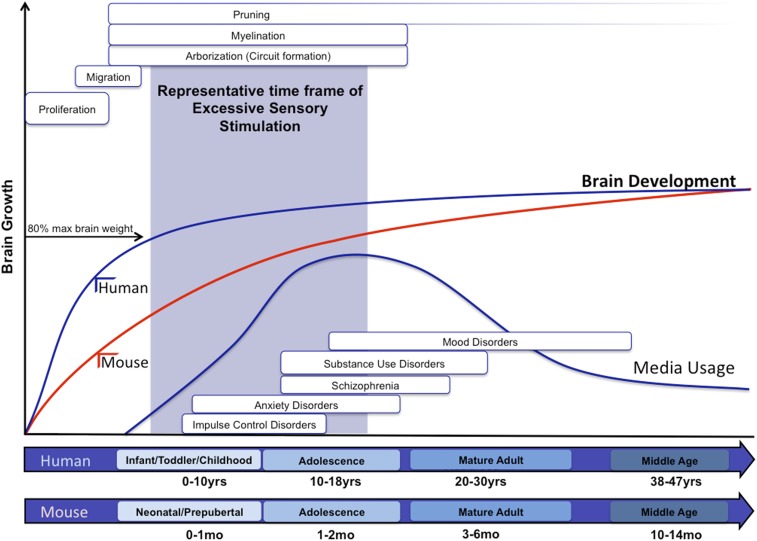
The fully developed and functional human brain takes more than 20 y to develop, and different areas have different developmental profiles (70–72), but the first few years of life are widely acknowledged to be the most crucial (73). The human brain triples in size in the first 3 y of life, a slope that is uniquely steep over the life span (Fig. 1). This initial phase of neurodevelopment is characterized by a proliferation phase that is associated with an increase in the number of neurons, spinogenesis, and dendritic and axonal growth (72, 74, 75). This initial phase leads to an overproduction of connections, and it is estimated that an infant has three times as many synapses as adolescents and adults (76). What follows is a “pruning” phase during which connectivity specializes: Connections that are functionally important are strengthened, while those that are not used are weakened (72). Various cellular mechanisms change in this biphasic manner. For example, the number of spines follows an inverted U-shaped trajectory, with a peak in spine density at the age of ∼3.5 mo. However, it is important to emphasize that all of these changes have different trajectories in different brain regions. These ultrastructural changes are complemented by dramatic changes in functional connectivity conditioned on their location (74, 77, 78). Insights into these changes have been gained through resting-state functional connectivity MRI (rs-fcMRI) studies. These studies investigate how fluctuations in the blood oxygen level-dependent signal of different regions of the brain are correlated with one another at rest, forming a number of specialized functional networks (e.g., the default mode network). Functional networks are differentially shaped in a region- and species-specific manner, involving the migration of neurons, myelination of axons, the formation of synapses, and the continuous synaptic pruning that occurs throughout the first 20 y of life (79, 80). The first structures to be functionally connected are primary sensorimotor and visual networks, while frontoparietal, executive control networks are still premature and form later during development (66). Early life functional network patterns are fundamentally distinct from adult networks and are reorganized throughout development (81–83). Developmental brain maturation trajectories are prominent enough to predict the age of an individual using patterns in rs-fcMRI (84).
Fig. 1.
Schematic illustrating the hypothesized relationship between human brain development and exposure to ESS. Typical cortical development involves proliferation, migration, arborization, and myelination. Proliferation and migration predominantly occur during prenatal stages, and arborization (circuit formation) and myelination continue through the first two postnatal decades. Synaptic pruning predominantly occurs in the early part of development but continues for years into adulthood. The brain grows drastically in size and complexity, which can be influenced by genetic and environmental factors. During these developmental stages, psychological disorders are developed. Media usage increases dramatically. The blue shaded area indicates a representative time frame in which these occurrences happen during mouse development, and when we used the ESS. Human and rodent (e.g., mouse and rat) developmental stages and corresponding time windows (years for humans and months for rodents) are represented in the x axis of the graph. Typical brain growth in weight is displayed for human (blue brain development line) and mice (red brain development line) in the y axis.
Of note, the trajectory of this brain development is profoundly influenced by experiences. Perhaps the most cited influence is the type of environment. Animals reared in a complex and interactive environment (the so-called “enriched environment”) show numerous changes in neurodevelopment compared with animals reared in an impoverished one devoid of social and environmental stimuli. The brain size, cortical thickness, complexity in dendritic branching, and spine density of animals exposed to an enriched environment are highly increased, as are the cognitive abilities (85–90). Furthermore, light tactile stimulation for the first 10–15 d of postnatal neurodevelopment results in significant changes in nervous system and behavior that are beneficial (91, 92). These changes are permanent, which is consistent with experiments performed by Mychasiuk et al. (93), indicating that enriched environment leads to a significant decrease in gene methylation in the frontal cortex and hippocampus, suggesting that early experiences result in epigenetic changes.
While the salubrious effects of stimulation on brain development and cognition are well established, what has not been adequately studied is to what extent the developing brain can be overstimulated. Is there good stimulation and bad stimulation? Can too much sensory stimulation during this complex process of neurodevelopment result in detrimental consequences?
Studying the Effects of Media Overuse
The pacing of shows designed for infants is extremely rapid compared with reality and even to shows designed for older children and adults (94). These formal features may be what keep infants engaged in the screen (27). Conceptually, this raises the concern that this excessive auditory and visual stimulation might condition the developing brain to expect an intensity of inputs that reality cannot provide, thus leading to inattention in later life. Put another way, is it possible that the highly interactive process of wiring the brain will adjust the sensory cortices to the fast-paced bombardment associated with some media? Moreover, will sensory overstimulation also affect other brain areas that are not directly affected by fast sensory stimulation? The “overstimulation hypothesis” was first tested in small experimental studies in the 1970s (95–97). The results were mixed with some finding the pacing of programs was associated with short-term deficits in attention while others did not. A more recent experimental study did find that a rapidly paced show (compared with a slowly paced one) diminished executive function at least briefly after viewing (98). There is increasing evidence that, in its extreme, excessive media usage can also lead to behavioral addiction, This seems to be particularly the case for internet gaming. Studies examining the neuronal consequences of internet gaming disorder reported numerous significant changes (99, 100), including alterations in resting-state EEG coherence (101), significant alterations in cortical thickness (102, 103), altered functional connectivity in the default mode network (104, 105), and significant associations with ADHD and other psychiatric disorders (106, 107). As they now stand, these findings are correlational and cannot establish causation. Nevertheless, exploring the consequences of excessive internet use and internet gaming has become a rapidly emerging field of research, with immense clinical and public health implications. However, all of these studies focus on preschoolers, school age children, and college students and therefore did not test the effects of viewing during the most critical window of brain development.
In a large observational study, we found that increased television viewing before the age of 3 was associated with increased risk of attentional problems at school age (21). Moreover, in a follow-up study, we found that the pacing of shows drove these effects with faster pacing having stronger associations with subsequent attentional problems (108). These findings may have important public health implications given that attentional capacity in early childhood is associated with improved outcomes in adulthood in several domains including the following: higher socioeconomic status, lower rates of substance use and incarceration, and lower divorce rates (2). Based on the existing literature, the American Academy of Pediatrics discourages television viewing before 2 y of age (109).
Developing an Animal Model for Excessive Rapidly Paced Media
All studies of infant television viewing and subsequent deficits in attention have been observational for logistical and ethical reasons, and although they controlled for many potential confounding factors, the possibility of residual confounding remains. Indeed, when trying to understand what aspects of electronic media use are detrimental and what aspects are beneficial, observational studies are inadequate to gain mechanistic insights into the potential contribution of these nongenetic factors to ADHD. Media use is an exceedingly complex stimulus. Dissecting its various components will be essential to understanding which aspects of it may be detrimental. One salient aspect of media is that it can deploy surreal pacing, producing scene changes that are unachievable in the “real” world, which raises important questions that need to be addressed mechanistically. Can the sheer sensory, nonnormative bombardment (i.e., sensory overstimulation) be sufficient to cause increased impulsivity, hyperactivity, or cognitive impairment? These questions can be mechanistically dissected and tested in animal models. Indeed, it took experimental proof in animal studies to convince the tobacco industry and politicians that cigarettes are highly addictive and cause lung cancer. At this point, we can only speculate that media use affects behavior, and any association with ADHD remains on relatively soft ground. Although the recent studies on internet gaming provide increasing evidence for a link between media use and ADHD as well as other neurological and psychiatric sequelae, the data are far from conclusive (110).
Consequently, we have developed a mouse model of what we have termed excessive sensory stimulation (ESS) to further advance the field, and dissect one particular aspect of media use. In humans, it will be impossible to isolate the “mere” sensory overstimulation aspect from cognitive involvement, and from the interactive potential of media use. Our model builds on the seminal studies in rodents that actively explored environmental influences on brain development and cognition. Rodents reared under enriched environmental conditions perform better on maze trials later in life (86, 87, 111). This improvement is associated with increased dendritic branching in the occipital and motor-sensory cortex (89, 111), increased size and complexity of the superior colliculus (89), and increased neurogenesis in the hippocampus (90, 112–116). Our research has tested the “opposite” hypothesis: that ESS during a similar period will subsequently diminish performance and adversely affect neurogenesis. In contrast to the enriched environment, ESS requires no active engagement, increased locomotor activity, or curiosity; it isolates the sheer bombardment of the senses from other aspects of media use. We brought a hypothesis based on observational studies in humans to the laboratory to experimentally validate it in a rodent model. While many studies test hypotheses in animal models and speculate about human implications, we in effect did the opposite.
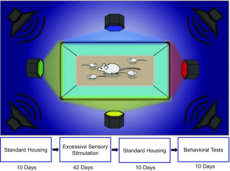
To test this condition, experimental mice received an ESS experience for 6 h per day for 42 d (Fig. 2). Speakers, connected to a precision amplification device, were mounted above standard mouse cages, and colored lights were positioned at all four walls. Audio from the “cartoon channel” was piped into the mouse cage at 70 dB. This level is typical for television watching and well below the 100–115 dB that are typically used for acoustic stress models in rodents (117, 118). A photorhythmic modulator was used to change colors and intensities in concordance with the audio, thereby simulating television that cannot be avoided (e.g., flashing lights on all four sides of the cage). We consider this excessive in the sense that it far exceeds any stimulation that mice would encounter under normative conditions in a vivarium of natural setting.
Fig. 2.
An illustration of the mouse excessive sensory stimulation (ESS) chamber and the experimental procedure.
Beginning at postnatal day 10 (P10), mice were divided into two groups: (i) The control group was reared according to approved and established protocols at the Seattle Children’s Research Institute vivarium. (ii) The experimental group was treated identically to the control group except that it was exposed to ESS for 6 h every night in the ESS chamber. Exposure lasted for 42 d, which is comparable to the length commonly used in enriched environment studies. Mice remained with their mother until weaning (P21) after which pups were housed in groups of up to five mice per cage. Following the exposure period, lights and speakers were removed, but mice remained in their familiar, regular mouse cages. Ten days later, mice were behaviorally tested using the light/dark latency test (LDL), the elevated plus maze (EPM), the open field test (OFT), the novel object recognition test (NORT), and the Barnes maze (BM). Table 1 summarizes the tests that were performed. In all cases, technicians blinded to research group made assessments.
Table 1.
Summary of behavioral tests performed on control and ESS mice
| Test name | Description |
| Light/dark latency test (LDL) | Mice are placed in light/dark box. Mouse behavior is tracked using VideoTrack (ViewPoint LS) to assess the time they spent in the light side of the chamber compared with the dark. |
| Elevated plus maze (EPM) | Mice are placed on EPM. Behavior is tracked to determine how much time they spent in the open arms compared with the closed. |
| Open-field test | Mice are placed in a large square box for 10 min. Behavior is tracked to determine how much time is spent on the inner edge of the box compared with the center of the chamber. Overall distance traveled in the chamber is also tracked. |
| Novel object recognition test | Mice are put in the same test apparatus as the open field. This time two identical objects are put in the chamber as well for an acquisition trial. After mice get familiarized with these objects, they are taken out for 1 h. One hour later, the mice were put back in the chamber for the test trial. For the test trial, one of the previous familiar objects remains in the chamber, but the other one is replaced with a new “novel” object. Time spent on each object is recorded. |
| Barnes maze | Mice are placed on a large elevated circle, which has 19 mock holes and 1 target hole, which leads to an escape hole underneath the table. Mice are placed in the middle of the circle and time to find the escape hole is measured. This is done for 4 training days. On the fifth day, the escape hole is blocked and number of pokes into the escape hole are measured. |
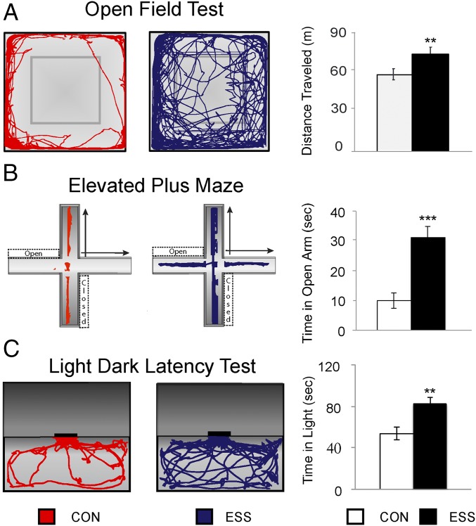
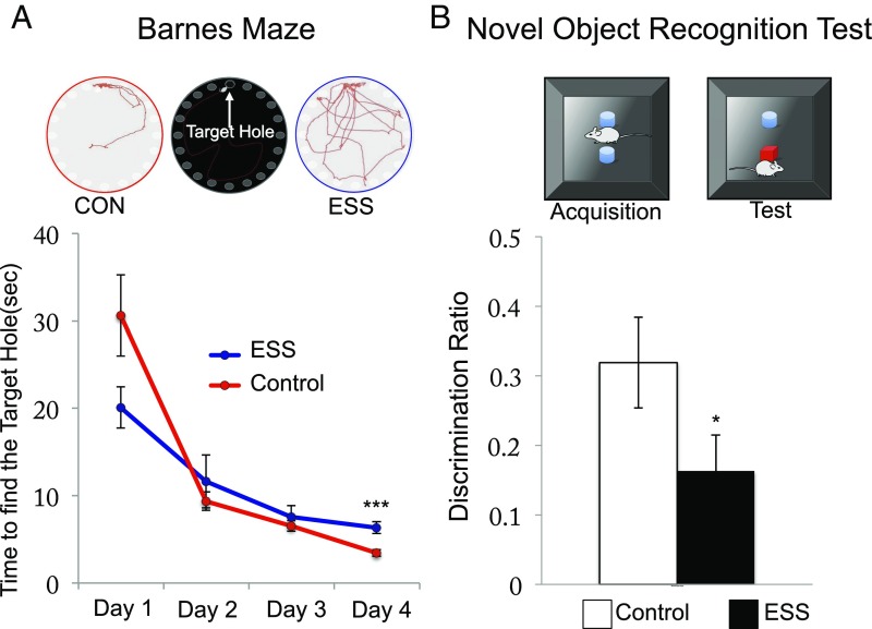
Interestingly, anxiety, learning, and memory were decreased, whereas risk-taking and motor activity levels were enhanced in ESS mice compared with controls (119) (Figs. 3 and 4). Specifically, ESS mice traveled greater distances in the open field, spent more time in the open on the elevated maze test, and spent more time in the lighted chamber compared with control mice. These findings can be interpreted as showing that mice are hyperkinetic and less risk averse. This is an important finding as it directly addresses the question raised earlier: Is the sensory overstimulation alone sufficient to have detrimental consequences resembling those of ADHD? Our data indeed suggest that, even without cognitive engagement and without social isolation, sensory overstimulation alone is sufficient to have detrimental consequences. Aside from numerous behavioral changes, we found significant neurobiological alterations in glutamatergic transmission in the nucleus accumbens and amygdala (120). A different group of investigators in Israel did a replication and extension study of our work (121). They exposed juvenile rats to 1 h daily of highly salient odors that were changed frequently, whereas control rats had consistent odors. They found that overstimulated rats performed more poorly on the five-choice serial reaction time task when auditory distractors were present. The five-choice serial reaction time task tests for impulsivity and attention and is considered analogous to the computer performance task used in humans to measure ADHD symptomology (122, 123). Notably, there findings are consistent with what is seen clinically in children with ADHD: They perform better when distractions are minimized. Importantly, while it can be argued that our ESS paradigm used types of stimulation that rodents would never encounter in the real world, Hadas et al. (124) used a paradigm of odors that in theory could exist in more naturalistic environments. This suggests that it is the intensity of the stimulation that drives the observed effects.
Fig. 3.
This figure highlights the results of (A) the open-field test (OFT), (B) elevated plus maze (EPM), and (C) light/dark latency (LDL) tests. A demonstrates illustrative examples of control (CON) (red) and excessive sensory stimulation (ESS) (blue) travel paths. These are quantified for each group indicating the overall distance traveled in the OFT [mean ± SEM; CON, 58.37 ± 1.94, n = 72; ESS, 66.12 ± 2.01; n = 72; t(142) = 2.62, P < 0.01]. B shows an illustrative example of the paths during the EPM for CON and ESS. Time spent in open arms is depicted in the bar graph [mean ± SEM; CON, 9.93 ± 2.11, n = 48; ESS, 31.03 ± 2.78; n = 61, t(105) = 3.39, P < 0.001]. C depicts the examples of CON and ESS path lengths during the LDL, and time in the light chamber is quantified in the bar graph [mean ± SEM; CON, 53.79 ± 4.17, n = 48; ESS, 82.39 ± 6.41, n = 61; t(105) = 2.62, P < 0.01]. Error bars in graphs represent the SEM of variability within each group. Note: **P < 0.01; ***P < 0.001. Adapted with permission from ref. 119.
Fig. 4.
Results of (A) Barnes maze (BM) test and (B) novel object recognition test (NORT). A shows differences in search strategies on the test day of the BM for control (CON) (red) and excessive sensory stimulation (ESS) (blue). Learning throughout the 4-d training trials is depicted in terms of the time to find the target hole for CON and ESS. ESS mice trend toward finding the target hole faster than CON on day 1 (effect of hyperactivity) [mean ± SEM; CON, 30.60 ± 4.64, n = 12; ESS, 20.08 ± 2.35, n = 10; t(16) = 1.80, P < 0.09] but spent significantly more time on day 4 to find the target hole [mean ± SEM; CON, 3.44 ± 0.39, n = 12; ESS, 6.33 ± 0.67, n = 10; t(15) = 5.24, P < 0.001]. B illustrates the NORT and the results of the discrimination ratio on the test trial. The discrimination ratio was calculated as follows: (time spent on the novel object – time spent on the familiar object)/total time. ESS mice spent less time with the novel object compared with CON [mean ± SEM; CON, 0.32 ± 0.07, n = 39; ESS, 0.16 ± 0.05; n = 42; t(70) = 1.99, P < 0.05]. Error bars in graphs represent the SEM of variability within each group. Significance was determined using a two-tailed t test. Note: *P < 0.05; ***P < 0.001. Adapted with permission from ref. 119.
These results provide experimental corroboration of observational data in humans. Indeed, this was hypothesis-driven research confirming findings from children in rodents rather than the reverse as is frequently the case. Indeed, it is important to note that the arguably most frequently used rodent model for ADHD, the so called hypertensive rat, was not created in a hypothesis-driven and/or mechanistic manner; rather the behavioral traits seen in this rodent were considered “ADHD-like.” (125–129) This means that the behavioral phenotype of these rats could be caused by a myriad of mechanisms that may or may not be relevant for understanding the etiology of ADHD. Nevertheless, as is the case for all animal models, the differences (and similarities) between what is observed in human infants and what is induced in rodents are worth considering in some detail.
First, there is the issue of cognitive engagement. It is unclear how much cognitive engagement infants have while watching television (130), but it is highly probable that mice have little to none when experiencing ESS. Thus, our findings cannot speak to the role of cognitive engagement itself. Instead, our findings support a much less intuitive but more important hypothesis: that the formal features of the medium are what present a risk, thereby making even potentially educational programming detrimental, not because of the content but because it overstimulates too many senses for too long too early in development. In other words, ESS, in and of itself and independent of any cognitive content, suffices to have significant behavioral and neurobiological consequences. This phenomenon has been observed in humans where even programs that have demonstrable educational benefits in preschoolers (e.g., Sesame Street) have been shown to result in decreased language when viewed by infants (131).
Second, some might argue that the control condition does not represent “normal” mouse developmental exposures since laboratory conditions are clearly different from what would be encountered in natural habitats. We propose two counterfactuals to this. The work on enriched environments that has been so highly impactful also used a similar control group (88, 115). In addition, laboratory-reared mice are not offered unlimited terrain to cover or predatory threats to avoid. Seen in this light, our findings are notable in that overstimulated mice have outcomes that are consistently worse than those reared in what might be deemed an understimulating one. In other words, relative sensory deprivation is better than sensory overload. Future experiments should build on these findings and directly compare ESS with EE mice.
Last, it might be argued that our findings are confounded by stress. While it should be noted that the observations in humans might also be induced by stress as isolating infants and bombarding their audio and visual senses may well be stressful, we do not believe that is operative in our model for several reasons. We have opted to keep the pups with their mother before weaning to minimize stress related to separation. In addition, the audio levels we use (70 dB) are well below acoustic stress levels (100–115 dB) for mice (132). Furthermore, our finding of increased risk taking (decreased anxiety) runs contrary to what has been found in stress models where increased anxiety has been repeatedly demonstrated (133–137). Moreover, stress alters appetite and weight gain, and there were no differences in body weights between mice exposed to the ESS paradigm and controls; finally, we have measured cortisol levels in experimental and control animals and found no significant differences (120).
In summary, our observations in humans have been at least provisionally confirmed in experimental studies in mice. ESS early in life can negatively impact cognitive function and behavior. These findings support the American Academy of Pediatrics recommendation that screen time—particularly when it involves fast-paced media—should be minimized for children under 2 (138). However, it should be noted that the age of 2 y is arbitrary, and given evidence that brain development continues until the early 20s, further research should be conducted to better clarify potential impacts throughout the pediatric life span.
Footnotes
The authors declare no conflict of interest.
This paper results from the Arthur M. Sackler Colloquium of the National Academy of Sciences, “Digital Media and Developing Minds,” held, October 14–16, 2015, at the Arnold and Mabel Beckman Center of the National Academies of Sciences and Engineering in Irvine, CA. The complete program and video recordings of most presentations are available on the NAS website at www.nasonline.org/Digital_Media_and_Developing_Minds.
This article is a PNAS Direct Submission.
References
- 1.Akinbami LJ, Liu X, Pastor PN, Reuben CA. Attention deficit hyperactivity disorder among children aged 5–17 years in the United States, 1998–2009. NCHS Data Brief. 2011;2011:1–8. [PubMed] [Google Scholar]
- 2.Moffitt TE, et al. A gradient of childhood self-control predicts health, wealth, and public safety. Proc Natl Acad Sci USA. 2011;108:2693–2698. doi: 10.1073/pnas.1010076108. [DOI] [PMC free article] [PubMed] [Google Scholar]
- 3.Christakis DA. Rethinking attention-deficit/hyperactivity disorder. JAMA Pediatr. 2016;170:109–110. doi: 10.1001/jamapediatrics.2015.3372. [DOI] [PubMed] [Google Scholar]
- 4.Cantwell DP. Attention deficit disorder: A review of the past 10 years. J Am Acad Child Adolesc Psychiatry. 1996;35:978–987. doi: 10.1097/00004583-199608000-00008. [DOI] [PubMed] [Google Scholar]
- 5.Reiff MI, Stein MT. Attention-deficit/hyperactivity disorder: Diagnosis and treatment. Adv Pediatr. 2004;51:289–327. [PubMed] [Google Scholar]
- 6.Hechtman L. Developmental, neurobiological, and psychosocial aspects of hyperactivity, impulsivity, and attention. In: Lewis M, editor. Child and Adolescent Psychiatry: A Comprehensive Textbook. 3rd Ed. Lippincott Williams and Wilkins; Philadelphia: 2002. pp. 366–387. [Google Scholar]
- 7.Acosta MT, Arcos-Burgos M, Muenke M. Attention deficit/hyperactivity disorder (ADHD): Complex phenotype, simple genotype? Genet Med. 2004;6:1–15. doi: 10.1097/01.gim.0000110413.07490.0b. [DOI] [PubMed] [Google Scholar]
- 8.Kent L. Recent advances in the genetics of attention deficit hyperactivity disorder. Curr Psychiatry Rep. 2004;6:143–148. doi: 10.1007/s11920-004-0054-4. [DOI] [PubMed] [Google Scholar]
- 9.Faraone SV, Biederman J. Nature, nurture, and attention deficit hyperactivity disorder. Dev Rev. 2000;20:568–581. [Google Scholar]
- 10.Jensen PS. ADHD: Current concepts on etiology, pathophysiology, and neurobiology. Child Adolesc Psychiatr Clin N Am. 2000;9:557–572, vii–viii. [PubMed] [Google Scholar]
- 11.Shastry BS. Molecular genetics of attention-deficit hyperactivity disorder (ADHD): An update. Neurochem Int. 2004;44:469–474. doi: 10.1016/j.neuint.2003.08.011. [DOI] [PubMed] [Google Scholar]
- 12.Barkley RA. International consensus statement on ADHD. J Am Acad Child Adolesc Psychiatry. 2002;41:1389. doi: 10.1097/00004583-200212000-00001. [DOI] [PubMed] [Google Scholar]
- 13.US Department of Health and Human Services . Mental Health: A Report of the Surgeon General. US Department of Health and Human Services, Substance Abuse and Mental Health Services Administration, National Institute of Mental Health; Rockville, MD: 1999. [Google Scholar]
- 14.Campbell SB. Attention-deficit/hyperactivity disorder: A developmental view. In: Sameroff AJ, Lewis M, Miller SM, editors. Handbook of Developmental Psychopathology. 2nd Ed. Kluwer Academic/Plenum Publishers; New York: 2000. pp. 383–401. [Google Scholar]
- 15.Glover V, O’Connor TG. Effects of antenatal stress and anxiety: Implications for development and psychiatry. Br J Psychiatry. 2002;180:389–391. doi: 10.1192/bjp.180.5.389. [DOI] [PubMed] [Google Scholar]
- 16.Campbell SB, Breaux AM, Ewing LJ, Szumowski EK. Correlates and predictors of hyperactivity and aggression: A longitudinal study of parent-referred problem preschoolers. J Abnorm Child Psychol. 1986;14:217–234. doi: 10.1007/BF00915442. [DOI] [PubMed] [Google Scholar]
- 17.Jacobvitz D, Sroufe LA. The early caregiver-child relationship and attention-deficit disorder with hyperactivity in kindergarten: A prospective study. Child Dev. 1987;58:1496–1504. doi: 10.1111/j.1467-8624.1987.tb03862.x. [DOI] [PubMed] [Google Scholar]
- 18.Rose Krasnor L, Rubin KH, Booth CL, Coplan R. The relation of maternal directiveness and child attachment security to social competence in preschoolers. Int J Behav Dev. 1996;19:309–325. [Google Scholar]
- 19.Shaw DS, Owens EB, Giovannelli J, Winslow EB. Infant and toddler pathways leading to early externalizing disorders. J Am Acad Child Adolesc Psychiatry. 2001;40:36–43. doi: 10.1097/00004583-200101000-00014. [DOI] [PubMed] [Google Scholar]
- 20.Stiefel I. Can disturbance in attachment contribute to attention deficit hyperactivity disorder? A case discussion. Clin Child Psychol Psychiatry. 1997;2:45–64. [Google Scholar]
- 21.Christakis DA, Zimmerman FJ, DiGiuseppe DL, McCarty CA. Early television exposure and subsequent attentional problems in children. Pediatrics. 2004;113:708–713. doi: 10.1542/peds.113.4.708. [DOI] [PubMed] [Google Scholar]
- 22.Rodriguez A. Maternal pre-pregnancy obesity and risk for inattention and negative emotionality in children. J Child Psychol Psychiatry. 2010;51:134–143. doi: 10.1111/j.1469-7610.2009.02133.x. [DOI] [PubMed] [Google Scholar]
- 23.Rodriguez A, et al. Maternal adiposity prior to pregnancy is associated with ADHD symptoms in offspring: Evidence from three prospective pregnancy cohorts. Int J Obes. 2008;32:550–557. doi: 10.1038/sj.ijo.0803741. [DOI] [PubMed] [Google Scholar]
- 24.Buss C, et al. Impaired executive function mediates the association between maternal pre-pregnancy body mass index and child ADHD symptoms. PLoS One. 2012;7:e37758. doi: 10.1371/journal.pone.0037758. [DOI] [PMC free article] [PubMed] [Google Scholar]
- 25.Chen Q, et al. Maternal pre-pregnancy body mass index and offspring attention deficit hyperactivity disorder: A population-based cohort study using a sibling-comparison design. Int J Epidemiol. 2014;43:83–90. doi: 10.1093/ije/dyt152. [DOI] [PMC free article] [PubMed] [Google Scholar]
- 26.Rivera HM, Christiansen KJ, Sullivan EL. The role of maternal obesity in the risk of neuropsychiatric disorders. Front Neurosci. 2015;9:194. doi: 10.3389/fnins.2015.00194. [DOI] [PMC free article] [PubMed] [Google Scholar]
- 27.Christakis DA, Zimmerman FJ. The Elephant in the Living Room: Make Television Work for Your Kids. Rodale; Emmaus, PA: 2006. [Google Scholar]
- 28.Zimmerman FJ, Christakis DA, Meltzoff AN. Television and DVD/video viewing in children younger than 2 years. Arch Pediatr Adolesc Med. 2007;161:473–479. doi: 10.1001/archpedi.161.5.473. [DOI] [PubMed] [Google Scholar]
- 29.Garrison M, Christakis D. A Teacher in the Living Room?: Educational Media for Babies, Toddlers, and Preschoolers. Kaiser Family Foundation; Menlo Park, CA: 2005. [Google Scholar]
- 30.Tong L, Xiong X, Tan H. Attention-deficit/hyperactivity disorder and lifestyle-related behaviors in children. PLoS One. 2016;11:e0163434. doi: 10.1371/journal.pone.0163434. [DOI] [PMC free article] [PubMed] [Google Scholar]
- 31.Montagni I, Guichard E, Kurth T. Association of screen time with self-perceived attention problems and hyperactivity levels in French students: A cross-sectional study. BMJ Open. 2016;6:e009089. doi: 10.1136/bmjopen-2015-009089. [DOI] [PMC free article] [PubMed] [Google Scholar]
- 32.Lagercrantz H. Connecting the brain of the child from synapses to screen-based activity. Acta Paediatr. 2016;105:352–357. doi: 10.1111/apa.13298. [DOI] [PubMed] [Google Scholar]
- 33.Goodhill GJ. Can molecular gradients wire the brain? Trends Neurosci. 2016;39:202–211. doi: 10.1016/j.tins.2016.01.009. [DOI] [PubMed] [Google Scholar]
- 34.Hassan BA, Hiesinger PR. Beyond molecular codes: Simple rules to wire complex brains. Cell. 2015;163:285–291. doi: 10.1016/j.cell.2015.09.031. [DOI] [PMC free article] [PubMed] [Google Scholar]
- 35.Sur M, Rubenstein JL. Patterning and plasticity of the cerebral cortex. Science. 2005;310:805–810. doi: 10.1126/science.1112070. [DOI] [PubMed] [Google Scholar]
- 36.McConnell MJ, et al. Intersection of diverse neuronal genomes and neuropsychiatric disease: The brain somatic mosaicism network. Science. 2017;356:eaal1641. doi: 10.1126/science.aal1641. [DOI] [PMC free article] [PubMed] [Google Scholar]
- 37.Schotzinger R, Yin X, Landis S. Target determination of neurotransmitter phenotype in sympathetic neurons. J Neurobiol. 1994;25:620–639. doi: 10.1002/neu.480250605. [DOI] [PubMed] [Google Scholar]
- 38.Glebova NO, Ginty DD. Growth and survival signals controlling sympathetic nervous system development. Annu Rev Neurosci. 2005;28:191–222. doi: 10.1146/annurev.neuro.28.061604.135659. [DOI] [PubMed] [Google Scholar]
- 39.Hao MM, et al. The emergence of neural activity and its role in the development of the enteric nervous system. Dev Biol. 2013;382:365–374. doi: 10.1016/j.ydbio.2012.12.006. [DOI] [PubMed] [Google Scholar]
- 40.Momose-Sato Y, Sato K. The embryonic brain and development of vagal pathways. Respir Physiol Neurobiol. 2011;178:163–173. doi: 10.1016/j.resp.2011.01.012. [DOI] [PubMed] [Google Scholar]
- 41.Dasen JS. Transcriptional networks in the early development of sensory-motor circuits. Curr Top Dev Biol. 2009;87:119–148. doi: 10.1016/S0070-2153(09)01204-6. [DOI] [PubMed] [Google Scholar]
- 42.Leighton AH, Lohmann C. The wiring of developing sensory circuits-from patterned spontaneous activity to synaptic plasticity mechanisms. Front Neural Circuits. 2016;10:71. doi: 10.3389/fncir.2016.00071. [DOI] [PMC free article] [PubMed] [Google Scholar]
- 43.Vitali I, Jabaudon D. Synaptic biology of barrel cortex circuit assembly. Semin Cell Dev Biol. 2014;35:156–164. doi: 10.1016/j.semcdb.2014.07.009. [DOI] [PubMed] [Google Scholar]
- 44.Ezkurdia I, et al. Multiple evidence strands suggest that there may be as few as 19,000 human protein-coding genes. Hum Mol Genet. 2014;23:5866–5878. doi: 10.1093/hmg/ddu309. [DOI] [PMC free article] [PubMed] [Google Scholar]
- 45.Hodgkin J. What does a worm want with 20,000 genes? Genome Biol. 2001;2:COMMENT2008. doi: 10.1186/gb-2001-2-11-comment2008. [DOI] [PMC free article] [PubMed] [Google Scholar]
- 46.Prevedel R, et al. Simultaneous whole-animal 3D imaging of neuronal activity using light-field microscopy. Nat Methods. 2014;11:727–730. doi: 10.1038/nmeth.2964. [DOI] [PMC free article] [PubMed] [Google Scholar]
- 47.Kaletta T, Hengartner MO. Finding function in novel targets: C. elegans as a model organism. Nat Rev Drug Discov. 2006;5:387–398. doi: 10.1038/nrd2031. [DOI] [PubMed] [Google Scholar]
- 48.Maffei A, Turrigiano G. The age of plasticity: Developmental regulation of synaptic plasticity in neocortical microcircuits. Prog Brain Res. 2008;169:211–223. doi: 10.1016/S0079-6123(07)00012-X. [DOI] [PubMed] [Google Scholar]
- 49.Turrigiano G. Too many cooks? Intrinsic and synaptic homeostatic mechanisms in cortical circuit refinement. Annu Rev Neurosci. 2011;34:89–103. doi: 10.1146/annurev-neuro-060909-153238. [DOI] [PubMed] [Google Scholar]
- 50.Turrigiano GG. The dialectic of Hebb and homeostasis. Philos Trans R Soc Lond B Biol Sci. 2017;372:20160258. doi: 10.1098/rstb.2016.0258. [DOI] [PMC free article] [PubMed] [Google Scholar]
- 51.Turrigiano GG, Nelson SB. Homeostatic plasticity in the developing nervous system. Nat Rev Neurosci. 2004;5:97–107. doi: 10.1038/nrn1327. [DOI] [PubMed] [Google Scholar]
- 52.Tyborowska A, et al. Early-life and pubertal stress differentially modulate grey matter development in human adolescents. Sci Rep. 2018;8:9201. doi: 10.1038/s41598-018-27439-5. [DOI] [PMC free article] [PubMed] [Google Scholar]
- 53.Miranda A, Sousa N. Maternal hormonal milieu influence on fetal brain development. Brain Behav. 2018;8:e00920. doi: 10.1002/brb3.920. [DOI] [PMC free article] [PubMed] [Google Scholar]
- 54.Geary DC. Evolutionary perspective on sex differences in the expression of neurological diseases. Prog Neurobiol. June 8, 2018 doi: 10.1016/j.pneurobio.2018.06.001. [DOI] [PubMed] [Google Scholar]
- 55.Singh G, Singh V, Sobolewski M, Cory-Slechta DA, Schneider JS. Sex-dependent effects of developmental lead exposure on the brain. Front Genet. 2018;9:89. doi: 10.3389/fgene.2018.00089. [DOI] [PMC free article] [PubMed] [Google Scholar]
- 56.Manto M, Perrotta G. Toxic-induced cerebellar syndrome: From the fetal period to the elderly. Handb Clin Neurol. 2018;155:333–352. doi: 10.1016/B978-0-444-64189-2.00022-6. [DOI] [PubMed] [Google Scholar]
- 57.Eskenazi B, et al. Prenatal exposure to DDT and pyrethroids for malaria control and child neurodevelopment: The VHEMBE cohort, South Africa. Environ Health Perspect. 2018;126:047004. doi: 10.1289/EHP2129. [DOI] [PMC free article] [PubMed] [Google Scholar]
- 58.Ramel SE, Georgieff MK. Preterm nutrition and the brain. World Rev Nutr Diet. 2014;110:190–200. doi: 10.1159/000358467. [DOI] [PubMed] [Google Scholar]
- 59.Gabbianelli R, Damiani E. Epigenetics and neurodegeneration: Role of early-life nutrition. J Nutr Biochem. 2018;57:1–13. doi: 10.1016/j.jnutbio.2018.01.014. [DOI] [PubMed] [Google Scholar]
- 60.Hertz-Picciotto I, Schmidt RJ, Krakowiak P. Understanding environmental contributions to autism: Causal concepts and the state of science. Autism Res. 2018;11:554–586. doi: 10.1002/aur.1938. [DOI] [PubMed] [Google Scholar]
- 61.Goldfeld S, et al. More than a snapshot in time: Pathways of disadvantage over childhood. Int J Epidemiol. June 5, 2018 doi: 10.1093/ije/dyy086. [DOI] [PubMed] [Google Scholar]
- 62.Liu S, et al. Effects of early comprehensive interventions on child neurodevelopment in poor rural areas of China: A moderated mediation analysis. Public Health. 2018;159:116–122. doi: 10.1016/j.puhe.2018.02.010. [DOI] [PubMed] [Google Scholar]
- 63.Jobe EM, Zhao X. DNA methylation and adult neurogenesis. Brain Plast. 2017;3:5–26. doi: 10.3233/BPL-160034. [DOI] [PMC free article] [PubMed] [Google Scholar]
- 64.Berson A, Nativio R, Berger SL, Bonini NM. Epigenetic regulation in neurodegenerative diseases. Trends Neurosci. 2018;41:587–598. doi: 10.1016/j.tins.2018.05.005. [DOI] [PMC free article] [PubMed] [Google Scholar]
- 65.Insel TR. Rethinking schizophrenia. Nature. 2010;468:187–193. doi: 10.1038/nature09552. [DOI] [PubMed] [Google Scholar]
- 66.Gao W, Lin W, Grewen K, Gilmore JH. Functional connectivity of the infant human brain: Plastic and modifiable. Neuroscientist. February 29, 2016 doi: 10.1177/1073858416635986. [DOI] [PMC free article] [PubMed] [Google Scholar]
- 67.Geschwind DH, Levitt P. Autism spectrum disorders: Developmental disconnection syndromes. Curr Opin Neurobiol. 2007;17:103–111. doi: 10.1016/j.conb.2007.01.009. [DOI] [PubMed] [Google Scholar]
- 68.Yaron A, Zheng B. Navigating their way to the clinic: Emerging roles for axon guidance molecules in neurological disorders and injury. Dev Neurobiol. 2007;67:1216–1231. doi: 10.1002/dneu.20512. [DOI] [PubMed] [Google Scholar]
- 69.Stoeckli ET. What does the developing brain tell us about neural diseases? Eur J Neurosci. 2012;35:1811–1817. doi: 10.1111/j.1460-9568.2012.08171.x. [DOI] [PubMed] [Google Scholar]
- 70.van Dyck LI, Morrow EM. Genetic control of postnatal human brain growth. Curr Opin Neurol. 2017;30:114–124. doi: 10.1097/WCO.0000000000000405. [DOI] [PMC free article] [PubMed] [Google Scholar]
- 71.Collin G, van den Heuvel MP. The ontogeny of the human connectome: Development and dynamic changes of brain connectivity across the life span. Neuroscientist. 2013;19:616–628. doi: 10.1177/1073858413503712. [DOI] [PubMed] [Google Scholar]
- 72.Elston GN, Fujita I. Pyramidal cell development: Postnatal spinogenesis, dendritic growth, axon growth, and electrophysiology. Front Neuroanat. 2014;8:78. doi: 10.3389/fnana.2014.00078. [DOI] [PMC free article] [PubMed] [Google Scholar]
- 73.Institute of Medicine . From Neurons to Neighborhoods: The Science of Early Childhood Development. National Academy Press; Washington, DC: 2000. [PubMed] [Google Scholar]
- 74.Huttenlocher PR. Basic neuroscience research has important implications for child development. Nat Neurosci. 2003;6:541. doi: 10.1038/nn0603-541. [DOI] [PubMed] [Google Scholar]
- 75.Huttenlocher PR, Dabholkar AS. Regional differences in synaptogenesis in human cerebral cortex. J Comp Neurol. 1997;387:167–178. doi: 10.1002/(sici)1096-9861(19971020)387:2<167::aid-cne1>3.0.co;2-z. [DOI] [PubMed] [Google Scholar]
- 76.Kolb B, Mychasiuk R, Gibb R. Brain development, experience, and behavior. Pediatr Blood Cancer. 2014;61:1720–1723. doi: 10.1002/pbc.24908. [DOI] [PubMed] [Google Scholar]
- 77.Hagmann P, et al. White matter maturation reshapes structural connectivity in the late developing human brain. Proc Natl Acad Sci USA. 2010;107:19067–19072. doi: 10.1073/pnas.1009073107. [DOI] [PMC free article] [PubMed] [Google Scholar]
- 78.Hagmann P, et al. Mapping the structural core of human cerebral cortex. PLoS Biol. 2008;6:e159. doi: 10.1371/journal.pbio.0060159. [DOI] [PMC free article] [PubMed] [Google Scholar]
- 79.Huttenlocher PR. Synaptic density in human frontal cortex–Ddevelopmental changes and effects of aging. Brain Res. 1979;163:195–205. doi: 10.1016/0006-8993(79)90349-4. [DOI] [PubMed] [Google Scholar]
- 80.Fair DA, et al. The maturing architecture of the brain’s default network. Proc Natl Acad Sci USA. 2008;105:4028–4032. doi: 10.1073/pnas.0800376105. [DOI] [PMC free article] [PubMed] [Google Scholar]
- 81.Elston GN. Cortex, cognition and the cell: New insights into the pyramidal neuron and prefrontal function. Cereb Cortex. 2003;13:1124–1138. doi: 10.1093/cercor/bhg093. [DOI] [PubMed] [Google Scholar]
- 82.Power JD, Fair DA, Schlaggar BL, Petersen SE. The development of human functional brain networks. Neuron. 2010;67:735–748. doi: 10.1016/j.neuron.2010.08.017. [DOI] [PMC free article] [PubMed] [Google Scholar]
- 83.Elston GN. Cortical heterogeneity: Implications for visual processing and polysensory integration. J Neurocytol. 2002;31:317–335. doi: 10.1023/a:1024182228103. [DOI] [PubMed] [Google Scholar]
- 84.Dosenbach NU, et al. Prediction of individual brain maturity using fMRI. Science. 2010;329:1358–1361. doi: 10.1126/science.1194144. [DOI] [PMC free article] [PubMed] [Google Scholar]
- 85.Greenough A, Nicolaides KH, Lagercrantz H. Human fetal sympathoadrenal responsiveness. Early Hum Dev. 1990;23:9–13. doi: 10.1016/0378-3782(90)90124-2. [DOI] [PubMed] [Google Scholar]
- 86.Greenough WT, Black JE, Wallace CS. Experience and brain development. Child Dev. 1987;58:539–559. [PubMed] [Google Scholar]
- 87.Greenough WT, Juraska JM, Volkmar FR. Maze training effects on dendritic branching in occipital cortex of adult rats. Behav Neural Biol. 1979;26:287–297. doi: 10.1016/s0163-1047(79)91278-0. [DOI] [PubMed] [Google Scholar]
- 88.Greenough WT, Madden TC, Fleischmann TB. Effects of isolation, daily handling, and enriched rearing on maze learning. Psychon Sci. 1972;27:279–280. [Google Scholar]
- 89.Fuchs JL, Montemayor M, Greenough WT. Effect of environmental complexity on size of the superior colliculus. Behav Neural Biol. 1990;54:198–203. doi: 10.1016/0163-1047(90)91422-8. [DOI] [PubMed] [Google Scholar]
- 90.Kuzumaki N, et al. Hippocampal epigenetic modification at the brain-derived neurotrophic factor gene induced by an enriched environment. Hippocampus. 2011;21:127–132. doi: 10.1002/hipo.20775. [DOI] [PubMed] [Google Scholar]
- 91.Kolb B, Gibb R. Tactile stimulation after frontal or parietal cortical injury in infant rats facilitates functional recovery and produces synaptic changes in adjacent cortex. Behav Brain Res. 2010;214:115–120. doi: 10.1016/j.bbr.2010.04.024. [DOI] [PubMed] [Google Scholar]
- 92.Richards S, Mychasiuk R, Kolb B, Gibb R. Tactile stimulation during development alters behaviour and neuroanatomical organization of normal rats. Behav Brain Res. 2012;231:86–91. doi: 10.1016/j.bbr.2012.02.043. [DOI] [PubMed] [Google Scholar]
- 93.Mychasiuk R, et al. Parental enrichment and offspring development: Modifications to brain, behavior and the epigenome. Behav Brain Res. 2012;228:294–298. doi: 10.1016/j.bbr.2011.11.036. [DOI] [PubMed] [Google Scholar]
- 94.Goodrich SA, Pempek TA, Calvert SL. Formal production features of infant and toddler DVDs. Arch Pediatr Adolesc Med. 2009;163:1151–1156. doi: 10.1001/archpediatrics.2009.201. [DOI] [PubMed] [Google Scholar]
- 95.Geist EA, Gibson M. The effect of network and public television programs on four and five year olds ability to attend to educational tasks. J Instr Psychol. 2000;27:250–261. [Google Scholar]
- 96.Anderson DR, Levin SR, Lorch EP. The effects of TV program pacing on the behavior of preschool children. AV Commun Rev. 1977;25:159–166. [Google Scholar]
- 97.Friedrich LK, Stein AH. Aggressive and prosocial television programs and the natural behavior of preschool children. Monogr Soc Res Child Dev. 1973;38:1–64. [PubMed] [Google Scholar]
- 98.Lillard AS, Peterson J. The immediate impact of different types of television on young children’s executive function. Pediatrics. 2011;128:644–649. doi: 10.1542/peds.2010-1919. [DOI] [PMC free article] [PubMed] [Google Scholar]
- 99.Kuss DJ, Pontes HM, Griffiths MD. Neurobiological correlates in internet gaming disorder: A systematic literature review. Front Psychiatry. 2018;9:166. doi: 10.3389/fpsyt.2018.00166. [DOI] [PMC free article] [PubMed] [Google Scholar]
- 100.Sussman CJ, Harper JM, Stahl JL, Weigle P. Internet and video game addictions: Diagnosis, epidemiology, and neurobiology. Child Adolesc Psychiatr Clin N Am. 2018;27:307–326. doi: 10.1016/j.chc.2017.11.015. [DOI] [PubMed] [Google Scholar]
- 101.Park S, et al. Longitudinal changes in neural connectivity in patients with internet gaming disorder: A resting-state EEG coherence study. Front Psychiatry. 2018;9:252. doi: 10.3389/fpsyt.2018.00252. [DOI] [PMC free article] [PubMed] [Google Scholar]
- 102.Wang Z, et al. Cortical thickness and volume abnormalities in internet gaming disorder: Evidence from comparison of recreational internet game users. Eur J Neurosci. June 8, 2018 doi: 10.1111/ejn.13987. [DOI] [PubMed] [Google Scholar]
- 103.Pan N, et al. Brain structures associated with internet addiction tendency in adolescent online game players. Front Psychiatry. 2018;9:67. doi: 10.3389/fpsyt.2018.00067. [DOI] [PMC free article] [PubMed] [Google Scholar]
- 104.Hong JS, Kim SM, Bae S, Han DH. Impulsive internet game play is associated with increased functional connectivity between the default mode and salience networks in depressed patients with short allele of serotonin transporter gene. Front Psychiatry. 2018;9:125. doi: 10.3389/fpsyt.2018.00125. [DOI] [PMC free article] [PubMed] [Google Scholar]
- 105.Dong G, Wang Z, Wang Y, Du X, Potenza MN. Gender-related functional connectivity and craving during gaming and immediate abstinence during a mandatory break: Implications for development and progression of internet gaming disorder. Prog Neuropsychopharmacol Biol Psychiatry. 2018;88:1–10. doi: 10.1016/j.pnpbp.2018.04.009. [DOI] [PubMed] [Google Scholar]
- 106.Lee D, Namkoong K, Lee J, Jung YC. Preliminary evidence of altered gray matter volume in subjects with internet gaming disorder: Associations with history of childhood attention-deficit/hyperactivity disorder symptoms. Brain Imaging Behav. May 11, 2018 doi: 10.1007/s11682-018-9872-6. [DOI] [PubMed] [Google Scholar]
- 107.Liu L, et al. The comorbidity between internet gaming disorder and depression: Interrelationship and neural mechanisms. Front Psychiatry. 2018;9:154. doi: 10.3389/fpsyt.2018.00154. [DOI] [PMC free article] [PubMed] [Google Scholar]
- 108.Zimmerman FJ, Christakis DA. Associations between content types of early media exposure and subsequent attentional problems. Pediatrics. 2007;120:986–992. doi: 10.1542/peds.2006-3322. [DOI] [PubMed] [Google Scholar]
- 109.Council on Communications and Media Media and young minds. Pediatrics. 2016;138:e20162591. doi: 10.1542/peds.2016-2591. [DOI] [PubMed] [Google Scholar]
- 110.van Rooij AJ, et al. A weak scientific basis for gaming disorder: Let us err on the side of caution. J Behav Addict. 2018;7:1–9. doi: 10.1556/2006.7.2018.19. [DOI] [PMC free article] [PubMed] [Google Scholar]
- 111.Greenough WT, Larson JR, Withers GS. Effects of unilateral and bilateral training in a reaching task on dendritic branching of neurons in the rat motor-sensory forelimb cortex. Behav Neural Biol. 1985;44:301–314. doi: 10.1016/s0163-1047(85)90310-3. [DOI] [PubMed] [Google Scholar]
- 112.Choi SH, et al. Non-cell-autonomous effects of presenilin 1 variants on enrichment-mediated hippocampal progenitor cell proliferation and differentiation. Neuron. 2008;59:568–580. doi: 10.1016/j.neuron.2008.07.033. [DOI] [PMC free article] [PubMed] [Google Scholar]
- 113.Schloesser RJ, Lehmann M, Martinowich K, Manji HK, Herkenham M. Environmental enrichment requires adult neurogenesis to facilitate the recovery from psychosocial stress. Mol Psychiatry. 2010;15:1152–1163. doi: 10.1038/mp.2010.34. [DOI] [PMC free article] [PubMed] [Google Scholar]
- 114.Goshen I, et al. Environmental enrichment restores memory functioning in mice with impaired IL-1 signaling via reinstatement of long-term potentiation and spine size enlargement. J Neurosci. 2009;29:3395–3403. doi: 10.1523/JNEUROSCI.5352-08.2009. [DOI] [PMC free article] [PubMed] [Google Scholar]
- 115.Nithianantharajah J, Hannan AJ. Enriched environments, experience-dependent plasticity and disorders of the nervous system. Nat Rev Neurosci. 2006;7:697–709. doi: 10.1038/nrn1970. [DOI] [PubMed] [Google Scholar]
- 116.Kempermann G, Kuhn HG, Gage FH. Experience-induced neurogenesis in the senescent dentate gyrus. J Neurosci. 1998;18:3206–3212. doi: 10.1523/JNEUROSCI.18-09-03206.1998. [DOI] [PMC free article] [PubMed] [Google Scholar]
- 117.Nithianantharajah J, Murphy M. Auditory specific fear conditioning results in increased levels of synaptophysin in the basolateral amygdala. Neurobiol Learn Mem. 2008;90:36–43. doi: 10.1016/j.nlm.2007.12.002. [DOI] [PubMed] [Google Scholar]
- 118.Romanski LM, LeDoux JE. Bilateral destruction of neocortical and perirhinal projection targets of the acoustic thalamus does not disrupt auditory fear conditioning. Neurosci Lett. 1992;142:228–232. doi: 10.1016/0304-3940(92)90379-l. [DOI] [PubMed] [Google Scholar]
- 119.Christakis DA, Ramirez JSB, Ramirez JM. Overstimulation of newborn mice leads to behavioral differences and deficits in cognitive performance. Sci Rep. 2012;2:546. doi: 10.1038/srep00546. [DOI] [PMC free article] [PubMed] [Google Scholar]
- 120.Ravinder S, et al. Excessive sensory stimulation during development alters neural plasticity and vulnerability to cocaine in mice. eNeuro. 2016;3:ENEURO.0199-16.2016. doi: 10.1523/ENEURO.0199-16.2016. [DOI] [PMC free article] [PubMed] [Google Scholar]
- 121.Hadas I, et al. Exposure to salient, dynamic sensory stimuli during development increases distractibility in adulthood. Sci Rep. 2016;6:21129. doi: 10.1038/srep21129. [DOI] [PMC free article] [PubMed] [Google Scholar]
- 122.Bari A, Dalley JW, Robbins TW. The application of the 5-choice serial reaction time task for the assessment of visual attentional processes and impulse control in rats. Nat Protoc. 2008;3:759–767. doi: 10.1038/nprot.2008.41. [DOI] [PubMed] [Google Scholar]
- 123.Robbins TW. The 5-choice serial reaction time task: Behavioural pharmacology and functional neurochemistry. Psychopharmacology. 2002;163:362–380. doi: 10.1007/s00213-002-1154-7. [DOI] [PubMed] [Google Scholar]
- 124.Hadas I, et al. Exposure to salient, dynamic sensory stimuli during development increases distractibility in adulthood. Sci Rep. 2016;6:21129. doi: 10.1038/srep21129. [DOI] [PMC free article] [PubMed] [Google Scholar]
- 125.Bayless DW, Perez MC, Daniel JM. Comparison of the validity of the use of the spontaneously hypertensive rat as a model of attention deficit hyperactivity disorder in males and females. Behav Brain Res. 2015;286:85–92. doi: 10.1016/j.bbr.2015.02.029. [DOI] [PubMed] [Google Scholar]
- 126.Kantak KM, et al. Advancing the spontaneous hypertensive rat model of attention deficit/hyperactivity disorder. Behav Neurosci. 2008;122:340–357. doi: 10.1037/0735-7044.122.2.340. [DOI] [PubMed] [Google Scholar]
- 127.Sagvolden T. Behavioral validation of the spontaneously hypertensive rat (SHR) as an animal model of attention-deficit/hyperactivity disorder (AD/HD) Neurosci Biobehav Rev. 2000;24:31–39. doi: 10.1016/s0149-7634(99)00058-5. [DOI] [PubMed] [Google Scholar]
- 128.Sagvolden T, Russell VA, Aase H, Johansen EB, Farshbaf M. Rodent models of attention-deficit/hyperactivity disorder. Biol Psychiatry. 2005;57:1239–1247. doi: 10.1016/j.biopsych.2005.02.002. [DOI] [PubMed] [Google Scholar]
- 129.Perry GM, Sagvolden T, Faraone SV. Intraindividual variability (IIV) in an animal model of ADHD–The spontaneously hypertensive rat. Behav Brain Funct. 2010;6:56. doi: 10.1186/1744-9081-6-56. [DOI] [PMC free article] [PubMed] [Google Scholar]
- 130.Robb MB, Richert RA, Wartella EA. Just a talking book? Word learning from watching baby videos. Br J Dev Psychol. 2009;27:27–45. doi: 10.1348/026151008x320156. [DOI] [PubMed] [Google Scholar]
- 131.Linebarger DL, Walker D. Infants’ and toddlers’ television viewing and language outcomes. Am Behav Sci. 2005;48:624–645. [Google Scholar]
- 132.Xie K, Kuang H, Tsien JZ. Mild blast events alter anxiety, memory, and neural activity patterns in the anterior cingulate cortex. PLoS One. 2013;8:e64907. doi: 10.1371/journal.pone.0064907. [DOI] [PMC free article] [PubMed] [Google Scholar]
- 133.Berridge CW, Dunn AJ. CRF and restraint-stress decrease exploratory behavior in hypophysectomized mice. Pharmacol Biochem Behav. 1989;34:517–519. doi: 10.1016/0091-3057(89)90551-0. [DOI] [PubMed] [Google Scholar]
- 134.Berridge CW, Dunn AJ. Restraint-stress-induced changes in exploratory behavior appear to be mediated by norepinephrine-stimulated release of CRF. J Neurosci. 1989;9:3513–3521. doi: 10.1523/JNEUROSCI.09-10-03513.1989. [DOI] [PMC free article] [PubMed] [Google Scholar]
- 135.Gilhotra N, Dhingra D. GABAergic and nitriergic modulation by curcumin for its antianxiety-like activity in mice. Brain Res. 2010;1352:167–175. doi: 10.1016/j.brainres.2010.07.007. [DOI] [PubMed] [Google Scholar]
- 136.Heinrichs SC, et al. Anti-stress action of a corticotropin-releasing factor antagonist on behavioral reactivity to stressors of varying type and intensity. Neuropsychopharmacology. 1994;11:179–186. doi: 10.1038/sj.npp.1380104. [DOI] [PubMed] [Google Scholar]
- 137.Dunn AJ, Berridge CW. Physiological and behavioral responses to corticotropin-releasing factor administration: Is CRF a mediator of anxiety or stress responses? Brain Res Brain Res Rev. 1990;15:71–100. doi: 10.1016/0165-0173(90)90012-d. [DOI] [PubMed] [Google Scholar]
- 138.Brown A. Council on Communications and Media Media use by children younger than 2 years. Pediatrics. 2011;128:1040–1045. doi: 10.1542/peds.2011-1753. [DOI] [PubMed] [Google Scholar]


