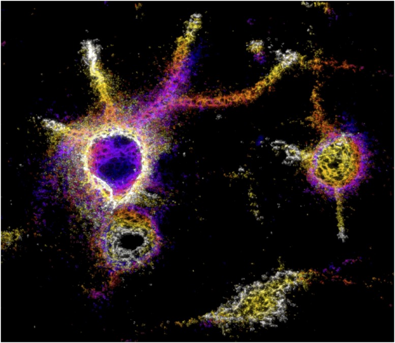
In the field of brain research, neurons have long held the center of attention. But in recent years, some researchers are starting to expand their focus beyond neurons to a long-overlooked structure called the perineuronal net.
Lattice-like elements called perineuronal nets wrap around certain neurons, sharing components with cartilage. Researchers constructed this image of perineuronal nets in the mouse visual cortex using a super-resolution microscope and a fluorescent marker that labels certain sugar-rich molecules. Image courtesy of Yaron Sigal, Xiaowei Zhuang, and Takao Hensch (Harvard University, Cambridge, MA).
Discovered in the late 1800s, this lattice-like element wraps around certain neurons and shares some components with cartilage. But far from providing mere structural support, growing evidence suggests that perineuronal nets, which are highly organized aggregates of proteins and sugar chains, help regulate the brain’s ability to make new connections and tweak existing ones. Researchers are now eager to understand how these nets may contribute to healthy brain function, as well as disease.
This modern revival stems from a chance meeting between two neuroscientists about 2 decades ago. While studying spinal cord injury in rats, James Fawcett at the University of Cambridge in the United Kingdom tested an enzyme that he hoped would help regrow severed nerves. It did, to a limited extent, but more notably, it appeared to encourage rapid rewiring and compensatory activity by spared nerves. Tommaso Pizzorusso, then at the Scuola Normale Superiore di Pisa in Italy, was intrigued by the finding. The two scientists decided to combine the treatment with studies of the developing rat visual system conducted by Pizzorusso’s group (1).
The enzyme, it turns out, was degrading perineuronal nets. When the researchers applied it to the visual cortex of adult rats, they found that neural circuits returned to a younger, more malleable or “plastic” state. In young rats and other animals, including humans, temporarily blocking one eye leads to lifelong disruption of the visual system because the brain’s circuits are actively wiring up in response to visual experience. In adults, blocking one eye normally has no effect. But with perineuronal nets gone, the researchers discovered that adult brains were once again vulnerable to wiring problems. The nets, they inferred, might somehow be limiting neural plasticity in adult brains.
The work, published in 2002, didn’t catch on right away. “At the beginning, I think that people were very surprised,” says Pizzorusso. Neuroscientists weren’t used to thinking about much other than the neurons themselves, and biochemists were hardly interested in whole neural circuits.
Flash forward 15 years, and a small but growing community of researchers is tackling this interdisciplinary intersection. Understanding the nets could mean a better understanding of learning and memory, as well as neuropsychiatric disorders, such as addiction or schizophrenia. Ultimately, many scientists hope that new treatments will emerge.
A Concept Reborn
In the 1890s, Camillo Golgi published the first descriptions of perineuronal nets in a series of pioneering microscopy studies of the nervous system. In the series, he observed a “delicate covering” that surrounded the cell bodies and extensions of neurons. This extensive sheath, Golgi argued, supported his theory that the entire nervous system was a single continuous network.
But Golgi’s rival, the Spanish neuroscientist Santiago Ramón y Cajal, dismissed the perineuronal nets as an experimental artifact. Ramón y Cajal championed the competing theory that the nervous system consisted of discrete cells that communicated across gaps (an idea that later proved correct). Although other scientists had observed the nets after Golgi did, Ramón y Cajal’s influence helped drive the structures into obscurity (2, 3).
The existence of perineuronal nets was not confirmed until the 1970s, aided by modern methods for staining tissue and labeling specific molecules. Biochemists took interest in these webs of proteins and sugar chains. But the structures remained largely unknown to neuroscientists until 2002, with Pizzorusso’s
“There’s a relatively new idea that perineuronal nets are a very dynamic structure.”
—Alain Prochiantz
and Fawcett’s collaboration. “It really rekindled interest in the idea that the space between neurons would actually be important for plasticity,” says neuroscientist Takao Hensch at Harvard University.
Building on that study, other researchers discovered that perineuronal nets regulate plasticity not only in the visual cortex but also in learning and memory circuits. In a seminal 2009 paper, scientists found that breaking down nets with an enzyme helped erase fearful memories in adult rats (4). Unlike juvenile rats, adults are often resilient to “extinction training” that neutralizes fearful associations, such as a musical tone paired to a shock on the foot. Adult rats are especially susceptible to relapse—a phenomenon thought to be related to post-traumatic stress disorder in humans. Dissolving perineuronal nets in the amygdala, a fear-processing center, appeared to return adult brains to a juvenile-like state in which fearful memories were quickly and fully erased with the right training. “This was a big step,” recalls Pizzorusso, now at the University of Florence. “It widened interest in the community.”
That growing audience included Nobel Prize-winning chemist Roger Tsien at the University of California, San Diego. In 2013, Tsien published a sweeping theory in PNAS proposing that the patterns of holes in perineuronal nets hold the “code” for very-long-term memories (5). These holes coincide with where underlying neurons form connections with other neurons, and Tsien proposed that they help stabilize the circuits underlying long-term memories. Tsien died in 2016 before he could fully test his ideas, leaving his staff research scientist Varda Lev-Ram to pursue his final experiments. But Tsien’s provocative publication has already given a boost to the field.
“This paper by Roger Tsien was an eye opener for many, since he was such a prominent figure,” says neuroscientist Marianne Fyhn at the University of Oslo. “Perineuronal nets had gone under the radar for many people.”
A Multitude of Functions
In recent years, researchers have documented a role for perineuronal nets in a number of brain regions and different forms of memory, including fear learning or object recognition (6–8). In general, these studies support the idea that the nets put the brakes on neural plasticity and help consolidate or maintain memories over time. But the precise rules appear to be complex. “It depends on the type of memory we’re talking about and the brain region,” says neuroscientist Daniela Carulli at the University of Turin in Italy. Researchers still have a lot of questions about how perineuronal nets form, how they change under different conditions, and how exactly they influence neural plasticity. But many intriguing clues have emerged.
In many brain regions, perineuronal nets are mostly found enveloping a class of neurons that produce a calcium-binding protein called parvalbumin (PV) (9). These PV neurons release inhibitory neurotransmitters that suppress the firing of other neurons, and their maturation appears to restrict neural plasticity in some regions of the developing brain (10). Together with Hensch’s group, Alain Prochiantz and his colleagues at the Collège de France in Paris showed in 2012 that perineuronal nets in the mouse visual cortex bind Otx2, a protein that regulates gene expression, and help shuttle it into PV neurons (11).
Their data suggest that, as Otx2 accumulates, it hits two key threshold levels: The first helps open the critical period, and the second closes it. Sustained high Otx2 levels appear necessary to keep the critical period closed in adulthood. “You can imagine that Otx2 maintains a stable circuit network,” explains Hensch. “If you remove the net, then suddenly Otx2 levels drop, and synapses are physically free to be rewired.”
Dynamic Structures
Emerging evidence suggests that perineuronal nets, even once established, are sensitive to changes, such as stress, immune responses, and even learning itself (12, 13). “There’s a relatively new idea that perineuronal nets are a very dynamic structure,” says Prochiantz. “They assemble and disassemble, and you have to keep this equilibrium between assembly and disassembly.” The molecular details of how perineuronal nets behave could help researchers understand how these structures participate in various brain disorders and how they might be manipulated to create new treatments.
Drug addiction has attracted intense interest because it is thought to involve persistent, unwanted memories linking drugs to pleasurable experiences. In a 2015 study, Barbara Sorg’s lab at Washington State University in Vancouver found that removing perineuronal nets in the prefrontal cortex, a decision-making center, made it harder for rats to acquire memories associated with cocaine. In animals that already exhibited these memories, removing the nets interfered with a process that helps stabilize and maintain the memories over time (14). Others have shown that removing perineuronal nets in combination with extinction training inhibits cocaine or heroin relapse in rats (15).
And perineuronal nets might be involved in other neuropsychiatric conditions, including autism, epilepsy, and mood disorders. At Harvard Medical School’s McLean Hospital, for example, Sabina Berretta compared postmortem brain samples from people with schizophrenia to healthy control subjects. In 2013, her team reported lower-than-normal levels of perineuronal nets in several emotional and sensory processing centers of the brain in schizophrenia cases (16, 17). By analyzing specimens of different ages, Berretta’s team also found that perineuronal nets increase during childhood, peaking in late adolescence or early adulthood, when schizophrenia typically manifests. “Their maturation and function maps really nicely to the time course of the disorder,” says Berretta. She suggests that problems with developing perineuronal nets might disrupt the brain maturation and rewiring that occur around adolescence.
For the most part, neuropsychiatric treatments involving perineuronal nets remain a distant prospect. But some researchers are laying the early groundwork. In 2015, Fawcett and his colleagues reported that using an enzyme to degrade perineuronal nets in part of the cerebral cortex helped restore memory for familiar objects in two mouse models of Alzheimer’s disease (18). The treatment didn’t halt neurodegeneration but appeared to boost plasticity in the remaining neurons.
The enzyme that Fawcett and many other researchers have used to digest perineuronal nets in animal studies is not feasible as a therapeutic—it can’t cross the blood-brain barrier and, hence, must be injected directly into the brain. The enzyme might also cause unwanted side effects because it targets a molecule found in other extracellular structures.
But Fawcett, Carulli, Hensch, and others are working on alternative ways to influence the nets. Perineuronal nets are aggregates of many different molecules. They include not only Otx2 but also Semaphorin 3A, a protein that repels neuronal connections during development. Researchers hope that by understanding the many different components that control perineuronal net function, they can develop more precise and effective therapeutics. Ultimately, their aim is to regulate plasticity in psychiatric conditions or to boost brain function after stroke, brain injury, or neurodegenerative conditions.
Perineuronal nets may not replace the neuron as the primary entity of interest, but their potential as a means of understanding various brain functions—and as an avenue toward therapies—is increasingly clear. “Controlling plasticity is a key to recovery of function in the damaged nervous system,” says Fawcett. “The perineuronal net is an extremely promising and tractable target for doing this.”
References
- 1.Pizzorusso T, et al. Reactivation of ocular dominance plasticity in the adult visual cortex. Science. 2002;298:1248–1251. doi: 10.1126/science.1072699. [DOI] [PubMed] [Google Scholar]
- 2.Spreafico R, De Biasi S, Vitellaro-Zuccarello L. The perineuronal net: A weapon for a challenge. J Hist Neurosci. 1999;8:179–185. doi: 10.1076/jhin.8.2.179.1834. [DOI] [PubMed] [Google Scholar]
- 3.Celio MR, Spreafico R, De Biasi S, Vitellaro-Zuccarello L. Perineuronal nets: Past and present. Trends Neurosci. 1998;21:510–515. doi: 10.1016/s0166-2236(98)01298-3. [DOI] [PubMed] [Google Scholar]
- 4.Gogolla N, Caroni P, Lüthi A, Herry C. Perineuronal nets protect fear memories from erasure. Science. 2009;325:1258–1261. doi: 10.1126/science.1174146. [DOI] [PubMed] [Google Scholar]
- 5.Tsien RY. Very long-term memories may be stored in the pattern of holes in the perineuronal net. Proc Natl Acad Sci USA. 2013;110:12456–12461. doi: 10.1073/pnas.1310158110. [DOI] [PMC free article] [PubMed] [Google Scholar]
- 6.Romberg C, et al. Depletion of perineuronal nets enhances recognition memory and long-term depression in the perirhinal cortex. J Neurosci. 2013;33:7057–7065. doi: 10.1523/JNEUROSCI.6267-11.2013. [DOI] [PMC free article] [PubMed] [Google Scholar]
- 7.Banerjee SB, et al. Perineuronal nets in the adult sensory cortex are necessary for fear learning. Neuron. 2017;95:169–179.e3. doi: 10.1016/j.neuron.2017.06.007. [DOI] [PMC free article] [PubMed] [Google Scholar]
- 8.Thompson EH, et al. Removal of perineuronal nets disrupts recall of a remote fear memory. Proc Natl Acad Sci USA. 2018;115:607–612. doi: 10.1073/pnas.1713530115. [DOI] [PMC free article] [PubMed] [Google Scholar]
- 9.van ’t Spijker HM, Kwok JCF. A sweet talk: The molecular systems of perineuronal nets in controlling neuronal communication. Front Integr Nuerosci. 2017;11:33. doi: 10.3389/fnint.2017.00033. [DOI] [PMC free article] [PubMed] [Google Scholar]
- 10.Bernard C, Prochiantz A. Otx2-PNN interaction to regulate cortical plasticity. Neural Plast. 2016;2016:7931693. doi: 10.1155/2016/7931693. [DOI] [PMC free article] [PubMed] [Google Scholar]
- 11.Beurdeley M, et al. Otx2 binding to perineuronal nets persistently regulates plasticity in the mature visual cortex. J Neurosci. 2012;32:9429–9437. doi: 10.1523/JNEUROSCI.0394-12.2012. [DOI] [PMC free article] [PubMed] [Google Scholar]
- 12.Sorg BA, et al. Casting a wide net: Role of perineuronal nets in neural plasticity. J Neurosci. 2016;36:11459–11468. doi: 10.1523/JNEUROSCI.2351-16.2016. [DOI] [PMC free article] [PubMed] [Google Scholar]
- 13.Berretta S, Pantazopoulos H, Markota M, Brown C, Batzianouli ET. Losing the sugar coating: Potential impact of perineuronal net abnormalities on interneurons in schizophrenia. Schizophr Res. 2015;167:18–27. doi: 10.1016/j.schres.2014.12.040. [DOI] [PMC free article] [PubMed] [Google Scholar]
- 14.Slaker M, et al. Removal of perineuronal nets in the medial prefrontal cortex impairs the acquisition and reconsolidation of a cocaine-induced conditioned place preference memory. J Neurosci. 2015;35:4190–4202. doi: 10.1523/JNEUROSCI.3592-14.2015. [DOI] [PMC free article] [PubMed] [Google Scholar]
- 15.Xue YX, et al. Depletion of perineuronal nets in the amygdala to enhance the erasure of drug memories. J Neurosci. 2014;34:6647–6658. doi: 10.1523/JNEUROSCI.5390-13.2014. [DOI] [PMC free article] [PubMed] [Google Scholar]
- 16.Mauney SA, et al. Developmental pattern of perineuronal nets in the human prefrontal cortex and their deficit in schizophrenia. Biol Psychiatry. 2013;74:427–435. doi: 10.1016/j.biopsych.2013.05.007. [DOI] [PMC free article] [PubMed] [Google Scholar]
- 17.Pantazopoulos H, Woo TU, Lim MP, Lange N, Berretta S. Extracellular matrix-glial abnormalities in the amygdala and entorhinal cortex of subjects diagnosed with schizophrenia. Arch Gen Psychiatry. 2010;67:155–166. doi: 10.1001/archgenpsychiatry.2009.196. [DOI] [PMC free article] [PubMed] [Google Scholar]
- 18.Yang S, et al. Perineuronal net digestion with chondroitinase restores memory in mice with tau pathology. Exp Neurol. 2015;265:48–58. doi: 10.1016/j.expneurol.2014.11.013. [DOI] [PMC free article] [PubMed] [Google Scholar]


