Abstract
The short chain fatty acid, 4-phenylbutyric acid (PBA), is used for the treatment of urea cycle disorders and sickle cell disease as an endoplasmic reticulum stress inhibitor. PBA is also known as a histone deacetylase inhibitor (HDACi). We report here the effect of combination therapy on HeLa cancer cells using PBA as the HDACi together with the anticancer drug, doxorubicin (DOX). We synthesized γ-4-phenylbutyrate–ε-caprolactone monomer which was polymerized to form poly(γ-4-phenylbutyrate–ε-caprolactone) (PPBCL) homopolymer using NdCl3·3TEP/TIBA (TEP = triethyl phosphate, TIBA = triisobutylaluminum) catalytic system. DOX-loaded nanoparticles were prepared from the PPBCL homopolymer using poly(ethylene glycol) as a surfactant. An encapsulation efficiency as high as 88% was obtained for these nanoparticles. The DOX-loaded nanoparticles showed a cumulative release of >95% of DOX at pH 5 and 37 °C within 12 h, and PBA release was monitored by 1H NMR spectroscopy. The efficiency of the combination therapy can notably be seen in the cytotoxicity study carried out on HeLa cells, where only ~20% of cell viability was observed after treatment with the DOX-loaded nanoparticles. This drastic cytotoxic effect on HeLa cells is the result of the dual action of DOX and PBA on the DNA strands and the HDAC enzymes, respectively. Overall, this study shows the potential of combination treatment with HDACi and DOX anticancer drug as compared to the treatment with an anticancer drug alone.
Graphical abstract
INTRODUCTION
4-Phenylbutyric acid (PBA) is a short chain fatty acid used mainly for the treatment of urea cycle disorders as an ammonia scavenger1–3 and as endoplasmic reticulum stress inhibitor.4–6 Its ability to activate β-globin transcription allowed it to be used for the treatment of sickle cell disease and thalassemia.7,8 In addition, PBA has been shown to have neuroprotective effects9 and is known as a histone deacetylase inhibitor (HDACi).3,10,11 The HDAC inhibitory effect of PBA is attributed to its anticancer activity. During gene transcription and upon HDAC inhibition, hyperacetylation of the histone residues in nucleosomes leads to enhanced differentiation, inhibits proliferation, and eventually causes cell death.3,10 As such, PBA is used in clinical trials as an anticancer agent.12,13 Yet, the effectiveness of HDACi as anticancer agents is limited due to their dose-limiting toxicity14 and due to acquired resistance.15 Therefore, combination therapy using an HDACi together with an anticancer drug would be more efficient in terms of potency to kill cancer cells.
The anthracycline, doxorubicin (DOX), is a widely used chemotherapeutic agent for the treatment of a wide variety of cancers. These include breast, lung, ovarian, Hodgkin’s and non-Hodgkin’s lymphoma, lung, gastric, sarcoma, and pediatric cancers.16,17 The side effects, such as cardiotoxicity caused by DOX, however, limits the use of an anticancer drug.17 One solution to circumvent this issue is the use of nanocarriers to encapsulate the aforementioned drug. Thereby, the drug will accumulate specifically in the tumor site either by the enhanced permeation and retention (EPR) effect or by active targeting which will reduce the side effects to some extent.
Among the different types of HDACis, hydroxamic acids,18,19 benzamides,20 and aliphatic acids21,22 have been conjugated to polymer backbones. However, these only focused on the delivery of the HDACi. The use of nanocarriers made from HDACi conjugated polymeric material will allow for the codelivery of the HDACi together with a more potent anticancer drug, such as DOX. The dual delivery of the HDACi and the anticancer drug is expected to have enhanced cytotoxicity to the cancer cells. Sustained release of the HDACi as well as the anticancer drug at the tumor site can be obtained by polymeric micelles/nanoparticles. Recently, we reported PBA conjugated poly(ethylene glycol)-b-poly(γ-4-phenylbutyrate–ε-caprolactone-ran-ε-caprolactone) (PEG-b-(PCL-ran-PPBCL)) copolymer for the delivery of DOX.23 The γ-4-phenylbutyrate–ε-caprolactone monomer was copolymerized with ε-caprolactone in the presence of PEG as the macro-initiator with tin(II) 2-ethylhexanoate (Sn(Oct)2) as the catalyst. The low incorporation of the PBA, into the polymeric backbone, resulted in reduced cytotoxicity to the human cervical cancer cells (HeLa).
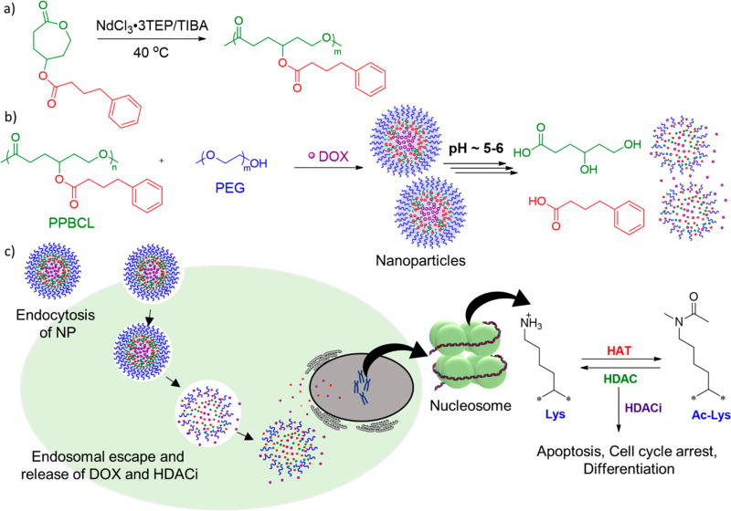
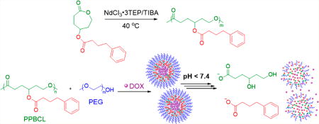
Herein, we report the synthesis of poly(γ-4-phenylbutyrate–ε-caprolactone) (PPBCL) with a newly developed catalytic system, NdCl3·3TEP/TIBA (TEP = triethyl phosphate, TIBA = triisobutylaluminum) (Scheme 1).24 The synthesized homopolymer was employed to prepare nanoparticles (NP) with PEG surfactant to encapsulate DOX. The formed empty and DOX-loaded NPs were then analyzed to determine the size, morphology, drug release behavior, cytotoxicity, and cellular uptake in HeLa cells (Scheme 1).
Scheme 1.
Schematic Diagram of (a) Synthesis of PPBCL Polymer with NdCl3·3TEP/TIBA Catalytic System, (b) Formation of DOX-Loaded NP Using PEG and Release of DOX and PBA upon the Introduction to Acidic Medium, and (c) Endocytosis and Subsequent Release of DOX and PBA in the Cancer Cell
MATERIALS AND METHODS
Materials
All commercially available chemicals and solvents were purchased from Sigma-Aldrich or Fisher Scientific. 4-Hydroxycyclohexanone and NdCl3·3TEP catalyst were synthesized according to previously published procedures.24,25
Analysis
Proton and carbon nuclear magnetic resonance (1H and 13C NMR, respectively) spectra were recorded in CDCl3 on a Bruker AVANCE III 500 spectrometer (500 MHz) at 25 °C unless otherwise mentioned. Multiplicities were given as s (singlet), d (doublet), t (triplet), q (quartet), and br (broad). Size exclusion chromatography (SEC) was carried out on a Viscotek VE 3580 system equipped with Viscotek columns (T6000M), connected to the refractive index (RI), low angle light scattering (LALS), right angle light scattering (RALS), and viscosity detectors. An SEC solvent/sample module (GPCmax) was used with HPLC grade THF as the eluent, and calibration was based on polystyrene standards. A PerkinElmer LS 50 BL luminescence spectrometer was used to collect fluorescence spectra at 25 °C. Dynamic light scattering (DLS) measurements were performed using a Malvern Zetasizer Nano ZS instrument equipped with a He–Ne laser (633 nm) and 173° backscatter detector. The morphology of empty and loaded nanoparticles was visualized by transmission electron microscopy (TEM) imaging performed on a Tecnai G2 Spirit Biotwin microscope operated at 120 keV, and images were analyzed using Gatan Digital Micrograph software. An Agilent UV/vis spectrophotometer was used to obtain absorbance spectra. BioTek Cytation 3 cell imaging multimode reader was used for cytotoxicity and cellular uptake measurements.
Synthesis of 4-Oxocyclohexanoyl Phenylbutanoate (2)
4-Hydroxycyclohexanone (2.00 g, 0.017 mol), phenylbutyric acid (3.17 g, 0.019 mol), 4-(dimethylamino)pyridine (DMAP) (0.21 g, 1.7 mmol), and dichloromethane (DCM) (60 mL) were cooled to 0 °C in a round-bottom flask. A solution of 1-ethyl-3-(3-(dimethylamino)-propyl)carbodiimide hydrochloride (EDCl) (4.04 g, 0.21 mol) in DCM (40 mL) was added dropwise to the cooled solution. The reaction was stirred overnight and was washed with a saturated solution of potassium carbonate and several times with water to remove any excess DMAP, EDCl, and other water-soluble products. The organic layer was dried over anhydrous magnesium sulfate, and the solvent was evaporated under reduced pressure to obtain the pure product as a pale yellow liquid (4.50 g, 97%). 1H NMR (δ = 7.26, CDCl3): δ = 1.98 (q, 2H), 2.04–2.08 (m, 4H), 2.33–2.39 (m, 4H), 2.5–2.56 (m, 2H), 2.67 (t, 2H), 5.15–5.19 (q, 1H), 7.17–7.21 (m, 3H), 7.27–7.31 (m, 2H).
Synthesis of γ-4-Phenylbutyrate–ε-caprolactone (3)
To a stirred solution of 4-oxocyclohexanoyl phenylbutanoate (4.5 g, 0.017 mol) in DCM (100 mL) at 0 °C was added a solution of 77% m-chloroperoxybenzoic acid (6.6 g, 0.029 mol) in DCM (100 mL). The reaction mixture was stirred overnight, and potassium carbonate (10.0 g in 20 mL of water) was added to the reaction mixture and further stirred vigorously for 4 h. The organic layer was separated, and the aqueous layer was washed with DCM (2 × 50 mL). The combined organic layers were dried over anhydrous magnesium sulfate and concentrated in vacuo. The product was isolated by flash chromatography with gradient elution using hexanes and ethyl acetate to yield a pale yellow oil (4.0 g, 83%). 1H NMR (δ = 7.26, CDCl3): δ = 1.94–2.13 (m, 6H), 2.35 (t, 2H), 2.5–2.55 (m, 1H), 2.66 (t, 2H), 2.85–2.91 (m, 1H), 4.11–4.16 (m, 1H), 4.39–4.44 (m, 1H), 5.09–****5.14 (m, 1H), 7.16–7.23 (m, 3H), 7.27–7.31 (t, 2H).
Synthesis of PPBCL
A 10 mL Schlenk flask was flushed with nitrogen for 10 min. Triisobutylaluminum (25% w/w) (0.02 mL, 0.014 mmol) and NdCl3·3TEP (0.01 mL, 0.0014 mmol) were added to the Schlenk flask and stirred for 1 min under a nitrogen environment. Monomer γ-4-phenylbutyrate–ε-caprolactone (0.2 g, 0.7 mmol) was added to the catalyst mixture, and the flask was immersed in an oil bath at 40 °C. The polymer was precipitated out in methanol after overnight stirring and further purified by washing the polymer with methanol a few times. Mn = 13 000 Da, PDI = 1.51. 1H NMR (δ = 7.26, CDCl3): δ = 1.79–2.01 (m, 6H), 2.29 (br s, 4H), 2.63 (t, 2H), 4.05 (t, 2H), 5.00 (m, 1H), 7.16–7.23 (m, 3H), 7.26–7.32 (m, 2H).
Preparation of Nanoparticles
A solvent evaporation method was employed for the preparation of nanoparticles with PPBCL:PEG in a 1:1 weight ratio. A stock solution of PPBCL (10 mg) and PEG (10 mg) in THF (5 mL) was prepared at a concentration of PPBCL of 2 mg/mL. A volume of 3 mL of this stock solution was added dropwise to deionized water (6 mL) under vigorous stirring. The solution was kept under stirring until THF was completely evaporated. The concentration of the final nanoparticles solution was 1 mg/mL with respect to PPBCL. The resulting solution was filtered through a 0.22 µm polyamide (PA) syringe filter to remove any polymer that has precipitated out to obtain the nanoparticles in DI water.
Determination of Critical Aggregation Concentration (CAC)
CAC was determined with the aid of pyrene, a hydrophobic fluorescent probe. For all polymer samples, PPBCL and PEG were used in a weight ratio of 1:1. A volume of 200 µL of pyrene in THF (6.0 × 10⁻⁵ M) was added to polymer solutions of various concentrations. This solution was added dropwise to deionized water (10 mL) under stirring. The solutions were stirred for 4 h for complete evaporation of THF and for the formation of nanoparticles. The concentrations of the resulting solutions were in the range of 1 g/L to 4 × 10⁻⁵ g/L. Fluorescence spectra of the solutions were recorded with the emission wavelength set at 390 nm. The intensity ratios of pyrene at the wavelengths of 337.5 and 334.5 nm were plotted against the logarithm of polymer concentration. CAC was determined as the value at the x coordinate where the two trend lines intersected.
Preparation of DOX-Loaded Nanoparticles
DOX-loaded nanoparticles were prepared in a similar manner as the empty nanoparticles. Briefly, 4 mg of DOX·HCl was neutralized with 3 equiv of triethylamine (2.9 µL), and a stock solution was prepared in THF (4 mL). A stock solution of PPBCL (10 mg) and PEG (10 mg) in THF (5 mL) was made so that the concentration of PPBCL was 2 mg/mL. A volume of 3 mL of this stock solution and 0.6 mL of the DOX stock solution were added dropwise to deionized water (6 mL) under vigorous stirring. The weight ratio between PPBCL and DOX was kept at 10:1. The resulting solution was filtered through a 0.22 µm PA syringe filter in order to remove any nonencapsulated aggregated drug molecules to obtain DOX-loaded nanoparticles in deionized water. Drug loading capacity (DLC) and encapsulation efficiency (EE) were determined using UV/vis absorbance spectra by diluting the nanoparticle solutions with DMSO in a 1:1 ratio. The absorbance at 485 nm was fitted to a pre-established calibration curve for DOX in DMSO/deionized water.
DLS and TEM Analysis
DOX-loaded and empty nanoparticles were prepared as stated above and were analyzed by DLS to obtain the hydrodynamic size. Size measurements were obtained in triplicate at 25 °C. Morphological studies were carried out by TEM where the samples were prepared by treating the copper mesh grid with 1 mg/mL aqueous nanoparticle solution for 2 min, followed by staining with 2% phosphotungstic acid for 30 s.
Release Studies for DOX
A 1 mg/mL solution of DOX-loaded nanoparticles in deionized water was prepared as stated before. A volume of 4 mL from the nanoparticle solution was transferred to Snakeskin dialysis tubing with 3500 Da molecular weight cutoff and was dialyzed against 15 mL of buffer (pH 7.4, 6.0, and 5.0). Aliquots of 2 mL were withdrawn at various time intervals and were replaced by the same volume. The samples taken were diluted with DMSO in a ratio of 1:1, and UV/vis spectra were recorded. DOX release was then determined by a previously established calibration plot for DOX in DMSO/DI water.
Release Studies for PBA
Empty nanoparticles were prepared in deionized water with a concentration of 2 mg/mL. A volume of 4 mL of the nanoparticle solution was transferred to Snakeskin dialysis tubing with 3500 Da molecular weight cutoff and was dialyzed against 15 mL of buffer (pH 6.0). Three samples were taken at the initial time, 12 h, and 24 h, and the buffer solution was allowed to evaporate. After complete evaporation of the buffer solution, D2O (0.5 mL) was added, and 1H NMR spectra were recorded at different time intervals.
Biological Studies
All cell culture experiments were carried out using RPMI-1640 medium with l-glutamine and sodium bicarbonate supplemented with 10% fetal bovine serum and 1% penicillin–streptomycin. Cells were incubated in a humidified environment with 5% CO2 at 37 °C.
Cytotoxicity Studies for Empty Nanoparticles
HeLa cells were grown in a 96-well plate at a cell density of 5000 cells per well with 100 µL of growth medium. After the cells were incubated for 24 h for cell adhesion, the growth medium was removed and washed with 100 µL of PBS. Volumes of 100 µL of empty nanoparticles prepared in PBS in concentrations of 0.2, 0.1, 0.06, 0.04, and 0.02 mg/mL were added to the cells with growth media (100 µL) and were incubated at 37 °C for 24 h. Cytotoxicity studies were performed using CellTiter Blue reagent using manufacturer recommended protocol (N = 4).
Cytotoxicity Studies for DOX Loaded Nanoparticles
HeLa cells were grown in a 96-well plate at a cell density of 5000 cells per well with 100 µL of growth medium. After the cells were incubated for 24 h for cell adhesion, the growth medium was removed and washed with 100 µL of PBS. 100 µL of DOX-loaded nanoparticles, free DOX, free PBA, and a combination of DOX and PBA, all prepared in PBS in concentrations of 0.2, 0.06, and 0.02 mg/mL, were added to the cells with growth media (100 µL) and were incubated at 37 °C for 24 h. The amount of free DOX to be added was calculated by DLC, and the amount of free PBA was calculated by the concentration of PPBCL used. Cytotoxicity studies were performed using CellTiter Blue reagent using manufacturer recommended protocol (N = 4). To determine IC50, 100 µL volumes of DOX-loaded NPs in concentrations of 1, 0.5, 0.25, 0.2, 0.1, 0.06, 0.02, 0.001, and 0.0001 mg/mL were added with growth media (100 µL) into 96-well plates with HeLa cells already incubated for 24 h. Cytotoxicity studies were performed using CellTiter Blue reagent using manufacturer recommended protocol (N = 5).
Cellular Uptake Studies
HeLa cells were grown in 35 mm clear bottom dishes at a cell density of 250 000 cells per dish with 2 mL growth media. DOX-loaded micelles in PBS at the concentration of 0.1 mg/mL (1 mL) were added to the cells along with growth media (1 mL). After an incubation period of 4 and 24 h, the media was removed, washed with PBS (2 mL × 3), fixed with 4% paraformaldehyde (1 mL, incubated for 10 min at room temperature), washed with PBS (2 mL × 2), treated with Triton X-100 (1 mL, incubated for 2 min at room temperature), washed with PBS (2 mL × 4), and counterstained the nucleus with 4′,6-diamidino-2-phenylindole (DAPI) according to the manufacturer’s recommended protocol.
RESULTS AND DISCUSSION
Synthesis and Characterization of PPBCL Homopolymer
Monomer γ-4-phenylbutyrate–ε-caprolactone was synthesized previously by our group with an overall yield of ~40%.23 In this report, we employed a new synthesis that increased the overall yield to ~60%. First, 4-hydroxycylohexanone was synthesized from 1,4-cyclohexanediol according to previous reports,25 and then a Steglich esterification was carried out followed by Baeyer–Villiger oxidation to form γ-4-phenylbutyrate–ε-caprolactone (Scheme 2). The previously reported methodology to synthesize γ-substituted-ε-caprolactone monomers involved the conjugation of the substituent first, followed by oxidation26–29 which could be a disadvantage in conjugating substituents that are prone to oxidation or in some cases that are hydrophilic.
Scheme 2.
Synthesis of γ-4-Phenylbutyrate–ε-Caprolactone
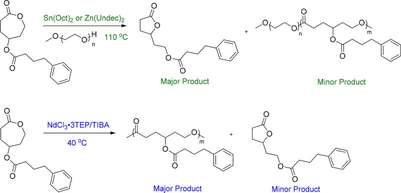
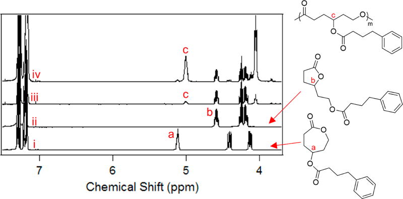
In our previous report, homopolymerization of γ-4-phenylbutyrate–ε-caprolactone was unachievable; thus, copolymerization with ε-caprolactone was carried out with PEG-OH as the hydrophilic block to synthesize PEG-b-(PCL-ran-PPBCL). Only 30 mol % of PBA was incorporated in the polymer; hence, only minimal activity of HDAC inhibition was observed in vitro studies.23 In an attempt to further increase the content of HDACi conjugated to the polymer backbone, exhaustive trials were carried out to obtain the homopolymer of γ-4-phenylbutyrate–ε-caprolactone. With the use of the commonly used catalyst, Sn(Oct)2, for ring-opening polymerizations,30–32 we observed that the major product obtained was not the homopolymer but the more thermodynamically stable butyrolactone analogue (Scheme 3). Therefore, further attempts were carried out with zinc undecylenate (Zn(Undec)2) and 1,8-diazabicyclo[5.4.0]undec-7-ene (DBU), since both of these catalysts have shown the ability to polymerize substituted or unsubstituted ε-caprolactones.33–35 Unfortunately, neither of these catalytic systems provided the desired homopolymer in good yields. By use of Zn(Undec)2 as the catalyst, the majority of the caprolactone monomer was converted into the butyrolactone analogue (Scheme 3 and Table 1). DBU, on the other hand, showed no polymerization of the caprolactone monomer. To confirm the structure of the formed transesterified product, we carried out the synthesis of the butyrolactone monomer (Scheme S1, Figures S1 and S2, see Supporting Information for details). By comparison of the 1H NMR spectra, we confirmed the formation of the butyrolactone analogue during the polymerization (Figure 1). With the goal of obtaining the homopolymer in high yields we carried out the polymerization with a catalytic system introduced by our group, NdCl3·3TEP/TIBA,24 which produced the homopolymer with only <20 mol % transesterified product (Table 1 and Figure 1). With Sn(Oct)2 and Zn(Undec)2, transesterification is more facilitated due to the higher temperatures employed (110 °C). The mild polymerization temperatures used with our catalytic system of NdCl3·3TEP/TIBA (40 °C) favored the formation of the desired homopolymer. Unfortunately, a major limitation of this catalytic system is the inability to use a macroinitiator such as PEG to synthesize amphiphilic block copolymers. Therefore, nanoparticles of PPBCL were formed using PEG as a surfactant. The synthesized PPBCL homopolymer was characterized by 1H NMR, 13C NMR (Figures S3 and S4), and SEC using triple-point calibration (Table 1).
Scheme 3.
Polymerization of γ-4-Phenylbutyrate–ε-Caprolactone Using Sn(Oct)2 and Zn(Undec)2 and NdCl3·3TEP/TIBA
Table 1.
Polymerization of γ-4-Phenylbutyrate–ε-Caprolactone with Different Catalytic Systems
| catalyst | % conv to polymer |
% conv to transesterified product |
Mn (g mol−1) |
PDI |
|---|---|---|---|---|
| Sn(Oct)2 | 32 | 68 | 6000 | 1.52 |
| NdCl3·3TEP/TIBA | 73 | 27 | 13000 | 1.51 |
| Zn(Undec)2 | yield of the polymer was extremely low; higher transesterification | |||
| DBU | no polymer or trans esterified product formed | |||
Figure 1.
1H NMR spectra for (i) γ-4-phenylbutyrate–ε-caprolactone and (ii) 2-(5-oxotetrahydrofuran-2-yl)ethyl 4-phenylbutanoate. 1H NMR spectra for the polymerization of γ-4-phenylbutyrate–ε-caprolactone with (iii) Sn(Oct)2 and (iv) NdCl3·3TEP/TIBA. All spectra are shown for the region 3.5–7.5 ppm.
Preparation and Characterization of Nanoparticles
Nanoparticles were formed with PEG as the surfactant, using a solvent evaporation method. THF was used as the organic solvent due to the miscibility with water and the moderate evaporation of the solvent that limits the formation of aggregates. Initially, different weight ratios between PPBCL and PEG were used to make the nanoparticles, and a study was performed to determine the size of the nanoparticles (Table 2). It was observed that irrespective of the weight ratios used, the size remained constant at ~100 nm. However, during the formation of the nanoparticles with the increasing weight ratios of PPBCL, the polymer started to precipitate out. Therefore, for all experiments in this study a weight ratio of 1:1 between PPBCL and PEG was employed.
Table 2.
Size Distribution of the PPBCL Empty Nanoparticles with the Variation of PEG Weight Ratio
| weight ratios of PEG:PPBCL | size (nm) | dispersity |
|---|---|---|
| 4:1 | 98.6 | 0.075 |
| 2:1 | 105.8 | 0.062 |
| 1:1 | 94.0 | 0.068 |
| 0.5:1 | 95.1 | 0.099 |
| 0.25:1 | 96.2 | 0.085 |
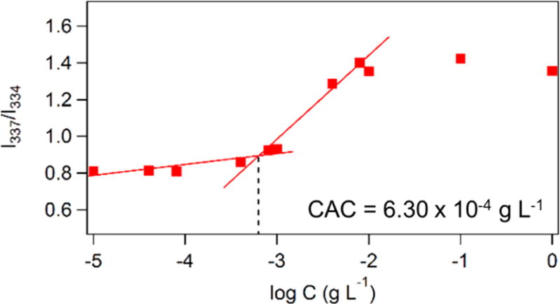
The critical aggregation concentration (CAC), which demonstrates the thermodynamic stability of the formed nanoparticles, was found to be 6.40 × 10⁻⁴ g/L as determined by using the fluorescent probe pyrene. Upon increasing the polymer concentration, the excitation of pyrene changed from 334 to 337 nm when the surrounding environment changed from hydrophilic to hydrophobic, respectively. This change was used to generate a plot of the intensity ratio of these two excitation wavelengths (I337/I334) versus the logarithm of the concentration of the polymer. CAC was determined at the intersection of the two slopes (Figure 2). The thermodynamic stability of the formed nanoparticles is comparable to the micelles that were reported previously with the terpolymer, PEG-b-(PCL-ran-PPBCL).23
Figure 2.
CAC plot for PPBCL obtained using the fluorescent probe pyrene.
DOX-loaded nanoparticles were prepared similarly to the empty nanoparticles (Scheme 3), by using a ratio of [polymer]: [DOX] = 10:1. Drug loading capacity (DLC) and encapsulation efficiency (EE) were estimated by using the following equations.
The calculated DLC was ~8.8%, while EE was ~88%. This represents a significant increase in DLC and EE as compared to our previously reported value of 5.1% DLC and 52% EE which was obtained for PEG-b-(PCL-ran-PPBCL).23 The increase in DOX loading is due to the high number of PBA units conjugated to the polymer backbone which resulted in hydrogen bonding and π-stacking interactions between DOX and the polymer backbone.
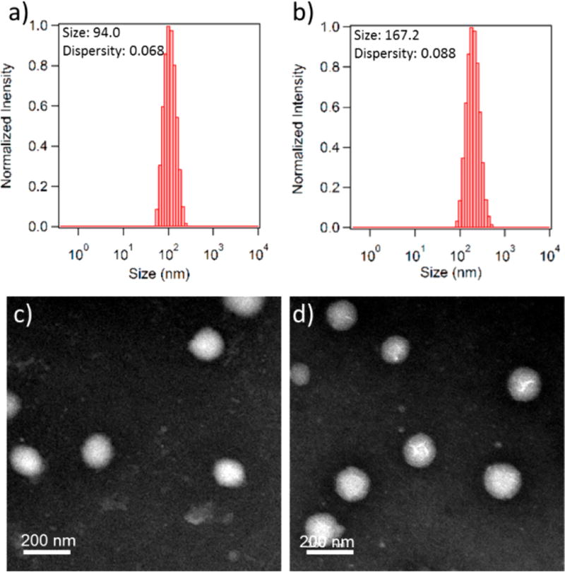
Size and morphology of the empty and DOX-loaded NPs were determined by DLS and TEM analysis. The size of empty NPs and DOX-loaded NPs as obtained from DLS was 94.0 and 167.2 nm, respectively (Figure 3a,b and Figure S5). An evident increase in size can be observed with DOX loading, and the size of the DOX-loaded NPs is similar in size to the DOX-loaded micelles obtained with PEG-b-(PCL-ran-PPBCL).23 TEM images showed the formation of spherical nanoparticles (Figure 3c,d), and the size distribution was comparable to that obtained from DLS analysis.
Figure 3.
DLS of (a) empty NPs (b) DOX-loaded NPs and TEM images of (c) empty NPs and (d) DOX-loaded NPs.
DOX and PBA Release
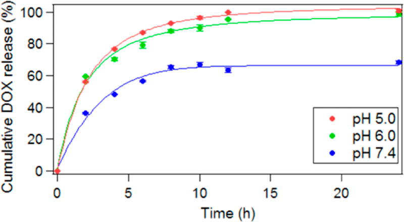
Cumulative DOX release studies were carried out using DOX-loaded NPs at three different pH conditions (pH = 5, 6, and 7.4) at the physiological temperature of 37 °C. Samples were withdrawn for 24 h, and each time a sample was taken, equal amounts of fresh buffer solution were added to maintain the floating conditions. Because of the increased number of ester linkages, a cumulative DOX release of ~90% was obtained for both pH 5 and 6 within 8 h (Figure 4), which is higher than the cumulative release obtained for the previously reported PEG-b-(PCL-ran-PPBCL) polymer.23 The release obtained at pH 5 and 6 was significantly higher than that measured at pH 7.4. In an acidic environment, the ester linkages in the polymer backbone of PPBCL will be cleaved, and thus the NPs will no longer hold the cargo, thereby releasing the DOX into the buffer.
Figure 4.
Cumulative DOX release for the DOX-loaded NP at 37 °C in different pH conditions of pH = 5, 6, and 7.4.
The release of the HDACi was monitored by 1H NMR analysis. The study was carried out using the empty NPs at pH 6 for 24 h. The pH 5 buffer was not used for the study due to the presence of potassium phthalate, which masks the 1H NMR peaks in the aromatic region of PBA. Samples withdrawn were evaporated to remove the buffer solution and were resuspended in D2O to obtain the 1H NMR spectra (Figure S6). The data analysis showed that in acidic environment the ester hydrolysis takes place promoting the release of DOX and PBA HDACi into the buffer solution (Scheme 1b).
In Vitro Cytotoxicity of the NPs
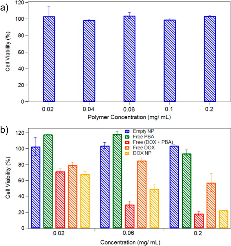
Cytotoxicity studies for empty and DOX-loaded NPs were carried out using CellTiter Blue reagent. The empty NPs with a polymer concentration in the range of 0.02–0.2 mg/mL were shown to be nontoxic to the HeLa cells (Figure 5a) as all concentrations of NPs the cells were >95% viable, which suggests the polymer by itself is nontoxic. Cytotoxicity of the DOX-loaded NPs was then compared to that of free DOX, free PBA, and free (DOX + PBA). Dosing for free DOX, free PBA, and free (DOX + PBA) was calculated based on the DLC of Dox and the degree of polymerization for PBA, for each concentration. When comparing the cytotoxicity of the DOX-loaded NPs to that of free PBA, and free DOX (Figure 5b), the DOX-loaded NPs showed a significantly higher cytotoxic effect to the HeLa cells. The free HDACi by itself did not cause cell death within the 24 h time frame. Enhanced cell death was observed with free (DOX + PBA) when compared with free DOX, due to the dual action of DOX and the HDACi. Furthermore, the cytotoxicity caused by the DOX-loaded NPs was similar to that of the free (DOX + PBA), confirming the release of the HDACi together with the DOX from the NPs. These cytotoxicity studies can be related to the release profiles discussed above, where within 24 h, in acidic environments, the release of PBA and >95% of DOX was observed. The cell viability of the DOX-loaded NPs within 24 h was ~20%, whereas the cell viability for the previously reported PEG-b-(PCL-ran-PPBCL) polymer was ~20% within 60 h.23 The enhanced cytotoxicity is most likely due to the increased amount of PBA and DOX. IC50 values calculated for the DOX-loaded NPs from the dose–response curve were found to be 0.045 mg/mL (Figure S7). The accelerated release of both DOX and PBA at conditions which mimics the endosomal pH conditions is desirable as these released molecules will then enter the nucleus where DOX will interact with the DNA strands while PBA inhibits the HDAC enzymes, causing an increased cellular death to the cancer cells.
Figure 5.
Cell viability on HeLa cells after 24 h of incubation time for (a) empty NPs and (b) DOX-loaded NPs, free DOX, free (DOX + PBA), and free PBA (N = 4).
Cellular Uptake of DOX-Loaded NPs
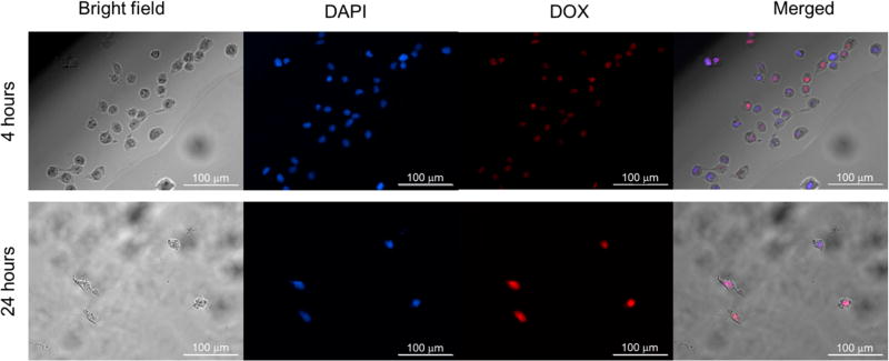
Cellular uptake studies on HeLa cells were performed using DOX-loaded NPs with a DOX concentration of 8.8 mg/L. The NPs were incubated with the cells for 4 and 24 h before fixing with paraformaldehyde and staining the nuclei with DAPI. The inherent fluorescent ability of DOX was used to determine the cellular uptake of DOX, and it was seen that DOX accumulated in the nucleus. The merged bright field and fluorescence images clearly showed the cellular uptake of DOX and the subsequent accumulation of DOX in the nucleus (Figure 6). With increasing incubation time up to 24 h, the intensity of DOX inside the nucleus increased as compared to 4 h. As observed from cytotoxicity studies, ~80% of HeLa cells were killed within 24 h of treatment with the DOX-loaded NPs, which can be related to the reduced cell density in cellular uptake studies performed at 24 h.
Figure 6.
Cellular uptake of DOX-loaded NPs into HeLa cells. NPs were incubated for 4 and 24 h at a DOX concentration of 8.8 mg/L.
CONCLUSION
In this study, a polycaprolactone containing a pendant PBA HDACi was successfully synthesized with the NdCl3·3TEP/TIBA catalytic system. Commonly utilized catalysts for the polymerization of caprolactone failed to produce the homopolymer of γ-4-phenylbutyrate–ε-caprolactone. The synthesized PPBCL polymer with PEG formed spherical NPs and successfully encapsulated DOX with an EE of 88%. These DOX-loaded NPs released both DOX and PBA at pH conditions that mimic the endosomal environment and also exhibited increased cytotoxicity (cell viability reduced to ~20%) when compared to free DOX due to the dual action of DOX and HDACi. Overall, this study shows the improvement of cytotoxicity by combination treatment with an HDACi and an anticancer drug as compared to the treatment with an anticancer drug alone.
Supplementary Material
Acknowledgments
Funding from NSF (CHE-1566059 and CHE-1609880), NIH (1R21EB019175-01A1), and Welch Foundation (AT-1740) is gratefully acknowledged.
Footnotes
ASSOCIATED CONTENT
- Synthesis and characterization of 2-(5-oxotetrahydrofuran-2-yl)ethyl 4-phenylbutanoate and 1H and 13C NMR of PPBCL, DLS comparison of empty and loaded nanoparticles, release study of PBA and dose-responsive curve for DOX-loaded NPs (PDF)
The authors declare no competing financial interest.
References
- 1.Brusilow SW, Maestri NE. Urea cycle disorders: diagnosis, pathophysiology, and therapy. Adv. Pediatr. 1996;43:127–170. [PubMed] [Google Scholar]
- 2.Burrage LC, Jain M, Gandolfo L, Lee BH, Nagamani SCS. Sodium phenylbutyrate decreases plasma branched-chain amino acids in patients with urea cycle disorders. Mol. Genet. Metab. 2014;113(1–2):131–135. doi: 10.1016/j.ymgme.2014.06.005. [DOI] [PMC free article] [PubMed] [Google Scholar]
- 3.Marks PA, Rifkind RA, Richon VM, Breslow R, Miller T, Kelly WK. Histone deacetylases and cancer: causes and therapies. Nat. Rev. Cancer. 2001;1(3):194–202. doi: 10.1038/35106079. [DOI] [PubMed] [Google Scholar]
- 4.Basseri S, Lhoták Š, Sharma AM, Austin RC. The chemical chaperone 4-phenylbutyrate inhibits adipogenesis by modulating the unfolded protein response. J. Lipid Res. 2009;50(12):2486–2501. doi: 10.1194/jlr.M900216-JLR200. [DOI] [PMC free article] [PubMed] [Google Scholar]
- 5.Yam GH-F, Gaplovska-Kysela K, Zuber C, Roth Jr. Sodium 4-Phenylbutyrate Acts as a Chemical Chaperone on Misfolded Myocilin to Rescue Cells from Endoplasmic Reticulum Stress and Apoptosis. Invest. Ophthalmol. Visual Sci. 2007;48(4):1683–1690. doi: 10.1167/iovs.06-0943. [DOI] [PubMed] [Google Scholar]
- 6.Kolb PS, Ayaub EA, Zhou W, Yum V, Dickhout JG, Ask K. The therapeutic effects of 4-phenylbutyric acid in maintaining proteostasis. Int. J. Biochem. Cell Biol. 2015;61:45–52. doi: 10.1016/j.biocel.2015.01.015. [DOI] [PubMed] [Google Scholar]
- 7.Collins AF, Pearson HA, Giardina P, McDonagh KT, Brusilow SW, Dover GJ. Oral sodium phenylbutyrate therapy in homozygous β thalassemia: A clinical trial. Blood. 1995;85(1):43–49. [PubMed] [Google Scholar]
- 8.Dover GJ, Brusilow S, Charache S. Induction of fetal hemoglobin production in subjects with sickle cell anemia by oral sodium phenylbutyrate. Blood. 1994;84(1):339. [PubMed] [Google Scholar]
- 9.Mimori S, Ohtaka H, Koshikawa Y, Kawada K, Kaneko M, Okuma Y, Nomura Y, Murakami Y, Hamana H. 4-Phenylbutyric acid protects against neuronal cell death by primarily acting as a chemical chaperone rather than histone deacetylase inhibitor. Bioorg. Med. Chem. Lett. 2013;23(21):6015–6018. doi: 10.1016/j.bmcl.2013.08.001. [DOI] [PubMed] [Google Scholar]
- 10.Mottamal M, Zheng S, Huang TL, Wang G. Histone Deacetylase Inhibitors in Clinical Studies as Templates for New Anticancer Agents. Molecules. 2015;20(3):3898–3941. doi: 10.3390/molecules20033898. [DOI] [PMC free article] [PubMed] [Google Scholar]
- 11.Miller AC, Cohen S, Stewart M, Rivas R, Lison P. Radioprotection by the histone deacetylase inhibitor phenylbutyrate. Radiat. Environ. Biophys. 2011;50(4):585. doi: 10.1007/s00411-011-0384-7. [DOI] [PubMed] [Google Scholar]
- 12.Warrell JRP, He L-Z, Richon V, Calleja E, Pandolfi PP. Therapeutic Targeting of Transcription in Acute Promyelocytic Leukemia by Use of an Inhibitor of Histone Deacetylase. J. Natl. Cancer Inst. 1998;90(21):1621–1625. doi: 10.1093/jnci/90.21.1621. [DOI] [PubMed] [Google Scholar]
- 13.Phuphanich S, Baker SD, Grossman SA, Carson KA, Gilbert MR, Fisher JD, Carducci MA. Oral sodium phenylbutyrate in patients with recurrent malignant gliomas: A dose escalation and pharmacologic study. Neuro Oncol. 2005;7(2):177–182. doi: 10.1215/S1152851704000183. [DOI] [PMC free article] [PubMed] [Google Scholar]
- 14.How J, Minden MD, Brian L, Chen EX, Brandwein J, Schuh AC, Schimmer AD, Gupta V, Webster S, Degelder T, Haines P, Stayner L-A, McGill S, Wang L, Piekarz R, Wong T, Siu LL, Espinoza-Delgado I, Holleran JL, Egorin MJ, Yee KWL. A phase I trial of two sequence-specific schedules of decitabine and vorinostat in patients with acute myeloid leukemia. Leuk. Lymphoma. 2015;56(10):2793–2802. doi: 10.3109/10428194.2015.1018248. [DOI] [PMC free article] [PubMed] [Google Scholar]
- 15.Robey RW, Chakraborty AR, Basseville A, Luchenko V, Bahr J, Zhan Z, Bates SE. Histone Deacetylase Inhibitors: Emerging Mechanisms of Resistance. Mol. Pharmaceutics. 2011;8(6):2021–2031. doi: 10.1021/mp200329f. [DOI] [PMC free article] [PubMed] [Google Scholar]
- 16.Cortés-Funes H, Coronado C. Role of anthracyclines in the era of targeted therapy. Cardiovasc. Toxicol. 2007;7(2):56–60. doi: 10.1007/s12012-007-0015-3. [DOI] [PubMed] [Google Scholar]
- 17.Thorn CF, Oshiro C, Marsh S, Hernandez-Boussard T, McLeod H, Klein TE, Altman RB. Doxorubicin pathways: pharmacodynamics and adverse effects. Pharmacogenet. Genomics. 2011;21(7):440–446. doi: 10.1097/FPC.0b013e32833ffb56. [DOI] [PMC free article] [PubMed] [Google Scholar]
- 18.el Bahhaj F, Denis I, Pichavant L, Delatouche R, Collette F, Linot C, Pouliquen D, Grégoire M, Héroguez V, Blanquart C, Bertrand P. Histone Deacetylase Inhibitors Delivery using Nanoparticles with Intrinsic Passive Tumor Targeting Properties for Tumor Therapy. Theranostics. 2016;6(6):795–807. doi: 10.7150/thno.13725. [DOI] [PMC free article] [PubMed] [Google Scholar]
- 19.Delatouche R, Denis I, Grinda M, Bahhaj FE, Baucher E, Collette F, Héroguez V, Grégoire M, Blanquart C, Bertrand P. Design of pH-responsive clickable prodrugs applied to histone deacetylase inhibitors: A new strategy for anticancer therapy. Eur. J. Pharm. Biopharm. 2013;85(3):862–872. doi: 10.1016/j.ejpb.2013.03.006. [DOI] [PubMed] [Google Scholar]
- 20.Denis I, el Bahhaj F, Collette F, Delatouche R, Gueugnon F, Pouliquen D, Pichavant L, Héroguez V, Grégoire M, Bertrand P, Blanquart C. Histone deacetylase inhibitor-polymer conjugate nanoparticles for acid-responsive drug delivery. Eur. J. Med. Chem. 2015;95:369–376. doi: 10.1016/j.ejmech.2015.03.037. [DOI] [PubMed] [Google Scholar]
- 21.Senevirathne SA, Boonsith S, Oupicky D, Biewer MC, Stefan MC. Synthesis and characterization of valproic acid ester prodrug micelles via an amphiphilic polycaprolactone block copolymer design. Polym. Chem. 2015;6(13):2386–2389. [Google Scholar]
- 22.Alfurhood JA, Sun H, Kabb CP, Tucker BS, Matthews JH, Luesch H, Sumerlin BS. Poly(N-(2-hydroxypropyl)-methacrylamide)-valproic acid conjugates as block copolymer nanocarriers. Polym. Chem. 2017;8:4983–4987. doi: 10.1039/C7PY00196G. [DOI] [PMC free article] [PubMed] [Google Scholar]
- 23.Senevirathne SA, Washington KE, Miller JB, Biewer MC, Oupicky D, Siegwart DJ, Stefan MC. HDAC inhibitor conjugated polymeric prodrug micelles for doxorubicin delivery. J. Mater. Chem. B. 2017;5(11):2106–2114. doi: 10.1039/C6TB03038F. [DOI] [PMC free article] [PubMed] [Google Scholar]
- 24.Kularatne RN, Yang A, Nguyen HQ, McCandless GT, Stefan MC. Neodymium catalyst for the polymerization of dienes and vinyl monomers. Macromol. Rapid Commun. 2017;38:1700427. doi: 10.1002/marc.201700427. [DOI] [PubMed] [Google Scholar]
- 25.Busto E, Martínez-Montero L, Gotor V, Gotor-Fernández V. Chemoenzymatic Asymmetric Synthesis of Serotonin Receptor Agonist (R)-Frovatriptan. Eur. J. Org. Chem. 2013;2013(19):4057–4064. [Google Scholar]
- 26.Hao J, Cheng Y, Ranatunga RJKU, Senevirathne S, Biewer MC, Nielsen SO, Wang Q, Stefan MC. A Combined Experimental and Computational Study of the Substituent Effect on Micellar Behavior of γ-Substituted Thermoresponsive Amphiphilic Poly(ε-caprolactone)s. Macromolecules. 2013;46(12):4829–4838. [Google Scholar]
- 27.Rainbolt EA, Washington KE, Biewer MC, Stefan MC. Towards smart polymeric drug carriers: self-assembling [gamma]-substituted polycaprolactones with highly tunable thermoresponsive behavior. J. Mater. Chem. B. 2013;1(47):6532–6537. doi: 10.1039/c3tb21488e. [DOI] [PubMed] [Google Scholar]
- 28.Hao J, Servello J, Sista P, Biewer MC, Stefan MC. Temperature-sensitive aliphatic polyesters: synthesis and characterization of [gamma]-substituted caprolactone monomers and polymers. J. Mater. Chem. 2011;21(29):10623–10628. [Google Scholar]
- 29.Trollsås M, Lee VY, Mecerreyes D, Löwenhielm P, Möller M, Miller RD, Hedrick JL. Hydrophilic Aliphatic Polyesters: Design, Synthesis, and Ring-Opening Polymerization of Functional Cyclic Esters. Macromolecules. 2000;33(13):4619–4627. [Google Scholar]
- 30.Storey RF, Sherman JW. Kinetics and Mechanism of the Stannous Octoate-Catalyzed Bulk Polymerization of ε-Caprolactone. Macromolecules. 2002;35(5):1504–1512. [Google Scholar]
- 31.Rainbolt EA, Miller JB, Washington KE, Senevirathne SA, Biewer MC, Siegwart DJ, Stefan MC. Fine-tuning thermoresponsive functional poly(ε-caprolactone)s to enhance micelle stability and drug loading. J. Mater. Chem. B. 2015;3(9):1779–1787. doi: 10.1039/c4tb02016b. [DOI] [PubMed] [Google Scholar]
- 32.Washington KE, Kularatne RN, Du J, Ren Y, Gillings MJ, Geng CX, Biewer MC, Stefan MC. Thermoresponsive starlike [gamma]-substituted poly(caprolactone)s for micellar drug delivery. J. Mater. Chem. B. 2017;5(28):5632–5640. [Google Scholar]
- 33.Washington KE, Kularatne RN, Du J, Gillings MJ, Webb JC, Doan NC, Biewer MC, Stefan MC. Synthesis of linear and star-like poly(ε-caprolactone)-b-poly{γ-2-[2-(2-methoxyethoxy)ethoxy]ethoxy-ε-caprolactone} amphiphilic block copolymers using zinc undecylenate. J. Polym. Sci., Part A. Polym. Chem. 2016;54(22):3601–3608. [Google Scholar]
- 34.Hao J, Granowski PC, Stefan MC. Zinc Undecylenate Catalyst for the Ring-Opening Polymerization of Caprolactone Monomers. Macromol. Rapid Commun. 2012;33(15):1294–1299. doi: 10.1002/marc.201200147. [DOI] [PubMed] [Google Scholar]
- 35.Kato K, Inoue K, Kudo M, Ito K. Synthesis of graft polyrotaxane by simultaneous capping of backbone and grafting from rings of pseudo-polyrotaxane. Beilstein J. Org. Chem. 2014;10:2573–2579. doi: 10.3762/bjoc.10.269. [DOI] [PMC free article] [PubMed] [Google Scholar]
Associated Data
This section collects any data citations, data availability statements, or supplementary materials included in this article.


