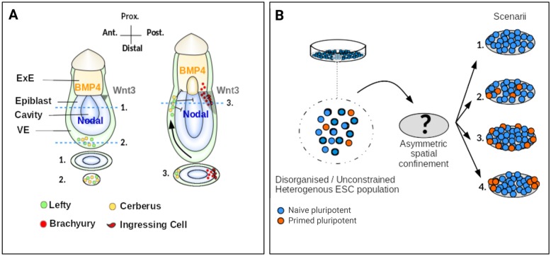
Fig. 1.
Methodological approach and tested hypotheses. (A) Schematic illustrating the emergence of AP polarity in the post-implantation mouse embryo. A sagittal section is drawn for each stage (top) as well as transverse sections (bottom) with numbered dashed lines indicating the positions of the represented sections. Note the ellipsoidal shape of the transverse sections. The black arrow represents the movement of the DVE cells towards the anterior side. (B) ESCs contain subpopulations with distinct expression profiles. Spatial confinement may (1) modify the balance of cell states, (2) have no apparent effect, (3) enable patterning via border effects in a symmetry-insensitive fashion or (4) enable patterning with geometry guiding spatial organisation. ExE, extra-embryonic ectoderm; VE, visceral endoderm.

