



Abstract
With the introduction of new biologics such as anti-TNF-alpha antibodies and other therapies in the treatment of inflammatory arthritis, capable of halting joint destruction and functional disability, there are new pressures on diagnostic and prognostic imaging. Early demonstration of pre-erosive inflammatory features and monitoring of the long-term effects of treatment are becoming increasingly important. Early detection of synovitis offers advantages in terms of allowing early instigation of therapy and may allow the identification of those patients displaying more aggressive disease who might benefit from early intervention with expensive DMARD therapy. Advanced imaging techniques such as ultrasound (US) and magnetic resonance imaging (MRI) have focussed on the demonstration and quantification of synovitis and allow early diagnosis of inflammatory arthropathies such as rheumatoid arthritis (RA) and psoriatic arthritis (PsA). Synovitis represents a potential surrogate measure of disease activity that can be monitored using either MRI or US; the techniques have, generally, focused on monitoring synovial volume or quality as assessed by its vascularity. However to achieve these goals, standardisation and validation of US and MRI are required to ensure accurate diagnosis, reproducibility and reliability. Each modality has different strengths and weaknesses and levels of validation. This article aims to increase the awareness of radiologists and rheumatologists about this field and to encourage them to participate and contribute to the ongoing development of these modalities. Without this collaboration, it is unlikely that these modalities will reach their full potential in the field of rheumatological imaging. This review is in two parts. The first part addresses the role of US and colour or power Doppler sonography (PDUS) in the detection and monitoring of synovitis in inflammatory arthropathies. The second part will look at advanced MR imaging and Dynamic contrast-enhanced MRI techniques and in particular how they are applied to the monitoring of the disease process. (www.actabiomedica.it)
Keywords: rheumatoid arthritis, psoriatic arthritis, synovitis, magnetic resonance imaging, ultrasound, clinical trials
Introduction
Synovitis (inflammation of the synovium) is a characteristic feature of chronic inflammatory arthritis, and is considered an important factor in the chronicity of the disease and the best predictive marker of joint damage (1, 2). Proliferation of the synovial tissue resulting in the formation of the pannus is an early event in the course of the rheumatic diseases and can be seen before destruction of cartilage and bone (3).
New blood vessel formation seems to be one of the many variables required in the pathogenesis of proliferative synovitis, of which rheumatoid arthritis (RA) is the prototype.
Under normal conditions, the synovium maintains a delicate balance between proangiogenic and antiangiogenic forces. In the inflammatory synovium, however, this balance is lost and there is an increase in proangiogenic factors, ultimately leading to endothelial cell proliferation and pannus growth. Accurate assessment of synovitis is essential in rheumatologic practice to make therapeutic decisions and to evaluate the response to treatment.
Ultrasonography (US) offers a non-invasive, reproducible, non-radiating, and relatively inexpensive method for detecting joint effusion and bursal fluid collection and may depict hyperplastic synovium and underlying erosive disease. However, it does not provide direct information about haemodynamic alterations, which may occur in soft tissue inflammation. Color Doppler US/Power Doppler US (CDUS/PDUS) is important in distinguishing complex effusion and pannus and in the assessment of vascular abnormalities at the synovial tissue. Several studies demonstrated that US, CDUS/PDUS and magnetic resonance imaging (MRI) are more sensitive than clinical examination in detecting synovitis (4-7) and in particular for large joints such as the shoulder and knee (8-10). An important issue consists of the possibility of identifying subclinical synovitis, being subclinical inflammation not an uncommon feature in rheumatic disease.
Ultrasound contrast agents enhance visualization of the small synovial vessels, can be used to estimate haemodynamic alterations and may have a role in assessing synovial activity and in distinguishing between inflammatory and non-inflammatory pannus. Estimation of the area under US contrast enhancement curves may help to produce a method of measuring synovial activity and in evaluating the efficacy of the therapeutic regimens.
MRI allows excellent viewing of all components of the joint simultaneously. MRI depicts soft tissue changes and damage to cartilage and bone and it is the only imaging modality that depicts bone marrow oedema, an MRI feature that is strongly associated with disease progression (11). It is an excellent tool to assess synovial swelling and volume. Dynamic MRI allows direct visualisation of the inflamed synovium in patients with arthritis and it may be considered the gold standard assessment technique (12, 13).
This review describes imaging modalities available and their main applications in patients with inflammatory arthritis and discusses the evidence and advantages supporting their use.
Imaging modalities
Ultrasonography
Ultrasound is an evolving technique and the rapid progress in ultrasound technology over the past ten years has dramatically increased its range of applications in rheumatology. US is used to detect, assess, and quantify both the inflammation of joints and structural damage caused by a variety of rheumatic diseases (14). Ultrasonic wave pulses hitting tissue interfaces produce echowaves that form US images. A large number of studies have clearly demonstrated that US examination of joints has a higher sensitivity for detecting synovitis than does physical examination (15-19).
US allows the visualization of the morphologic structures of the joints and surrounding tissue, such as tendons, and the differentiation of fluid and solid structures and between synovial inflammation and tenosynovitis, bursitis and other soft tissue lesions that can mimic clinical synovitis, but does not provide colour maps of tissue and direct information about haemodynamic alterations, which may occur in soft tissue inflammation. By US minimal changes of volume within a joint due to effusion or synovitis can be detected more sensitive than with clinical examination (20), especially in subclinical alterations.
To ensure that musculoskeletal ultrasonography findings are comparable, it is important to use a standardized examination procedure and to define typical pathologies (21, 22). According to EULAR (European League Against Rheumatism) and OMERACT (Outcome Measures in Rheumatology Clinical Trials) Ultrasound Task Force (23) effusion is defined as the abnormal presence of hypoechoic or anechoic intra-articular material that is displaceable and compressible, but does not exhibit a Doppler signal, while synovial hypertrophy or proliferation is represented by the abnormal presence of hyperechoic intra-articular tissue that is nondisplaceable and poorly compressible, and might also exhibit a Doppler signal. An inflammatory status at joint level is characterized by the presence of synovial effusion and/or hypertrophy and by the increase of local vascularization. In the presence of synovial hypertrophy, the application of PD and CD techniques can help in differentiating between effusion and pannus and between active and inactive inflammation (24).
Synovitis, either proliferative or exudative, can be ultrasonographically graded. Its quantification via grayscale ultrasound usually uses a semiquantitative scale with three levels of intensity, indicating mild, moderate or marked synovial changes (Figure 1) (25, 26).
Figure 1.
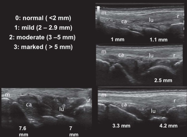
Rheumatoid arthritis. Dorsal longitudinal scan of the wrist showing joint cavity widening due to intrarticular fluid and synovial hypertrophy. Semiquantitative scoring systems in gray scale ultrasound on a scale of 0–3 (0, none; 1, mild; 2, moderate; 3, marked). Abbreviations: r, radius; lu, lunate bone; ca, capitate bone; m, third metacarpal bone
Spectral Doppler
Doppler ultrasonography is primarily used for the hemodynamic assessment of blood vessels. Doppler technique is based on the physical phenomenon, which consists of a change in the frequency of a sound wave resulting from motion of either the source or the receiver and reflect, giving us information about the movement of objects such as red blood cells in vessels. Spectral Doppler allows us to measure different blood flow parameters, some of which, such as the resistive index (RI), can be used to assess synovial inflammation.
In the cerebral and renal arteries, high diastolic flow is normal because low peripheral resistance is essential for continuous high perfusion. Conversely, normal blood flow in musculoskeletal tissues is characterized by high resistance, because the diastolic velocity has been considered to be zero, so the RI, which is the ratio between the systolic peak minus the end diastolic flow and the systolic peak, has a value of 1 in normal conditions; a decrease in the RI is registered in case of inflammation or neo-angiogenesis (27) (Figure 2).
Figure 2.
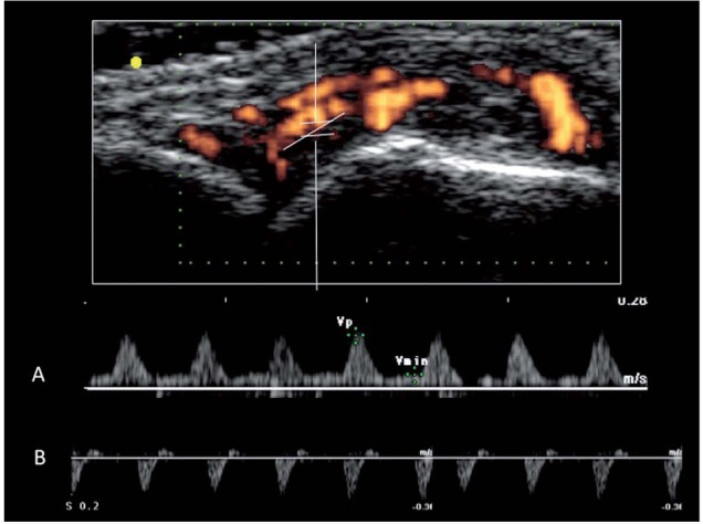
A. Spectral Doppler sonogram of the wrist joint of a rheumatoid arthritis patient. A persistent flow during the diastole and a reduced resistive index (RI), due to an abnormal vascularisation, can be observed. B. Example of a spectral Doppler sonogram in normal conditions. Physiological flow in musculoskeletal tissues is characterized by high resistance, because the diastolic velocity has been considered to be zero, so the RI has a value of 1
The inflammatory process is characterized by an increased perfusion and permeability of vessels and neovascularization and consequently an increased of the diastolic velocity and decreased RI at the level of inflamed synovium. Therefore, cut-off levels of RI are necessary to differentiate pathological flow from synovitis – like changes that can be documented also in healthy subjects. In addition, cut-off levels of RI are necessary to differentiate pathological flow from normal perfusion that can be found in joints of healthy control subjects. Terslev et al. (28) have reported a cut-off level of RI in healthy subjects ranging from 0.83 to 0.90. In another study Terslev et al. (29) reported a mean RI of 0.80 at the wrist and metacarpophalangeal joints in healthy subjects, which was higher than the values in the range of 0.4–0.76 reported in the literature for patients with rheumatoid disease (28).
More recently, we found a significant difference between mean RI at the wrist and finger joints in patients with rheumatoid disease and healthy subjects (0.72 versus 0.86, respectively) (30). We, also, observed a significant difference in mean RI between patients with early onset and those with long-standing of disease (0.71 versus 0.74). Inflammation leads to increased blood flow in synovial and peri articular tissue, at tendon insertions, and in tendon sheaths, which can be detected by CD ultrasonography. In addition, in clinical practice, spectral Doppler can be useful to distinguish real flow versus artefacts in some occasional doubtful cases.
In inflammatory arthritis, color or power Doppler signals within synovial hypertrophy is the main pathologic marker of inflammatory activity (31, 32).
Color Doppler Ultrasound
CD mode can detect pathological synovial blood flow, which reflects the joint inflammatory activity (33). CD ultrasound generates a real-time map of colored pixels in the gray scale setting, showing sites of blood flow in the area of interest defined by the Doppler box.
The color of the signal indicates the direction of blood flow, with red spots generated by blood flow directed toward the probe and blue ones away from it. The flow direction is arbitrarily assigned the colour red or blue, indicating flow toward or away from the transducer. No signal is displayed if the direction of blood flow is perpendicular to the ultrasound beam.
CD gives any information about the number of moving cells. The CDUS technique is affected by the direction of blood flow, whereas the images obtained by PDUS are not direction-coded. PDUS is, therefore, able to detect a very slow blood flow rate.
A number of studies have shown the ability of CD to detect synovial vascularization in patients with chronic inflammatory arthritis, to differentiate between normal and abnormal blood flow and between inactive and active pannus, and to assess changes in synovial blood flow induced by different treatments. However, CD findings can be found at the wrist and metacarpophalangeal joints in healthy subjects (28, 30). Terslev et al. (28) scanned 324 joints in 27 healthy subjects, and found color Doppler signals in 15 of 27 wrists, 17 of 27 first carpometacarpal joints, 10 of 135 metacarpophalangeal joints, and 1 of 135 proximal interphalangeal joints. Previously, we observed color Doppler signals in 45 of the 430 joints of healthy subjects and the positive Doppler findings were most common in the wrists, followed by metacarpophalangeal joints and proximal interphalangeal joints (30).
Several methods have been proposed for scoring intra-articular color Doppler signals (34). The two main methods are the semiquantitative scoring system, which assesses the amount of color Doppler signals using a four-point grading scale, and the quantitative scoring system, which counts color pixels in synovial tissue using dedicated post-processing software. The semiquantitative method allows for more rapid assessment, and relies on the skills and experience of the sonographer, whereas the quantitative method, with computer-assisted measurement of color pixels 12 on the basis of either an absolute number of pixels, or a percentage of an area covered with pixels, in a region of interest, has the potential to be more reproducible, but requires more time (35). In a recent study, Terslev et al. (36) compared these two methods when scoring at the wrists of 46 rheumatoid patients in the dorsal view and a high correlation and comparable inter-reader agreement was found between these scoring systems.
Power Doppler Ultrasound
Power Doppler encodes the amplitude of the Doppler signal resulting from the volume of blood present, regardless of direction and speed, thereby detecting flow also in case of perpendicularity of the flow to the US beam and enables sensitive assessment of low-velocity flow in small vessels of the synovial tissue, thus being more appropriate for the analysis of neo-angiogenesis (37, 38). Recent data suggested that PD can provide more accurate data than grey scale for synovitis in RA (39). Even if, using last-generation ultrasound equipment, substantial difference between color and power Doppler with regard to detection of slow flow was not found (40), the majority of studies investigating the role of ultrasound in patients with chronic inflammatory arthritis have been performed using power Doppler.
A number of studies showed the validity and reliability of power Doppler in the assessment of vascularized synovium in joints and tendon sheaths in patients with chronic inflammatory arthritis and a strong correlation was found between the qualitative estimates of blood flow obtained by PDUS and synovial blood vessel density in a histological tissue section (41-43). PDUS has also been validated against other modalities including contrast-enhanced MRI scanning (44), considered as the gold standard assessment technique. Walther et al. (43) compared PD and synovial histopathology of the knee joint in 23 patients (10 affected by RA, 13 by OA), who were undergoing total knee arthroplasty. They evaluated both grey-scale and PD synovitis, quantifying them on a 4-point scale, according to the Newman score (45) and adding an automatic quantification of red pixels a sign of vascularization. Both the qualitative and the quantitative estimation of vascularization correlated with the histopathological findings, leading to the conclusion that PD is a valid tool for the detection and quantification of synovial vascularization(43). One year later, a similar work was done on the hip using the same protocol (24 patients, 15 with OA and 9 with RA); the results showed a good correlation between histological findings and PD in the detection of synovial vascularity (46).
When contrast-enhanced MRI (CE-MRI) has been used for assessing the validity of PD, a good correlation between the two techniques has been shown (47). Szkudlarek et al. (44) showed a good correlation between power Doppler signals and contrast-agent enhancement in MRI for visualizing active synovitis and the sensitivity and specificity of CD/PD Doppler ultrasonography compared with MRI were 89% and 98%, respectively, for the assessment of inflammation in metacarpophalangeal joints in patients with RA.
Several studies have evaluated the validity of power Doppler in the assessment of synovitis in patients with inflammatory arthritis (48). Kaoru Takase-Minegishi et al. (49) in their systematic review and meta-analysis of studies evaluating the diagnostic test accuracy of US for synovitis detected by MRI as the reference standard for wrist, metacarpophalangeal joints, proximal interphalangeal joints and knee joints of patients with RA, found that US, especially power Doppler US, is a valid and reproducible technique for detecting synovitis in the wrist and finger joints of these patients .
In a number of studies, the presence of synovial Doppler signal has shown diagnostic value in relation to the development of chronic arthritis or RA in patients with early undifferentiated synovitis (50-52). Some PDUS signs can help to distinguish RA from other causes of arthritis. Gutierrez et al. (53) found that in early psoriatic arthritis hypoechoic swelling surrounding the extensor digitorum tendon and peritendinous PD signal seem to predominate compared with intra-articular PD signal in early RA (Figure 3). This study supports the hypothesis that the extra-articular tissue involvement seems to be characteristic of spondyloarthritis rather than RA (54). Additionally, it was noted that the peritenon extensor tendon inflammation pattern was frequently observed in patients with short time of disease duration. According to this observation, it is believed that the inflammatory process in psoriatic arthritis (PsA) could begin at the soft tissue level surrounding the extensor digitorum tendon, successively involving the synovial membrane (55), whereas an isolate synovitis could be detected in the late stages of PsA disease. These preliminary results suggest a relevant potential role for PDUS in the differential diagnosis between RA and PsA.
Figure 3.
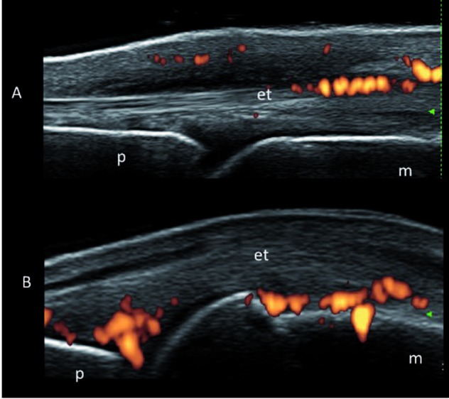
A: Psoriatic arthritis: Metacarpophalangeal joint on dorsal longitudinal scan showing theperitenon extensor tendon inflammation pattern. B. Rheumatoid arthritis. Metacarpophalangeal joint. Dorsal longitudinal scan showing intra-articular power Doppler signals prevalent at fat pad level. Abbreviations: et, extensor digitorum tendon; m, metacarpal bone; p, proximal phalanx. Adapted by Gutierrez et al. (50)
PDUS has several potential roles in gout diagnosis. It has shown diagnostic value in detecting early changes in the soft tissues in cases of gout and can be used especially when the clinical, laboratory and radiographic studies are negative or inconclusive. Some studies comparing the sensitivity and specificity of US versus X-ray showed that US is more sensitive that X-ray, because the sonographic changes are present at earlier stages, in comparison to typical X-ray signals (56, 57). PDUS allows the visualization of the characteristic aspects for the diagnosis of gout (such as the “double contour signal”, defined as an irregular linear hyperechoic layer on the superficial margin of the anechoic hyaline cartilage and parallel to the bone cortex, without a posterior acoustic shade, the bright hyperechoic foci, referable to microtophi and hyperechoic areas in synovial tissue, and tophi within joints, soft-tissue structures such as tendons, ligaments and bursae, and also invading into bone) as well as joint effusion, synovial hypertrophy and hypervascularity. PDUS allows the visualization of the changes in the inflammatory process of gout. The evaluation of gout by PDUS demonstrates increased flow in the acute phase of the podagra crisis, and generally, PD shows no flow when the patient is out of the gout crisis, but it has been observed that painful periarticular areas in patients with a known diagnosis of gout can exhibit hyperechoic tophi and flow with PD, even without the classic signs of crisis (58). Furthermore, PDUS has been demonstrated to be useful in the evidence that synovitis is frequently present in osteoarthritis. Despite osteoarthritis traditionally being viewed as a non-inflammatory disease, studies have shown a high prevalence of synovitis in both non-erosive and erosive osteoarthritis. The difference in prevalence of power Doppler activity across non-erosive and erosive osteoarthritis suggests more “active” synovitis with more neoangiogenesis in erosive osteoarthritis (59, 60) .
Several methodical papers have confirmed the potential of PDUS for the measurement of disease activity in patients with RA (61-64). Different semiquantitative systems have been proposed for scoring intra-articular power Doppler signals. The most frequently used are semiquantitative scoring systems that score the Doppler information on a scale of 0–3 (0, no intra-articular PD signal; 1, mild: singles vessel signal; 2, moderate : confluent vessel signals in less than half of the synovium area; 3, severe: vessel signals in more half of the synovium area), with increasing scores indicating increasing amounts of color in the synovium (65, 66) (Figure 4). This system is easily applicable in clinical practice and clinical trials, and is reliable and responsive to therapeutic interventions.
Figure 4.
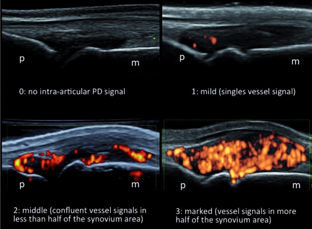
Rheumatoid arthritis. Metacarpophalangeal joint. Dorsal longitudinal scan showing intra-articular power Doppler signal. Semiquantitative scoring systems that mark the Doppler information on a scale of 0-3 (0, none; 1, mild; 2, moderate; 3, marked). Abbreviations: et, extensor digitorum tendon; m, metacarpal bone; p, proximal phalanx
Although PDUS is useful for the assessment of synovitis, it is a flexible and sonographer-dependent examination and settings of US machines and the scanning technique of the sonographer can greatly influence the visualization of synovial vascularity and reproducibility is a major problem (67). Several studies demonstrated that appropriate training in scanning technique and reading of PDUS images could improve and stabilize the reproducibility and reliability of scoring trials (68-71). A quantitative method to measure pixel counts of synovial vascularity in the region of interest has been proposed (72, 73).
More recently in the MEDUSA project, a computer aided diagnostic system that supports an assessment of synovitis severity, has been proposed (74). Quantitative assessment of synovial Doppler signal has the advantages of being more objectives than semiquantitative assessment. Schmid et al. (75) compare subjective estimation with grading system for synovial PDUS (using the semi-quantitative score from 0 to 3 proposed by Szkudlarek et al. and Naredo et al.) and computerized quantification of synovial perfusion in active RA and concluded that electronic measurement of the maximum colour fraction in the synovium may replace semi-quantitative scores, and is more sensitive to evaluate the change for follow-up RA trials (65, 76, 77). In addition, PDUS may be considered an important outcome measure, in fact high vascularity of synovitis and persistent synovitis, evaluated by PDUS, even in the absence of clinical symptoms, are predictive markers for the development of severe erosive RA (78, 79). Inpatients with RA in clinical remission, the presence of a power Doppler signal at the wrist and metacarpophalangeal joints was found to be associated with risk of relapse and structural progression (80). Other studies detected radiographic evidence of newly developed bone erosions in RA patients in persistent remission, suggesting that a residual subclinical inflammation may be missed using only clinical and laboratory data and therefore the use of imaging techniques has been claimed to state true disease remission (81, 82).
Vreju (83) scanned the metacarpophalangeal joints of twenty-four patients with RA in clinical remission according to EULAR criteria and found a positive correlation between the US bone erosions and PDUS signs of subclinical synovitis. Therefore this data support the hypothesis that in patients with RA joint damage and bone erosions are considered the result of persistent synovitisand that bone erosions may occur also in patients achieving clinical remission. Brown et al. (82), in asymptomatic RA patients, using ultrasonography, observed synovial hypertrophy and abnormal power Doppler signal in 73% and 43% of patients, respectively, while MRI revealed synovitis and bone marrow oedema in 96% and 46% of these patients. Therefore, its use in the clinical setting of remission may reveal active vascularized synovium and therefore influence therapeutic decision-making.
PDUS can help to differentiate patients with aggressive disease in early RA, allowing the targeting of expensive therapies to those with a poor prognosis (84). Many longitudinal studies have demonstrated the ability of PDUS to assess changes in synovial blood flow induced by different treatments, including intra-articular corticosteroid injections, systemic corticosteroid therapy, synthetic and biologic DMARDs and therefore it could be considered a useful tool for therapy monitoring in patients with chronic arthritis (85-89). In our previous study we evaluated 20 patients with clinically active synovitis of a small joint unresponsive to systemic drug treatment. They underwent a sonographic guided intralesional injection with triamcinolone acetonide. Joint cavity widening and PD signals were evaluated and graded on a semiquantitative scale ranging from 1 to 4. Clinical and sonographic follow up examinations were carried out 2 weeks after the injection with triamcinolone acetonide. The results of this study support the view that US and PDS may be regarded as useful adjunctive tools for assessing short-term soft tissue changes after intra-articular injection treatment with triamcinolone acetonide in the small joints of patients with chronic synovitis (90).
Contrast-Enhanced Color/Power Doppler
An ultrasound contrast agent consists of a suspension of relatively uniform, highly reflective, stabilized gas-filled spheres, which enhance the Doppler signal, increasing its sensitivity for detection of low velocity blood flow at the level of the small vessels.
In contrast to MRI contrast medium, the US contrast agent stays inside the lumen of the vessel and does not diffuse in the extraarterial tissue. No extravasation into the surrounding tissues is found, indicating that ultrasound contrast medium acts as an intravascular contrast agent.
First-generation ultrasound contrast agents, for example, galactose palmitic acid (Levovist®, SheringAG, Berlin, Germany), increase the sensitivity of the color signals. The more recent type of US contrast agents consists of stabilised microbubbles of a sulphur hexafluoride gas (SonoVue®, Bracco, Milan, Italy). They provide a higher sensitivity and allow the delineation of weak intraarticular blood flow. This method depicts the blood flow in a grey scale image that provides only inaccurate information on the anatomic structures around the perfused areas (Figure 5).
Figure 5.
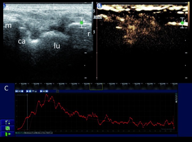
Rheumatoid arthritis. (A) Dorsal longitudinal scan of the wrist showing moderate joint cavity widening mainly due to an increased amount of synovial hypertrophy. (B) After intravenous injection of an ultrasound contrast agent (SonoVue), multiple intra-articular microbubbles, representing the degree of vascularity of the synovial membrane due to joint inflammation, could be detected. (C) The corresponding contrast enhancement curve after administration of SonoVue.Abbreviations: r, radius; lu, lunate bone; ca, capitate bone; m, third metacarpal bone. Adapted by Filippucci et al. (29)
The use of contrast-enhanced US (CEUS) seems to provide significantly higher sensitivity than PDUS in the identification of abnormal vascularization in joint inflammation, allowing a more exact measurement of the synovitis, as well as a quantitative assessment of inflammation by using the analysis of time-intensity curves. Analysis of time-intensity curves in a region of interest allows quantification of synovial inflammation. The area under the curve, the slope of the ascending and descending curve, measurement of flow rate, assessment of vascular volume, and estimate of mean velocity are all aspects allowing detailed evaluation of synovial inflammation.
In 2005 in a multicenter study of the International Arthritis Contrast Ultrasound study group, comparing contrast-enhanced ultrasound versus gray scale and power Doppler ultrasound for detection of joint vascularity in patients with rheumatoid disease, a total of 113 joints were examined and contrast-enhanced ultrasound was found to improve assessment of vascularized synovial proliferation and the differentiation of active synovitis from inactive intra-articular thickening (91). A number of other studies have investigated contrast-enhanced power and/or color Doppler ultrasound in the assessment of synovitis (92, 93). Most of these have been performed in patients with RA and showed that contrast agents amplify detection of the color signal and facilitate differentiation between active and inactive synovitis (94, 95). Other studies reported that contrast enhanced color Doppler US significantly improves the detection of intra-articular vascularization (94) and may increase the detection of even minor perfusion (96). There is evidence showing that administration of a contrast agent significantly improves the detection of color and power Doppler signals at the knee and finger joints in patients with RA (94, 97).
Previously, we employed PDS with ultrasound contrast agent to evaluate the degree of vascularization of the synovial membrane of the knee of 41 patients with RA, as well as to correlate the values of the area under the time–intensity curves obtained after administration of Levovist to the clinical and laboratory findings of disease activity (98). The results showed that the area under the curves (AUC) correlated with the degree of knee inflammation, being significantly higher in patients with clinically active synovitis compared with those with inactive synovitis. In addition, the mean value of the area under the curves was weakly correlated with the number of swollen joints, whereas it showed a stronger correlation with the composite indexes of disease activity, such as disease activity score and chronic arthritis systemic index. Therefore, the development and introduction of microbubbles ultrasound contrast and new ultrasound images may be considered a very promising technique for evaluating and measuring the degree of knee joint inflammation in RA and the time-intensity curves may have a clinical potential for both diagnostic and therapeutic purposes (98). In this regard, PDS with an intravenous ultrasound contrast agent has been shown to be able to detect changes in synovial perfusion after intra-articular steroid injection and may be an additional useful method in the evaluation of the therapeutic response.
In our previous study, to evaluate the ability of PDUS with ultrasound contrast agent to assess the synovial perfusion changes induced by intra-articular steroid injection therapy, we studied eighteen RA patients with a history and signs of active knee synovitis. Gray-scale US and PDS with an intravenous ultrasound contrast agent (Levovist) examinations were carried out before and 3 weeks after the intra-articular steroid injection. The calculation of the time–intensity curves provided a quantitative estimation of the synovial perfusion. The comparison between baseline and follow-up median values of the AUC showed a statistically significant reduction of blood flow in synovial pannus after intra-articular steroid therapy. These data confirmed the ability of PDUS with ultrasound contrast agent to evaluate the therapeutic response (99).
The main issues limiting the use of ultrasound contrast media are the relatively high running costs involved, the relatively short duration of examination, and the need for optimally designed bubbles for near-field investigation at higher frequencies.
Magnetic Resonance Imaging (MRI)
MRI is a multiplanar, non-ionizing and non-invasive imaging technique and is considered the reference imaging modality in the assessment of soft tissue inflammation, in form of synovitis, tenosynovitis, enthesitis and bursitis, as well as joint damage (in terms of bone erosion, cartilage loss, and tendon rupture) in chronic arthritis (100-105). It has advanced our understanding of many types of arthritis, both with respect to inflammatory processes and articular damage. According to the Recommendation of the Arthritis Subcommittee of the European Society in Musculoskeletal Radiology (106), MRI allows the:
assessment of peripheral joints for active inflammation in the form of effusion, synovitis, bone marrow oedema, as well as the subsequent structural lesions, such as articular surface damage and cortical bone erosions,
assessment of active inflammatory lesions and structural changes in the sacroiliac joints,
assessment of inflammatory and post-inflammatory lesions of the vertebral joints, i.e. assessment of the inflammatory activity, aseptic spondylodiscitis, atlanto- axial/atlanto-occipital structural lesions,
assessment of tenosynovitis and enthesopathic lesions,
confirmation of clinical diagnosis based on imaging characteristics and/or location of lesions,
qualitative, semi-quantitative and quantitative measurements of active inflammation and chronic joint damage.
As far as rheumatic diseases are concerned, MRI surpasses plain radiography and US with its ability to visualise the bone marrow involvement. Also, early inflammatory features within soft tissues (joints, tendons sheaths and bursae, muscles) that are not seen on plain radiography in detail and are inaccessible to ultrasound (e.g. of the spine, sacroiliac joints) or their assessment in ultrasound is limited (e.g. hip and gleno- humeral joints) are well seen in MRI. Although clinical examination has been a cornerstone in identifying and monitoring disease progression in RA and PsA patients, MRI has been shown to be more sensitive than clinical examination for identifying synovitis (Figure 6). The more important indications for MRI in patients with musculoskeletal rheumatic diseases include early diagnosis of inflammation, confirmation of the presence of clinically active changes and post inflammatory structural lesions, disease follow-up including monitoring of therapy response and identification of disease complications.
Figure 6.

Rheumatoid arthritis. Ankle joint. (A) Sagittal STIR image showing tibial-talus joint cavity widening. (B) Axial T1 fat sat images before and (C, D) after paramagnetic contrast showing the synovial thickening characterized by intense contrast enhancement, compatible with synovitis. Tenosynovitis of the posterior tibial and flexor digitorum longus tendons and (D) intense contrast enhancement, at the level of the medial malleolus of tibia, compatible with osteitis, can also be observed
MRI can identify bone erosions earlier than conventional radiography and allows the evaluation of bone marrow edema/osteitis, due to the presence of an inflammatory infiltrate within subchondral trabecular bone, and synovitis, which may be important precursors to bone erosions in RA (107) (Figure 7). Signs of inflammation may be detected by MRI also in PsA, and findings such as synovitis, tenosynovitis and bone marrow oedema (BMO) document the presence of an inflammatory process, although not specific for PsA (108). In early PsA disease, MRI of wrists and hands detected diaphyseal BMO and/or enthesitis in more than 70% of PsA patients, whereas these features were absent in a matched group of RA patients (109, 110). Given these properties, MRI has been proposed as a diagnostic tool for individuals with suspected inflammatory athritis and has been proposed as a means to improve rheumatologists’ ability to diagnose early RA and predict which patients will likely develop progressive disease and thus should receive more aggressive treatment (111).
Figure 7.

Juvenile idiopathic arthritis. (A) Sagittal T1 fat-sat image of ankle, before paramagnetic contrast showing moderate talo-calcaneal joint widening due to intrarticular fluid and synovial hypertrophy . (B-C) Sagittal T1 fat-sat images, after paramagnetic contrast, showing synovitis in the talo-calcaneal joint . (B) Contrast enhancement in the superior part of the talar body and calcaneous, compatible with osteitis, can also be observed
Currently its use is not extended to all affected patients due to long examination times, its elevated cost, limited availability, need for contrast medium to increase specificity, and contraindications in certain patients (112). In addition, MR images are in many cases non-specific and require differentiation from other pathologies manifested by the same spectrum of changes. The main indications for MRI in patients with musculoskeletal rheumatic diseases include (106):
early diagnosis of inflammation in both soft tissues and bone marrow, before destructive lesions develop (cysts, erosions, cartilage damage);
confirmation of the presence of clinically active changes and post-inflammatory structural lesions;
quantitative assessment of synovitis;
prognostic value in early RA and PsA;
disease follow-up, including monitoring of therapy response,
Diagnostic value of MRI in early diagnosis and synovial assessment
There are two main ways in which MRI can assist the clinician in this respect. First, the presence of subclinical synovitis can be confirmed, allowing for example, the patient with non-specific hand and/or wrist pain to be differentiated from the patient with true inflammatory synovitis. The second way by which MRI can assist in making a diagnosis of RA is by revealing erosions that comprise one of the seven ACR 1987 diagnostic criteria (113). Synovial thickening is the earliest pathologic abnormality in RA and it is secondarily responsible for bone and cartilage damage (114).
Studies comparing normal individuals with RA patients have shown that while minor synovial enhancement may be seen in normal wrists, significant thickening and inflammation of the synovial membrane is confined to those with inflammatory arthritis (115). Where there is a clinical diagnosis of RA, >90% have MRI evidence of synovitis and 70–80% have tenosynovitis (116). The synovial membrane, or synovium, which can be identified macroscopically as a connective tissue that lines normal joint cavities, bursae, and tendinous sheaths is usually too thin to be visible on MR images. The thickening of synovial tissue caused by the inflammatory process may be identified at MR imaging (117) and is also characterized by increased vascularity, which is a result of angiogenesis (118). MRI signs of synovitis include increased synovial volume, increased water content, contrast enhancement or a combination of them (119-121). The MRI data suggested that the level of synovial thickness was critical in determining the amount of bone damage. In fact, several studies have shown that synovial volume correlates with synovial inflammatory activity and with joint swelling and tenderness and is predictive of clinical disease activity (122-125).
The OMERACT group defines synovitis as an area in the synovial compartment with increased contrast enhancement whose thickness exceeds the width of the normal synovium (126, 127). Synovitis has intermediate to low signal intensity on T1-weighted images and high signal intensity on T2-weighted images, due to the increased water content.
The use of fat-suppressed T2-weighted MR imaging more clearly delineates disease extent. Synovitis and joint fluid are usually difficult to differentiate on unenhanced MR images; however, on T2-weighted images synovitis has a lower signal intensity than does joint effusion (128). The use of intravenous gadolinium-based contrast material is necessary to estimate the degree of synovial inflammation and for differentiation between active synovitis and fibrotic pannus (Figure 8). Fibrotic pannus, which is usually present in end-stage RA, appears relatively hypovascular after the intravenous administration of gadolinium-based contrast material. Sugimoto et al. (129) have shown that the “bilateral joint hyperuptake” criteria, evaluated through MR after the infusion of paramagnetic contrast (Gd- DTPA), increases the sensitivity of the ACR criteria for RA from 77% to 96%. The combination of this with positive biologic markers such as anti-citrullinated peptide antibodies or rheumatoid factor (RF), with the MRI parameters (symmetric synovitis, bone edema, erosions) has a sensitivity and a specificity for the detection of early RA of 82.5% and 84.8% respectively (130). Østergaard et al. (131) demonstrated that assessment of synovitis using the short tau inversion recovery (STIR) or T2 fat-saturated images is less accurate and reproducible than using T1-weighted post-contrast images.
Figure 8.

Undifferentiated arthritis in an 11year old patient, with pain and swelling at the ankle. (A) Axial T1 fat-sat image of ankle after paramagnetic contrast and (B) sagittal T1 fat-sat image, after paramagnetic contrast showing, at the medial and posterior regions of the ankle, extensive area, characterized by intense contrast enhancement, compatible with synovitis. (C) Coronal STIR, (D) axial DWI, (E) coronal DWI, (F) Whole body MRI images showing synovitis in the medial and posterior regions ok ankle
Contrast-enhanced T1-weighted images are considered more sensitive and specific in the assessment of acute synovitis, which has been shown to enhance rapidly and intensely after the intravenous administration of gadolinium-based contrast material, unlike joint effusion, which does not enhance in the early phase. The delay between contrast administration and scanning is important as the volume of enhancing synovitis increases initially before stabilizing after about 4 min. After 6–11 min contrast reaches the synovial fluid, obscuring the synovium/fluid interface.
The use of fat suppression increases the contrast between the inflamed synovium and adjacent structures on contrast-enhanced T1weighted images (132). Dynamic contrast enhanced MRI (DCE-MRI) involves the acquisition of sequential images in rapid succession every few seconds during and after the intravenous administration of contrast agent. This allows the time-course of the synovial enhancement to be analysed and measurements made from the enhancement curve are sensitive to various physiological parameters, including synovial perfusion and capillary permeability.
The region of interest (ROI) of inflamed synovium from which the enhancement curve is determined may be chosen in different ways. Then it is possibile to measure several parameters from the enhancement curve, that include the early enhancement rate, the maximum enhancement and the late or static enhancement. The early enhancement rate has been shown to correlate with erosions (133-135), erosive progression (136) and effects of treatment (137). There is a better correlation with histology (125) and response to treatment (138) than the static enhancement. The enhancement rate correlates with other imaging measures of synovial volume, erosion, vascularity, capillary permeability and metabolic activity. The rate and magnitude of synovial enhancement on sequential MR images after bolus intravenous injection of gadolinium have been shown to correlate with the histological severity of inflammation in the synovium and with clinical markers of disease activity (139).
Van de Sande et al. (140) showed that the shape analysis of DCE-MRI might have potential as a diagnostic biomarker in early arthritis patients. They observed a significantly higher percentage of Type 4 TIC shape (fast initial enhancement followed by a quick washout phase) in the RA patient group compared with the non-RA group. There is evidence showing correlations between perfusion imaging and histological synovial inflammation (125, 141). Vordenbäumen et al. (141) compared dynamic contrast-enhanced magnetic resonance imaging and histological synovitis of the second MCP joints in 9 RA patients. The results of this study demonstrated that synovitis of MCP joints measured by maximum enhancement on dynamic MRI strongly correlates to histological inflammation within the same joint.
These data underscore the validity of dynamic contrast-enhanced MRI for the assessment of the degree of synovitis in small joints. Relevant studies have mostly focused on the knee, where synovial tissue can be most readily obtained. Konig et al. (142) evaluated inflammation in the knees in 20 RA patients using dynamic enhanced MRI and compared these results with histological data obtained from biopsy of the synovial membrane performed arthroscopically or operatively in 12 patients. Their preliminary findings revealed hyperintense signal on T2 weighted images, correlating with “remarkable” enhancement on post contrast-enhanced T1weighted images in three patients, and this appearance matched hypervascular pannus in a histological assessment. In contrast, hypointense lesions on MRI correlated with histologic evidence of fibrous pannus with a “burnt out” appearance (117).
Gaffney et al. (143), compared the intensity of rheumatoid synovitis in the knee, measured using the initial rate of enhancement on postcontrast MRI scans, with a score for inflammation calculated from multiple synovial biopsy samples taken from the suprapatellar bursa. They found a strong correlation between the enhancement-rate and a composite histologic inflammatory score quantifying polymorphonuclear cellular infiltration, hyperemia, and fibrin deposition. Similar results were obtained by Tamai et al. (144), who compared the enhancement-ratio with a histologic score for inflammation on synovial biopsy specimens taken from the knee in 10 patients. In their study, enhancement post-contrast was greater in regions with a high degree of fibrin exudation, cellular infiltration, villous hypertrophy, vascular proliferation, and infiltration by granulation tissue but not in regions affected by fibrosis. A similar work was done by Østergaard et al. (124) on the knees of 17 RA patients and 25 with osteoarthritis (OA). The volume of synovial membrane was determined using a manual outlining method to determine its boundaries on consecutive slices, and this surrogate measure of synovitis was then compared with a histologic assessment of inflammation, again from biopsy specimens taken from the knee arthroscopically.
A strong correlation was found between this MRI synovial volume score and a composite histologic inflammation score. Synovial membrane volumes were also shown to be higher in RA and PsA than OA knees. Even though MRI will probably only rarely be able to assign specific diagnoses alone, it can be a very useful addition to the differential diagnostic process. MRI may be valuable for diagnosing specific arthritides, including early RA, in patients with undifferentiated arthritides, but the sensitivity and specificity, and so on, of MRI are not yet known. The few studies that employ MR for the differential diagnosis of RA with other rheumatic diseases, such as systemic lupus erythematosus, Sjögrens Syndrome or PsA don’t find statistically significant differences regarding synovitis, erosions, or tendinous alterations (145, 146). The different parameters evaluated by MRI are employed as predictive markers of erosions, allowing the clinician to select patients with a worse prognosis and establish a rapid and aggressive therapeutic strategy. Cimmino et al. (146) using dynamic MRI of the wrist, observed that the rate of increase in enhancement following contrast injection did not differ between PsA and RA patients when they were matched for disease activity, but in both groups it was higher than in normal control individuals. The authors concluded that dynamic MRI cannot be used diagnostically to differentiate PsA from RA. Others have measured PsA synovitis on static magnetic resonance scans. Savnik et al. (147) noted that the volume of synovial membrane was increased but did not change significantly over 1 year, contrasting with RA patients, in whom it fell in response to therapy. Jevtic et al. (148) described MRI of the finger joints in a group of patients with PsA, three with Reiter’s syndrome and with RA. Although in some PsA patients synovitis was observed to conform to a typical rheumatoid pattern, in others there was inflamed tissue extending far beyond the joint capsule, involving neighbouring structures such as thickened collateral ligaments and surrounding periarticular soft tissue. McGonagle and colleagues (149) went on to describe in greater detail the MRI features of enthesitis that may be seen in association with synovitis in PsA, and postulated that true PsA can be distinguished from RA with concomitant psoriasis on these grounds.
Although histopathological studies have suggested that inflamed synovial membrane of PsA differs in certain subtle ways from rheumatoid synovium with less lining layer hyperplasia, more subsynovial oedema and a greater number of synovial vessels per square millimetre, on MRI, PsA synovitis appears indistinguishable from that of RA (150). In an other study, Cimmino et al. (151) studied 8 patients affected by acute gout arthritis involving the wrist and MRI was performed during the acute attack. They found a high frequency of synovitis and bone lesions of the wrist, but they were difficult to differentiate from those of RA.
Synovitis is also a feature of OA, and angiogenesis, inflammation, and innervation are important processes in the pathophysiology contributing to the symptoms and radiological progression of OA. The different pathogenesis of OA and RA may reflect a difference in the mechanisms of inflammation and angiogenesis, and this could possibly be expressed in DCE-MRI. However, only a few studies have evaluated synovitis in OA using MRI and these studies have mainly estimated the volume of knee joint synovitis (124, 152). Kirkhus et al. (153) to investigate the utility of the DCE-MRI to differentiate between finger-joint synovitis in established OA and RA, studied 19 patients (11 with RA e 8 with OA) and 6 healthy individuals without any symptoms or known disease. All subjects underwent DCE-MRI of one hand and from the signal intensity curves, the three parameters: endothelial transfer constant, elimination rate constant from extracellular space back to plasma and elimination rate constant from plasma by renal excretion, were calculated. The results of this study showed that DCE-MRI with derived pharmacokinetic parameters can provide useful information in differentiating synovitis in hand OA from synovitis in RA, and it is also possible that the method can be used to estimate the degree of synovial inflammation.
MRI synovitis is a reasonable indicator of true synovial inflammation, but changes resembling mild synovitis or small bone erosions are occasionally found in the MCP and wrist joints of healthy controls. Ejbjerg et al. (115) studied 28 healthy individuals using contrast-enhanced MRI of the wrist and MCP joints. Low grade of synovitis was found in 9% of the MCP joints and in 10% of the wrist joints, but almost half of these changes were observed in 3 subjects who had elevated CRP levels and therefore could possibly have had subclinical inflammation. Dynamic MRI revealed only minimal synovial enhancement compared with levels in RA patients that were 30-fold higher. Furthermore, lesions suggesting erosions in healthy individuals are usually small, single and do not present gadolinium uptake. The results of this study give an insight into the specificity of MRI synovitis and indicate that while low-grade enhancement can occur in normal joints, most RA joints exhibit much greater degrees of membrane thickening and enhancement, which is strongly indicative of disease.
Quantitative assessment of synovitis
Quantifying total synovial volume (hypertrophy) and the synovial volume could be a marker for disease activity (122, 154-156). Synovitis can be assessed with quantitative or semiquantitative methods. With quantitative methods, inflammatory activity can be estimated by quantifying the volume of synovial tissue or evaluating the increase in early synovial signal intensity at dynamic contrast-enhanced MR imaging. The volume of inflamed synovium can be quantified on contrast-enhanced MR images with use of manual or computer-assisted techniques. Visualization of the inflamed synovium for volume measurement requires intravenous contrast to reliably exclude other tissues. The analysis of gadolinium-enhanced MRI can provide objective information regarding the severity of inflammation. DCE-MRI is an alternative quantitative method to measure synovitis by administering gadolinium-based contrast agents (GBCA) intravenously and collecting sequential images of the joint in a time course (157). The enhancement curve generated by DCE-MRI can be used to estimate physiological parameters, such as Ktrans, the volume transfer constant of GBCA between blood plasma and the synovium. This endpoint is related to capillary permeability and vascularity in the synovium, and correlates strongly with histological measures of inflammation (158).
DCE-MRI has previously been used to evaluate synovial inflammatory activity in patients with RA in the knees showing that the steepness of the dynamic curves correlates better with histological synovial vascularity and inflammatory cell infiltrate than measures of the corresponding post-contrast-enhancing synovial volumes (124, 159). The steepness of the dynamic curve in the synovium has also been shown to be very sensitive to change after intra-articular steroid injections in knee joints with arthritis (160-162) and recently the same observation was published for BME in wrists of RA patients starting anti-TNF-a treatment (163).
In RA the most frequently used method is the OMERACT Rheumatoid Arthritis Magnetic Resonance Imaging Score (RAMRIS)(126, 163), which allows assessment of synovitis in three wrist regions: the distal radioulnar joint; the radiocarpal joint; and the intercarpal, carpometacarpal, and metacarpophalangeal joints. Joints are assessed on a scale from 0 to 3. A score of 0 is normal, whereas scores of 1 (mild), 2 (moderate), or 3 (severe) reflect the presumed maximum volume of enhancing tissue in the synovial compartment (126). In table 1 definitions, basic sequences, anatomic planes and areas and RAMRIS System Scores as proposed by OMERACT Task Force are reported.
Table 1.
Definition, Basic Sequences, Anatomic Planes and Areas, and RAMRIS System Scores as Proposed by the OMERACT Task Force*

Both RAMRIS and DCE-MRI are valid measures for detecting treatment effect, but they are not interchangeable and may reflect somewhat different biological processes related to joint inflammation. Further, scoring MRI at the wrist is a complex task, requiring a detailed knowledge of the 3-dimensional anatomy of the carpal bones and joints as well as an understanding of the different sequences used, the signal characteristics of the tissues being imaged, and potential sources of error that are intrinsic to the modality itself. The latter include phenomena such as partial voluming, where signal from two sharply contrasting areas is “averaged” in the intervening zone, homogeneity of fat saturation, and variations in Gd-DTPA uptake, which need to be interpreted by a trained reader. This makes MRI scoring considerably more difficult than scoring radiographs to assess joint damage (164).
Cyteval et al. (165) developed a simplified MR imaging scoring method (Simplified Rheumatoid Arthritis Magnetic Resonance Imaging Score [SAMIS]) for assessing wrist and metacarpal joint damage in RA. SAMIS assessed only one hand and was based on the radiographic Simple Erosion Narrowing Score, thus reducing the number of study areas from 116 to 36. Erosions were scored with a scale from 1 to 10. Edema and synovitis were, respectively, scored on scales from 0 to 1 and 0 to 2. The simplified score is closely correlated with the MR imaging score standard RAMRIS, requires less examination time than RAMRIS (5 minutes vs 20 minutes) and shows good or excellent intra- and interobserver agreement (165).
The international MRI in arthritis group of OMERACT has developed the Psoriatic Arthritis Magnetic Resonance Image Score (PsAMRIS) for the evaluation of inflammatory and destructive changes in PsA hands (166). This is the most validated assessment system available and has a documented good intrareader and inter-reader reliability for status scores of all parameters (167). The responsiveness of the PsAMRIS was excellent for tenosynovitis (hand), synovitis (foot), and periarticular inflammation (hand and foot) (168).
Prognostic value of MRI
The ability of MRI to assess both detailed changes in bone structure (i.e. erosions) and synovial inflammation combined with its multiplanar capability is well established (169, 170). MRI can identify bone erosions earlier than conventional radiography (171) and can detect bone marrow edema and synovitis, which may be important precursors to erosive disease (172) (Figure 9). Given these properties, MRI has been proposed as a diagnostic tool among individuals with suspected inflammatory arthritis and as a prognostic tool among those with known RA. The ability to predict aggressive disease when a patient first presents is an important clinical goal, because this would allow potent and potentially toxic disease-suppressing medication to be targeted to patients who are most in need. This predictive ability has become even more desirable from a medico-economic viewpoint with the advent of anti–tumor necrosis factor alpha therapies, which have powerful antierosive effects (173).
Figure 9.

Rheumatoid arthritis. (A, B, C) Coronal STIR images showing joint effusion in the carpal joints, carpal-metacarpal joints and the distal radio-ulnar joint. Bone edema of the carpal bones and the metacarpal bases, with multiple erosion involving the carpal bones can be observed. An erosion of the head of the third metacarpal can be also observed. (D, E, F) Axial STIR images showing tenosynovitis of the extensor carpi ulnaris tendon, of the extensor digitorum and indicis tendons, of the extensor pollicis brevis and abduttor pollicis longus tendons. (F) Axial STIR images showing tenosynovitis of the flexor superficialis and profundus tendons. (G) Coronal Gradient T1 image showing multiple erosions in the carpal bones
Substantial efforts have been exerted to identify patients with poor prognosis at the time of diagnosis. Suter et al. (174) performed a systematic review of published studies assessing the diagnostic and prognostic capability of MRI findings in undifferentiated inflammatory arthritis and early RA, respectively. It found that are currently inadequate to justify widespread use of this technology for these purposes, although MRI bone edema may be predictive of progression in certain RA populations (175, 176). Duer-Jensen et al. (177) in a large, prospective follow-up study of patients with undifferentiated arthritis (UA), found the OMERACT MRI summary score for bone edema in the MTP and wrist joints to be an independent predictor of future development of RA. These finding that bone edema is the most important MRI predictor of the subsequent disease course in UA is consistent with the fact that several studies in patients with early RA have shown that bone edema revealed on MRI is the most important predictor for future progression of erosions (172, 178-180). The mechanism underlying the association between bone edema and erosion remains to be determined, but bone edema in this context could represent an intraosseous cellular infiltrate capable of eroding cartilage and bone from the subchondral aspect of the joint. Evidence for such an infiltrate has been described in animal models of RA, in which TNF-responsive mesenchymal cells were identified within enlarged bony canals connecting bone marrow to synovium (181). This is consistent with MRI evidence indicating that bone oedema is a pre-erosive change, increasing the risk of bone erosion more than 6-fold after 6 yrs according to one study (182). Haavardsholm et al. (183) and Hetland et al. (184) reproduced these findings at 1 and 2 years, respectively. Bone edema thus seems to be a very important predictive feature in both RA and pre-RA and MRI scans performed at the first presentation of RA can be used to help predict future radiographic damage (107). As in RA, the histopathological correlate of MRI bone oedema has not been defined in PsA, but Bollow and coworkers (185) found some evidence of osteitis in subcortical bone in their biopsy study of sacroiliac joints in SpA patients (including two with PsA).
To the rheumatologist, synovitis may appear to be the most important indicator of aggressive disease. In fact, the MRI evidence disputes this and data from the CIMESTRA study failed to show MRI synovitis to be an independent predictor of erosions at all. However, a number of groups have shown that a high score for all MRI disease activity and damage features combined (including synovitis, bone oedema, tenosynovitis and erosions) is the best indicator of poor prognosis (186, 187). Therefore, there was marked variation among studies regarding the MRI classification criteria used to diagnose RA, considerable variability in methodological quality and overall, sensitivity and specificity of MRI findings varied broadly (range 20–100% for sensitivity and 0–100% for specificity), even for comparable MRI definitions of RA.
The utility of MRI to predict radiographic progression among individuals with no baseline radiographic erosions or to predict clinical outcomes such as remission remains undefined. The increased sensitivity of MRI for erosions that arise from the direct paired-bone comparisons is evident (188-190), but must be tempered by the increased false-positive rate for MRI erosions in healthy controls (115, 191). Conaghan et al. (192) consider that the intensity of gadolinium uptake, that is, the synovial volume reinforced after paramagnetic contrast infusion (synovitis) and total synovial volume (hypertrophy), are predictors of erosions at the beginning of the disease.
Additionally, the quantitative measurement of the synovial volume that is responsible for the uptake of gadolinium in MRI of the manual measurement of the total volume (hypertrophy) seem to be disease activity markers and are correlated with progression of the erosions (147, 193). Studies are warranted on the prognostic value of MRI findings in PsA.
The utility of MRI to monitor response to therapy
A number of groups have investigated MRI features as ‘imaging biomarkers’ for measuring therapeutic responses (Figure 10). Changes in MRI synovitis were first assessed at the knee by Østergaard et al. (194) who demonstrated a 50% reduction in synovial membrane and effusion volumes during the week after intra-articular steroid injection. The modern treatment strategy involves early and aggressive treatment with frequent clinical follow-up aiming at reaching a target of clinical remission in patients with early RA and at least a state of low disease activity in patients with longstanding RA (195). This treat-to-target strategy has been shown to slow the destructive progression and prevent functional loss (196, 197). However, it has been demonstrated that, despite optimal control of clinical and laboratory findings, erosive disease progression may continue to progress for subsets of the disease population (20-30% of patients who reach the treatment target of clinical remission still show progressive erosive joint damage) (189, 198) These data suggest that diagnostic imaging may complement pharmacotherapeutic decision-making (199, 200). Early pharmacologic treatment of RA with conventional and biologic disease-modifying anti-rheumatic drugs (DMARDs) has proven to improve clinical (201-203) and radiological outcomes (204). Zikou et al. (205) evaluated the volume of synovial pannus in 13 patients pre - and post-adalimumab and found this to fall by 86% after 1 yr, correlating with other inflammatory markers. Haavardsholm et al. (206) showed a combined MRI inflammation score incorporating synovitis, tenosynovitis and bone oedema to be the most sensitive measure of response to anti-TNF agents.
Figure 10.
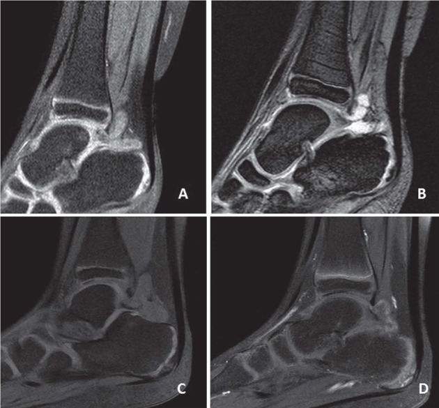
Juvenile idiopathic arthritis. Sagittal T1 fat-sat images of ankle. (A) Pre-gadolinium and (B) post-gadolinium images showing synovitis of the talo-calcanear joint at baseline and (C, D) after 3 months of treatment illustrating a marked reduction of synovitis
MRI depicts the pathological changes in all tissues involved in RA and shows greater sensitivity in detecting inflammatory and destructive changes than both clinical examination and x-ray (207, 208). Haavardsholm et a.l (206) has shown that MRI measures of inflammation provide superior responsiveness to conventional measures of disease activity in patients with RA treated with anti-TNFa medication. This superior sensitivity of MRI compared to conventional clinical examinations and radiographs, combined with the knowledge that MRI detected BME is a predictor of subsequent radiographic progression (175, 176) has generated the hypothesis that adding MRI to the conventional clinical and laboratory examinations and intensifying treatment in the presence of subclinical MRI-BME will reduce radiographic erosive progression and improve the patient’s functional level. The use of MRI as a treatment guide may be valuable in individualizing the treatment, so patients assessed as being at high risk of erosive progression can receive appropriately intensive treatment so disease progression can be avoided. Further, MRI measures as a structural end point in a clinical trial setting would impact the efficiency of the study design.
Whole-body MRI
Whole-body MRI is a novel imaging method, which allows MRI of the whole body in one scanning session, but at the cost of lower image resolution than conventional MRI.
With its comprehensive examination, whole-body MRI was first introduced in the specialities of oncology (209) and angiology (210). However, it may also prove useful in systemic musculoskeletal disorders, such as SpA, in the evaluation of both axial and peripheral abnormalities, including enthesitis (211).
Whole-body and conventional MRI showed a very good correlation for the detection of inflammatory lesions in the SI joints in patients with established and active SpA. Whole-body MRI is a fascinating new imaging modality also for RA. Using this technique most relevant joints can be assessed for synovitis, bone-marrow oedema and erosions, as well as axial and enthesial pathology. The major drawback with the technique has been its insufficient resolution, notably in peripheral joints, which are frequently affected in these patients. So much work is necessary to improve and evaluate this technique.
Whole body MRI is frequently used in the pediatric field, both for the assessment of patients with juvenile idiopathic arthritis, but also for other diffuse musculoskeletal disorders. Whole-body MRI for evaluation of juvenile idiopathic arthritis is advantageous in that it allows for assessment of both disease activity and extent (Figure 8). Studies in the pediatric population are limited, but whole-body MRI has been shown to be superior to clinical examination in detecting arthritis in the hips, sacroiliac joints and spine (212). Consequently, whole-body MRI may play an important role alongside clinical exam and radiography as an objective tool for assessing active disease activity and guiding therapy in juvenile spondyloarthropathy patients. However, a recommendation to use whole-body instead of conventional MRI in daily routine seems to be premature. Further studies need to address the comparative performance for assessing inflammatory lesions in the spine and the clinical relevance of the additional information on inflammation in the anterior chest wall and the hip and shoulder girdles. Complementary to analyzing status scores, the validation process of whole-body MRI needs to also compare responsiveness and reliability of change scores.
Conclusion and future application
US and MRI can be used both in clinical practice and clinical trials, for multiple purposes, including establishing or confirming a diagnosis of inflammatory joint disease, determining the extent of the disease, monitoring change in inflammation and structural damage, assessing therapeutic efficacy and potentially prognostication.
Within the field of imaging in inflammatory arthropathiess, large and exciting advances have been made during the last decade. However to achieve these goals, standardisation and validation of US and MRI are required to ensure accurate diagnosis, reproducibility and reliability. The introduction of volumetric probes (VP), with the automatic acquisition of three-dimensional (3D) data sets, allows for sensitive, rapid, and less operator-dependent acquisition of Doppler images in patients with chronic inflammatory arthritis. Such probes have a transducer inside, which moves electronically when the operator presses the acquisition button, and acquires all the echoes under the footprint of the volumetric probe. The stored 3D datasets can be explored using a dedicated software that generates sectional views of the three main planes (longitudinal, transverse and coronal, which share only one point) and 3D reconstructions. Both B-mode and CD or PD Doppler mode can be used in volumetric scanning. 3D PDUS has been proven to provide a good imaging reproduction of the synovial blood flow representing a complete vascular tree inside and on the verge of the synovial tissue (213). Naredo et al. (214) suggest that volumetric PDUS can be used in multicenter open-label cohort studies on patients with RA. The added value of this technology over conventional US could be to minimize assessment biases and reduce acquisition variability. 3D US techniques have been tested, particularly in RA, for their capacity to record the full extent of inflammation within a joint and to visualize the size and surface appearance of erosive lesions. In terms of sensitivity for the detection of synovitis and bony lesions, there is good to excellent correlation between results from this method and those from two-dimensional US (215). Therefore, 3D imaging is predestined to generate a more precise quantification of the vascularity and thereby affords the benefit of detecting small changes in the acquired volume in the monitoring of clinical and therapeutic strategies (215). The major disadvantage of 3D ultrasonography include the long image-acquisition time, the long size of the 3D dataset, which depends on the area of the probe footprint, and the static feature of the acquired images, which leaves the interpretation of imaging findings without the advantages provided by dynamic examination (216).
MRI is increasingly used in RA trials and practice. The capacity of MRI to detect early changes in soft and synovial tissue, bone and extraarticular makes this technique a useful tool in the study of RA. Possible future applications of MRI would be to examine the capacity of this technique to allow for the diagnosis of different inflammatory arthropathies based on anatomical localization of the structural lesions or the type of lesion, such as enthesis in the case of SpA (217). On the other hand, knowing the sensitivity and specificity of MRI as a diagnostic or classification criteria at the beginning of the disease, and lastly, the capacity of this modality to help in the selection of patients with worse or better prognosis or even as responders to therapy or not. In recent years MRI have increasingly been used as outcome measures in clinical trials of RA. Several studies have shown that MRI measures of synovitis, tenosynovitis, bone marrow oedema and erosions are valid and reliable, but less is known about the responsiveness of these measures during an intervention with a potent therapeutic agent (218, 219).
The technical advances in the development of equipment of dedicated MRI, with a larger resolution for imaging and even portability, new technological advantages such as the introduction of 3D MRI sequences with isotropic voxels, which allows for the multi-planar reconstruction permits significant reduction of scanning time without loss of image quality, as well as the adequate training to interpret results, are hopeful proposals for the application of MRI in the diagnosis, follow-up and prognosis in patients with inflammatory arthropathies. A simplified MRI volumetric data acquisition may provide gross estimates of disease activity when the threshold is set properly. Nonetheless, there are still many questions and more studies are needed to allow for some answers to these and other questions.
Practice points
US is a non-invasive, reproducible, non-radiating, and relatively inexpensive technique used to detect, assess, and quantify both the inflammation of joints and structural damage caused by a variety of rheumatic diseases.
PDUS is more sensitive than clinical examination in detecting inflammation in joints, tendon sheaths, tendons, entheses in patients with chronic inflammatory arthritis and allow monitoring of disease activity.
The use of CEUS seems to provide significantly higher sensitivity than PDUS in the identification of abnormal vascularization in joint inflammation, allowing a more exact measurement of the synovitis, as well as a quantitative assessment of inflammation by using the analysis of time-intensity curves.
MRI is a sensitive, accurate, non-invasive tool that allows simultaneous assessment of all the components of diarthrodial joints and offers the rheumatologist a chance to visualize joint inflammation as well as damage.
MRI is proving to be a useful tool with which to investigate disease processes in inflammatory arthritis such as RA and has the potential for clinical use in determining the prognosis and targeting aggressive therapy to patients with the most destructive disease.
Whole-body multi-joint is MRI allows a ‘snapshot’ of inflammation in systemic musculoskeletal disorders, such as spondyloarthritis, in the evaluation of both axial and peripheral abnormalities, including enthesitis.
Future research in established and new MRI methods will increase the value of MRI to diagnose, monitor, and establish a prognosis in inflammatory arthropathies.
References
- 1.Weber AJ, De Bandt M. Angiogenesis: general mechanisms and implications for rheumatoid arthritis. Joint Bone Spine. 2000;67:366–83. [PubMed] [Google Scholar]
- 2.Walsh DA. Angiogenesis and arthritis. Rheumatology (Oxford) 1999;38:103–12. doi: 10.1093/rheumatology/38.2.103. [DOI] [PubMed] [Google Scholar]
- 3.Harris ED. Jr. Rheumatoid arthritis. Pathophysiology and implications for therapy. N Engl J Med. 1990;322:1277–89. doi: 10.1056/NEJM199005033221805. [DOI] [PubMed] [Google Scholar]
- 4.Szkudlarek M, Narvestad E, Klarlund M, Court-Payen M, Thomsen HS, Ostergaard M. Ultrasonography of the metatarsophalangeal joints in rheumatoid arthritis: comparison with magnetic resonance imaging, conventional radiography, and clinical examination. Arthritis Rheum. 2004;50:2103–12. doi: 10.1002/art.20333. [DOI] [PubMed] [Google Scholar]
- 5.Scheel AK, Hermann KG, Ohrndorf S, Werner C, Schirmer C, Detert J, Bollow M, Hamm B, Muller GA, Burmester GR, Backhaus M. Prospective 7 year follow up imaging study comparing radiography, ultrasonography, and magnetic resonance imaging in rheumatoid arthritis finger joints. Ann Rheum Dis. 2006;65:595–600. doi: 10.1136/ard.2005.041814. [DOI] [PMC free article] [PubMed] [Google Scholar]
- 6.Spadaro A, Lubrano E. Psoriatic arthritis: imaging techniques. Reumatismo. 2012;64:99–106. doi: 10.4081/reumatismo.2012.99. [DOI] [PubMed] [Google Scholar]
- 7.Caranci F, Brunese L, Reginelli A, Napoli M, Fonio P, Briganti F. Neck Neoplastic Conditions in the Emergency Setting: Role of Multidetector Computed Tomography. Semin Ultrasound CT MRI. 2012;33:443–448. doi: 10.1053/j.sult.2012.06.011. [DOI] [PubMed] [Google Scholar]
- 8.Naredo E, Aguado P, De Miguel E, Uson J, Mayordomo L, Gijon-Banos J, Martin-Mola E. Painful shoulder: comparison of physical examination and ultrasonographic findings. Ann Rheum Dis. 2002;61:132–6. doi: 10.1136/ard.61.2.132. [DOI] [PMC free article] [PubMed] [Google Scholar]
- 9.Kane D, Balint PV, Sturrock RD. Ultrasonography is superior to clinical examination in the detection and localization of knee joint effusion in rheumatoid arthritis. J Rheumatol. 2003;30:966–71. [PubMed] [Google Scholar]
- 10.Zappia M, Castagna A, Barile A, Chianca V, Brunese L, Pouliart N. Imaging of the coracoglenoid ligament: a third ligament in the rotator interval of the shoulder. Skelet Radiol. 2017;46:1101–1111. doi: 10.1007/s00256-017-2667-9. [DOI] [PubMed] [Google Scholar]
- 11.van Steenbergen HW, van Nies JA, Huizinga TW, Reijnierse M, van der Helm-van Mil AH. Subclinical inflammation on MRI of hand and foot of anticitrullinated peptide antibody-negative arthralgia patients at risk for rheumatoid arthritis. Arthritis Res Ther. 2014;16:R92. doi: 10.1186/ar4536. [DOI] [PMC free article] [PubMed] [Google Scholar]
- 12.Li X, Liu X, Du X, Ye Z. Diagnostic performance of three-dimensional MR maximum intensity projection for the assessment of synovitis of the hand and wrist in rheumatoid arthritis: A pilot study. Eur J Radiol. 2014;83:797–800. doi: 10.1016/j.ejrad.2014.02.004. [DOI] [PubMed] [Google Scholar]
- 13.Lubrano E, Marchesoni A, Olivieri I, D’Angelo S, Palazzi C, Scarpa R, Ferrara N, Parsons WJ, Brunese L, Helliwell PS, Spadaro A. The radiological assessment of axial involvement in psoriatic arthritis. J Rheumatol. 2012;39:54–56. doi: 10.3899/jrheum.120244. [DOI] [PubMed] [Google Scholar]
- 14.Grassi W, Salaffi F, Filippucci E. Ultrasound in rheumatology. Best Pract Res Clin Rheumatol. 2005;19:467–85. doi: 10.1016/j.berh.2005.01.002. [DOI] [PubMed] [Google Scholar]
- 15.Rezaei H, Torp-Pedersen S, af Klint E, Backheden M, Kisten Y, Gyori N, van Vollenhoven RF. Diagnostic utility of musculoskeletal ultrasound in patients with suspected arthritis--a probabilistic approach. Arthritis Res Ther. 2014;16:448. doi: 10.1186/s13075-014-0448-6. [DOI] [PMC free article] [PubMed] [Google Scholar]
- 16.Mandl P, Balint PV, Brault Y, Backhaus M, D’Agostino MA, Grassi W, van der Heijde D, de Miguel E, Wakefield RJ, Logeart I, Dougados M. Metrologic properties of ultrasound versus clinical evaluation of synovitis in rheumatoid arthritis: results of a multicenter, randomized study. Arthritis Rheum. 2012;64:1272–82. doi: 10.1002/art.33491. [DOI] [PubMed] [Google Scholar]
- 17.Wakefield RJ, Green MJ, Marzo-Ortega H, Conaghan PG, Gibbon WW, McGonagle D, Proudman S, Emery P. Should oligoarthritis be reclassified? Ultrasound reveals a high prevalence of subclinical disease. Ann Rheum Dis. 2004;63:382–5. doi: 10.1136/ard.2003.007062. [DOI] [PMC free article] [PubMed] [Google Scholar]
- 18.Karim Z, Wakefield RJ, Quinn M, Conaghan PG, Brown AK, Veale DJ, O’Connor P, Reece R, Emery P. Validation and reproducibility of ultrasonography in the detection of synovitis in the knee: a comparison with arthroscopy and clinical examination. Arthritis Rheum. 2004;50:387–94. doi: 10.1002/art.20054. [DOI] [PubMed] [Google Scholar]
- 19.Grassi W, De Angelis R, Lamanna G, Cervini C. The clinical features of rheumatoid arthritis. Eur J Radiol. 1998;27(1):S18–24. doi: 10.1016/s0720-048x(98)00038-2. [DOI] [PubMed] [Google Scholar]
- 20.Klauser AS, Moriggl B, Duftner C, Smekal V, Pallwein L, Mur E, Schirmer M. Sonography of synovial and erosive inflammatory changes. Radiologe. 2006;46:365–75. doi: 10.1007/s00117-006-1361-z. [DOI] [PubMed] [Google Scholar]
- 21.Backhaus M, Burmester GR, Gerber T, Grassi W, Machold KP, Swen WA, Wakefield RJ, Manger B. Working Group for Musculoskeletal Ultrasound in the ESCoICSiTT. Guidelines for musculoskeletal ultrasound in rheumatology. Ann Rheum Dis. 2001;60:641–9. doi: 10.1136/ard.60.7.641. [DOI] [PMC free article] [PubMed] [Google Scholar]
- 22.Pinto A, Pinto F, Faggian A, Rubini G, Caranci F, Macarini L, Genovese EA, Brunese L. Sources of error in emergency ultrasonography. Crit Ultrasound J. 2013;5:1–5. doi: 10.1186/2036-7902-5-S1-S1. [DOI] [PMC free article] [PubMed] [Google Scholar]
- 23.Wakefield RJ, Balint PV, Szkudlarek M, Filippucci E, Backhaus M, D’Agostino MA, Sanchez EN, Iagnocco A, Schmidt WA, Bruyn GA, Kane D, O’Connor PJ, Manger B, Joshua F, Koski J, Grassi W, Lassere MN, Swen N, Kainberger F, Klauser A, Ostergaard M, Brown AK, Machold KP, Conaghan PG, Group OSI. Musculoskeletal ultrasound including definitions for ultrasonographic pathology. J Rheumatol. 2005;32:2485–7. [PubMed] [Google Scholar]
- 24.Iagnocco A, Epis O, Delle Sedie A, Meenagh G, Filippucci E, Riente L, Scire CA, Montecucco C, Bombardieri S, Grassi W, Valesini G. Ultrasound imaging for the rheumatologist. XVII. Role of colour Doppler and power Doppler. Clin Exp Rheumatol. 2008;26:759–62. [PubMed] [Google Scholar]
- 25.Dougados M, Jousse-Joulin S, Mistretta F, D’Agostino MA, Backhaus M, Bentin J, Chales G, Chary-Valckenaere I, Conaghan P, Etchepare F, Gaudin P, Grassi W, van der Heijde D, Sellam J, Naredo E, Szkudlarek M, Wakefield R, Saraux A. Evaluation of several ultrasonography scoring systems for synovitis and comparison to clinical examination: results from a prospective multicentre study of rheumatoid arthritis. Ann Rheum Dis. 2010;69:828–33. doi: 10.1136/ard.2009.115493. [DOI] [PubMed] [Google Scholar]
- 26.Weidekamm C, Koller M, Weber M, Kainberger F. Diagnostic value of high-resolution B-mode and doppler sonography for imaging of hand and finger joints in rheumatoid arthritis. Arthritis Rheum. 2003;48:325–33. doi: 10.1002/art.10784. [DOI] [PubMed] [Google Scholar]
- 27.Rubin JM. Spectral Doppler US. Radiographics. 1994;14:139–50. doi: 10.1148/radiographics.14.1.8128046. [DOI] [PubMed] [Google Scholar]
- 28.Terslev L, Torp-Pedersen S, Qvistgaard E, von der Recke P, Bliddal H. Doppler ultrasound findings in healthy wrists and finger joints. Ann Rheum Dis. 2004;63:644–8. doi: 10.1136/ard.2003.009548. [DOI] [PMC free article] [PubMed] [Google Scholar]
- 29.Terslev L, Torp-Pedersen S, Qvistgaard E, Kristoffersen H, Rogind H, Danneskiold-Samsoe B, Bliddal H. Effects of treatment with etanercept (Enbrel, TNRF:Fc) on rheumatoid arthritis evaluated by Doppler ultrasonography. Ann Rheum Dis. 2003;62:178–81. doi: 10.1136/ard.62.2.178. [DOI] [PMC free article] [PubMed] [Google Scholar]
- 30.Carotti M, Salaffi F, Morbiducci J, Ciapetti A, Bartolucci L, Gasparini S, Ferraccioli G, Giuseppetti GM, Grassi W. Colour Doppler ultrasonography evaluation of vascularization in the wrist and finger joints in rheumatoid arthritis patients and healthy subjects. Eur J Radiol. 2012;81:1834–8. doi: 10.1016/j.ejrad.2010.01.005. [DOI] [PubMed] [Google Scholar]
- 31.Andersen M, Ellegaard K, Hebsgaard JB, Christensen R, Torp-Pedersen S, Kvist PH, Soe N, Romer J, Vendel N, Bartels EM, Danneskiold-Samsoe B, Bliddal H. Ultrasound colour Doppler is associated with synovial pathology in biopsies from hand joints in rheumatoid arthritis patients: a cross-sectional study. Ann Rheum Dis. 2014;73:678–83. doi: 10.1136/annrheumdis-2012-202669. [DOI] [PubMed] [Google Scholar]
- 32.Takase K, Ohno S, Takeno M, Hama M, Kirino Y, Ihata A, Ideguchi H, Mochida Y, Tateishi U, Shizukuishi K, Nagashima Y, Aoki I, Ishigatsubo Y. Simultaneous evaluation of long-lasting knee synovitis in patients undergoing arthroplasty by power Doppler ultrasonography and contrast-enhanced MRI in comparison with histopathology. Clin Exp Rheumatol. 2012;30:85–92. [PubMed] [Google Scholar]
- 33.Perrotta FM, Astorri D, Zappia M, Reginelli A, Brunese L, Lubrano E. An ultrasonographic study of enthesis in early psoriatic arthritis patients naive to traditional and biologic DMARDs treatment. Rheumatol Int. 2016;36:1579–1583. doi: 10.1007/s00296-016-3562-8. [DOI] [PubMed] [Google Scholar]
- 34.Torp-Pedersen S, Christensen R, Szkudlarek M, Ellegaard K, D’Agostino MA, Iagnocco A, Naredo E, Balint P, Wakefield RJ, Torp-Pedersen A, Terslev L. Power and color Doppler ultrasound settings for inflammatory flow: impact on scoring of disease activity in patients with rheumatoid arthritis. Arthritis Rheumatol. 2015;67:386–95. doi: 10.1002/art.38940. [DOI] [PubMed] [Google Scholar]
- 35.Schmidt WA. Technology Insight: the role of color and power Doppler ultrasonography in rheumatology. Nat Clin Pract Rheumatol. 2007;3:35–42. doi: 10.1038/ncprheum0377. quiz 59. [DOI] [PubMed] [Google Scholar]
- 36.Terslev L, Ellegaard K, Christensen R, Szkudlarek M, Schmidt WA, Jensen PS, Bliddal H, Torp-Pedersen S. Head-to-head comparison of quantitative and semi-quantitative ultrasound scoring systems for rheumatoid arthritis: reliability, agreement and construct validity. Rheumatology (Oxford) 2012;51:2034–8. doi: 10.1093/rheumatology/kes124. [DOI] [PubMed] [Google Scholar]
- 37.Burns PN. Principles of Doppler and color flow. Radiol Med. 1993;85:3–16. [PubMed] [Google Scholar]
- 38.Newman JS, Adler RS, Bude RO, Rubin JM. Detection of soft-tissue hyperemia: value of power Doppler sonography. AJR Am J Roentgenol. 1994;163:385–9. doi: 10.2214/ajr.163.2.8037037. [DOI] [PubMed] [Google Scholar]
- 39.D’Agostino MA, Terslev L, Wakefield R, Ostergaard M, Balint P, Naredo E, Iagnocco A, Backhaus M, Grassi W, Emery P. Novel algorithms for the pragmatic use of ultrasound in the management of patients with rheumatoid arthritis: from diagnosis to remission. Ann Rheum Dis. 2016;75:1902–1908. doi: 10.1136/annrheumdis-2016-209646. [DOI] [PubMed] [Google Scholar]
- 40.Torp-Pedersen ST, Terslev L. Settings and artefacts relevant in colour/power Doppler ultrasound in rheumatology. Ann Rheum Dis. 2008;67:143–9. doi: 10.1136/ard.2007.078451. [DOI] [PubMed] [Google Scholar]
- 41.Breidahl WH, Stafford Johnson DB, Newman JS, Adler RS. Power Doppler sonography in tenosynovitis: significance of the peritendinous hypoechoic rim. J Ultrasound Med. 1998;17:103–7. doi: 10.7863/jum.1998.17.2.103. [DOI] [PubMed] [Google Scholar]
- 42.Baeten D, Demetter P, Cuvelier C, Van Den Bosch F, Kruithof E, Van Damme N, Verbruggen G, Mielants H, Veys EM, De Keyser F. Comparative study of the synovial histology in rheumatoid arthritis, spondyloarthropathy, and osteoarthritis: influence of disease duration and activity. Ann Rheum Dis. 2000;59:945–53. doi: 10.1136/ard.59.12.945. [DOI] [PMC free article] [PubMed] [Google Scholar]
- 43.Walther M, Harms H, Krenn V, Radke S, Faehndrich TP, Gohlke F. Correlation of power Doppler sonography with vascularity of the synovial tissue of the knee joint in patients with osteoarthritis and rheumatoid arthritis. Arthritis Rheum. 2001;44:331–8. doi: 10.1002/1529-0131(200102)44:2<331::AID-ANR50>3.0.CO;2-0. [DOI] [PubMed] [Google Scholar]
- 44.Szkudlarek M, Court-Payen M, Strandberg C, Klarlund M, Klausen T, Ostergaard M. Power Doppler ultrasonography for assessment of synovitis in the metacarpophalangeal joints of patients with rheumatoid arthritis: a comparison with dynamic magnetic resonance imaging. Arthritis Rheum. 2001;44:2018–23. doi: 10.1002/1529-0131(200109)44:9<2018::AID-ART350>3.0.CO;2-C. [DOI] [PubMed] [Google Scholar]
- 45.Newman JS, Laing TJ, McCarthy CJ, Adler RS. Power Doppler sonography of synovitis: assessment of therapeutic response--preliminary observations. Radiology. 1996;198:582–4. doi: 10.1148/radiology.198.2.8596870. [DOI] [PubMed] [Google Scholar]
- 46.Walther M, Harms H, Krenn V, Radke S, Kirschner S, Gohlke F. Synovial tissue of the hip at power Doppler US: correlation between vascularity and power Doppler US signal. Radiology. 2002;225:225–31. doi: 10.1148/radiol.2251011272. [DOI] [PubMed] [Google Scholar]
- 47.Terslev L, Torp-Pedersen S, Savnik A, von der Recke P, Qvistgaard E, Danneskiold-Samsoe B, Bliddal H. Doppler ultrasound and magnetic resonance imaging of synovial inflammation of the hand in rheumatoid arthritis: a comparative study. Arthritis Rheum. 2003;48:2434–41. doi: 10.1002/art.11245. [DOI] [PubMed] [Google Scholar]
- 48.Ziswiler HR, Tamborrini G. Musculoskeletal ultrasound II - <<why are bats better than physicians>>. Praxis (Bern 1994) 2011;100:1297–302. doi: 10.1024/1661-8157/a000702. [DOI] [PubMed] [Google Scholar]
- 49.Takase-Minegishi K, Horita N, Kobayashi K, Yoshimi R, Kirino Y, Ohno S, Kaneko T, Nakajima H, Wakefield RJ, Emery P. Diagnostic test accuracy of ultrasound for synovitis in rheumatoid arthritis: systematic review and meta-analysis. Rheumatology (Oxford) 2017 doi: 10.1093/rheumatology/kex036. [DOI] [PubMed] [Google Scholar]
- 50.Freeston JE, Wakefield RJ, Conaghan PG, Hensor EM, Stewart SP, Emery P. A diagnostic algorithm for persistence of very early inflammatory arthritis: the utility of power Doppler ultrasound when added to conventional assessment tools. Ann Rheum Dis. 2010;69:417–9. doi: 10.1136/ard.2008.106658. [DOI] [PubMed] [Google Scholar]
- 51.Salaffi F, Ciapetti A, Gasparini S, Carotti M, Filippucci E, Grassi W. A clinical prediction rule combining routine assessment and power Doppler ultrasonography for predicting progression to rheumatoid arthritis from early-onset undifferentiated arthritis. Clin Exp Rheumatol. 2010;28:686–94. [PubMed] [Google Scholar]
- 52.Filer A, de Pablo P, Allen G, Nightingale P, Jordan A, Jobanputra P, Bowman S, Buckley CD, Raza K. Utility of ultrasound joint counts in the prediction of rheumatoid arthritis in patients with very early synovitis. Ann Rheum Dis. 2011;70:500–7. doi: 10.1136/ard.2010.131573. [DOI] [PMC free article] [PubMed] [Google Scholar]
- 53.Gutierrez M, Filippucci E, Salaffi F, Di Geso L, Grassi W. Differential diagnosis between rheumatoid arthritis and psoriatic arthritis: the value of ultrasound findings at metacarpophalangeal joints level. Ann Rheum Dis. 2011;70:1111–4. doi: 10.1136/ard.2010.147272. [DOI] [PubMed] [Google Scholar]
- 54.Ritchlin CT. Pathogenesis of psoriatic arthritis. Curr Opin Rheumatol. 2005;17:406–12. doi: 10.1097/01.bor.0000167752.93543.76. [DOI] [PubMed] [Google Scholar]
- 55.Benjamin M, Moriggl B, Brenner E, Emery P, McGonagle D, Redman S. The “enthesis organ” concept: why enthesopathies may not present as focal insertional disorders. Arthritis Rheum. 2004;50:3306–13. doi: 10.1002/art.20566. [DOI] [PubMed] [Google Scholar]
- 56.Rettenbacher T, Ennemoser S, Weirich H, Ulmer H, Hartig F, Klotz W, Herold M. Diagnostic imaging of gout: comparison of high-resolution US versus conventional X-ray. Eur Radiol. 2008;18:621–30. doi: 10.1007/s00330-007-0802-z. [DOI] [PubMed] [Google Scholar]
- 57.Thiele RG, Schlesinger N. Diagnosis of gout by ultrasound. Rheumatology (Oxford) 2007;46:1116–21. doi: 10.1093/rheumatology/kem058. [DOI] [PubMed] [Google Scholar]
- 58.Dalbeth N, Doyle AJ. Imaging of gout: an overview. Best Pract Res Clin Rheumatol. 2012;26:823–38. doi: 10.1016/j.berh.2012.09.003. [DOI] [PubMed] [Google Scholar]
- 59.Vlychou M, Koutroumpas A, Alexiou I, Fezoulidis I, Sakkas LI. High-resolution ultrasonography and 3.0 T magnetic resonance imaging in erosive and nodal hand osteoarthritis: high frequency of erosions in nodal osteoarthritis. Clin Rheumatol. 2013;32:755–62. doi: 10.1007/s10067-013-2166-x. [DOI] [PubMed] [Google Scholar]
- 60.Haugen IK, Boyesen P, Slatkowsky-Christensen B, Sesseng S, Bijsterbosch J, van der Heijde D, Kvien TK. Comparison of features by MRI and radiographs of the interphalangeal finger joints in patients with hand osteoarthritis. Ann Rheum Dis. 2012;71:345–50. doi: 10.1136/annrheumdis-2011-200028. [DOI] [PubMed] [Google Scholar]
- 61.Qvistgaard E, Rogind H, Torp-Pedersen S, Terslev L, Danneskiold-Samsoe B, Bliddal H. Quantitative ultrasonography in rheumatoid arthritis: evaluation of inflammation by Doppler technique. Ann Rheum Dis. 2001;60:690–3. doi: 10.1136/ard.60.7.690. [DOI] [PMC free article] [PubMed] [Google Scholar]
- 62.Fukuba E, Yoshizako T, Kitagaki H, Murakawa Y, Kondo M, Uchida N. Power Doppler ultrasonography for assessment of rheumatoid synovitis: comparison with dynamic magnetic resonance imaging. Clin Imaging. 2013;37:134–7. doi: 10.1016/j.clinimag.2012.02.008. [DOI] [PubMed] [Google Scholar]
- 63.Kiris A, Ozgocmen S, Kocakoc E, Ardicoglu O. Power Doppler assessment of overall disease activity in patients with rheumatoid arthritis. J Clin Ultrasound. 2006;34:5–11. doi: 10.1002/jcu.20175. [DOI] [PubMed] [Google Scholar]
- 64.Naredo E, Bonilla G, Gamero F, Uson J, Carmona L, Laffon A. Assessment of inflammatory activity in rheumatoid arthritis: a comparative study of clinical evaluation with grey scale and power Doppler ultrasonography. Ann Rheum Dis. 2005;64:375–81. doi: 10.1136/ard.2004.023929. [DOI] [PMC free article] [PubMed] [Google Scholar]
- 65.Szkudlarek M, Court-Payen M, Jacobsen S, Klarlund M, Thomsen HS, Ostergaard M. Interobserver agreement in ultrasonography of the finger and toe joints in rheumatoid arthritis. Arthritis Rheum. 2003;48:955–62. doi: 10.1002/art.10877. [DOI] [PubMed] [Google Scholar]
- 66.Stone M, Bergin D, Whelan B, Maher M, Murray J, McCarthy C. Power Doppler ultrasound assessment of rheumatoid hand synovitis. J Rheumatol. 2001;28:1979–82. [PubMed] [Google Scholar]
- 67.Albrecht K, Muller-Ladner U, Strunk J. Quantification of the synovial perfusion in rheumatoid arthritis using Doppler ultrasonography. Clin Exp Rheumatol. 2007;25:630–8. [PubMed] [Google Scholar]
- 68.Koski JM, Saarakkala S, Helle M, Hakulinen U, Heikkinen JO, Hermunen H, Balint P, Bruyn GA, Filippucci E, Grassi W, Iagnocco A, Luosujarvi R, Manger B, De Miguel E, Naredo E, Scheel AK, Schmidt WA, Soini I, Szkudlarek M, Terslev L, Uson J, Vuoristo S, Ziswiler HR. Assessing the intra- and inter-reader reliability of dynamic ultrasound images in power Doppler ultrasonography. Ann Rheum Dis. 2006;65:1658–60. doi: 10.1136/ard.2005.051250. [DOI] [PMC free article] [PubMed] [Google Scholar]
- 69.Naredo E, Moller I, Moragues C, de Agustin JJ, Scheel AK, Grassi W, de Miguel E, Backhaus M, Balint P, Bruyn GA, D’Agostino MA, Filippucci E, Iagnocco A, Kane D, Koski JM, Mayordomo L, Schmidt WA, Swen WA, Szkudlarek M, Terslev L, Torp-Pedersen S, Uson J, Wakefield RJ, Werner C. Ultrasound EWGfM. Interobserver reliability in musculoskeletal ultrasonography: results from a “Teach the Teachers” rheumatologist course. Ann Rheum Dis. 2006;65:14–9. doi: 10.1136/ard.2005.037382. [DOI] [PMC free article] [PubMed] [Google Scholar]
- 70.Wakefield RJ, D’Agostino MA, Iagnocco A, Filippucci E, Backhaus M, Scheel AK, Joshua F, Naredo E, Schmidt WA, Grassi W, Moller I, Pineda C, Klauser A, Szkudlarek M, Terslev L, Balint P, Bruyn GA, Swen WA, Jousse-Joulin S, Kane D, Koski JM, O’Connor P, Milutinovic S, Conaghan PG, Group OU. The OMERACT Ultrasound Group: status of current activities and research directions. J Rheumatol. 2007;34:848–51. [PubMed] [Google Scholar]
- 71.Scheel AK, Schmidt WA, Hermann KG, Bruyn GA, D’Agostino MA, Grassi W, Iagnocco A, Koski JM, Machold KP, Naredo E, Sattler H, Swen N, Szkudlarek M, Wakefield RJ, Ziswiler HR, Pasewaldt D, Werner C, Backhaus M. Interobserver reliability of rheumatologists performing musculoskeletal ultrasonography: results from a EULAR “Train the trainers” course. Ann Rheum Dis. 2005;64:1043–9. doi: 10.1136/ard.2004.030387. [DOI] [PMC free article] [PubMed] [Google Scholar]
- 72.Fukae J, Tanimura K, Atsumi T, Koike T. Sonographic synovial vascularity of synovitis in rheumatoid arthritis. Rheumatology (Oxford) 2014;53:586–91. doi: 10.1093/rheumatology/ket311. [DOI] [PubMed] [Google Scholar]
- 73.Barile A, Bruno F, Mariani S, Arrigoni F, Brunese L, Zappia M, Splendiani A, Di Cesare E, Masciocchi C. Follow-up of surgical and minimally invasive treatment of Achilles tendon pathology: a brief diagnostic imaging review. Musculoskeletal Surg. 2017;101:51–61. doi: 10.1007/s12306-017-0456-1. [DOI] [PubMed] [Google Scholar]
- 74.Cupek R, Ziebinski A. Automated assessment of joint synovitis activity from medical ultrasound and power doppler examinations using image processing and machine learning methods. Reumatologia. 2016;54:239–242. doi: 10.5114/reum.2016.63664. [DOI] [PMC free article] [PubMed] [Google Scholar]
- 75.Schmidt WA, Schonau V, Reiche BE, Oberdorfer PD, Ohrndorf S, Backhaus M. Grading of ultrasound Doppler signals in synovitis: does it need an update. Rheumatology (Oxford) 2015;54:1897–903. doi: 10.1093/rheumatology/kev205. [DOI] [PubMed] [Google Scholar]
- 76.Barile A, Arrigoni F, Bruno F, Guglielmi G, Zappia M, Reginelli A, Ruscitti P, Cipriani P, Giacomelli R, Brunese L, Masciocchi C. Computed Tomography and MR Imaging in Rheumatoid Arthritis. Radiol Clin North Am. 2017 doi: 10.1016/j.rcl.2017.04.006. [DOI] [PubMed] [Google Scholar]
- 77.Naredo E, Rodriguez M, Campos C, Rodriguez-Heredia JM, Medina JA, Giner E, Martinez O, Toyos FJ, Ruiz T, Ros I, Pujol M, Miquel X, Garcia L, Aznar JJ, Chamizo E, Paez M, Morales P, Rueda A, Tuneu R, Corominas H, de Agustin JJ, Moragues C, Minguez D, Willisch A, Gonzalez-Cruz I, Aragon A, Iglesias G, Armas C, Pablo Valdazo J, Vargas C, Calvo-Alen J, Juan-Mas A, Salvador G, Puigdollers A, Galindez E, Garrido N, Salaberri J, Raya E, Salles M, Diaz C, Cuadra JL, Garrido J. Ultrasound Group of The Spanish Society of R. Validity, reproducibility, and responsiveness of a twelve-joint simplified power doppler ultrasonographic assessment of joint inflammation in rheumatoid arthritis. Arthritis Rheum. 2008;59:515–22. doi: 10.1002/art.23529. [DOI] [PubMed] [Google Scholar]
- 78.Hama M, Uehara T, Takase K, Ihata A, Ueda A, Takeno M, Shizukuishi K, Tateishi U, Ishigatsubo Y. Power Doppler ultrasonography is useful for assessing disease activity and predicting joint destruction in rheumatoid arthritis patients receiving tocilizumab--preliminary data. Rheumatol Int. 2012;32:1327–33. doi: 10.1007/s00296-011-1802-5. [DOI] [PMC free article] [PubMed] [Google Scholar]
- 79.Dougados M, Devauchelle-Pensec V, Ferlet JF, Jousse-Joulin S, D’Agostino MA, Backhaus M, Bentin J, Chales G, Chary-Valckenaere I, Conaghan P, Wakefield RJ, Etchepare F, Gaudin P, Grassi W, Heijde D, Mariette X, Naredo E, Szkudlarek M. The ability of synovitis to predict structural damage in rheumatoid arthritis: a comparative study between clinical examination and ultrasound. Ann Rheum Dis. 2013;72:665–71. doi: 10.1136/annrheumdis-2012-201469. [DOI] [PMC free article] [PubMed] [Google Scholar]
- 80.Nguyen H, Ruyssen-Witrand A, Gandjbakhch F, Constantin A, Foltz V, Cantagrel A. Prevalence of ultrasound-detected residual synovitis and risk of relapse and structural progression in rheumatoid arthritis patients in clinical remission: a systematic review and meta-analysis. Rheumatology (Oxford) 2014;53:2110–8. doi: 10.1093/rheumatology/keu217. [DOI] [PubMed] [Google Scholar]
- 81.Haavardsholm EA, Lie E, Lillegraven S. Should modern imaging be part of remission criteria in rheumatoid arthritis. Best Pract Res Clin Rheumatol. 2012;26:767–85. doi: 10.1016/j.berh.2012.10.004. [DOI] [PubMed] [Google Scholar]
- 82.Brown AK, Quinn MA, Karim Z, Conaghan PG, Peterfy CG, Hensor E, Wakefield RJ, O’Connor PJ, Emery P. Presence of significant synovitis in rheumatoid arthritis patients with disease-modifying antirheumatic drug-induced clinical remission: evidence from an imaging study may explain structural progression. Arthritis Rheum. 2006;54:3761–73. doi: 10.1002/art.22190. [DOI] [PubMed] [Google Scholar]
- 83.Vreju FA, Filippucci E, Gutierrez M, Di Geso L, Ciapetti A, Ciurea ME, Salaffi F, Grassi W. Subclinical ultrasound synovitis in a particular joint is associated with ultrasound evidence of bone erosions in that same joint in rheumatoid patients in clinical remission. Clin Exp Rheumatol. 2016;34:673–8. [PubMed] [Google Scholar]
- 84.Sudol-Szopinska I, Jans L, Teh J. Rheumatoid arthritis: what do MRI and ultrasound show. J Ultrason. 2017;17:5–16. doi: 10.15557/JoU.2017.0001. [DOI] [PMC free article] [PubMed] [Google Scholar]
- 85.Naredo E, Moller I, Cruz A, Carmona L, Garrido J. Power Doppler ultrasonographic monitoring of response to anti-tumor necrosis factor therapy in patients with rheumatoid arthritis. Arthritis Rheum. 2008;58:2248–56. doi: 10.1002/art.23682. [DOI] [PubMed] [Google Scholar]
- 86.Albrecht K, Grob K, Lange U, Muller-Ladner U, Strunk J. Reliability of different Doppler ultrasound quantification methods and devices in the assessment of therapeutic response in arthritis. Rheumatology (Oxford) 2008;47:1521–6. doi: 10.1093/rheumatology/ken318. [DOI] [PubMed] [Google Scholar]
- 87.Filippucci E, Iagnocco A, Salaffi F, Cerioni A, Valesini G, Grassi W. Power Doppler sonography monitoring of synovial perfusion at the wrist joints in patients with rheumatoid arthritis treated with adalimumab. Ann Rheum Dis. 2006;65:1433–7. doi: 10.1136/ard.2005.044628. [DOI] [PMC free article] [PubMed] [Google Scholar]
- 88.Saleem B, Brown AK, Keen H, Nizam S, Freeston J, Wakefield R, Karim Z, Quinn M, Hensor E, Conaghan PG, Emery P. Should imaging be a component of rheumatoid arthritis remission criteria? A comparison between traditional and modified composite remission scores and imaging assessments. Ann Rheum Dis. 2011;70:792–8. doi: 10.1136/ard.2010.134445. [DOI] [PubMed] [Google Scholar]
- 89.Larche MJ, Seymour M, Lim A, Eckersley RJ, Petavy F, Chiesa F, Rioja I, Lukey PT, Binks M, McClinton C, Dolan K, Taylor PC. Quantitative power Doppler ultrasonography is a sensitive measure of metacarpophalangeal joint synovial vascularity in rheumatoid arthritis and declines significantly following a 2-week course of oral low-dose corticosteroids. J Rheumatol. 2010;37:2493–501. doi: 10.3899/jrheum.100322. [DOI] [PubMed] [Google Scholar]
- 90.Filippucci E, Farina A, Carotti M, Salaffi F, Grassi W. Grey scale and power Doppler sonographic changes induced by intra-articular steroid injection treatment. Ann Rheum Dis. 2004;63:740–3. doi: 10.1136/ard.2003.007971. [DOI] [PMC free article] [PubMed] [Google Scholar]
- 91.Klauser A, Demharter J, De Marchi A, Sureda D, Barile A, Masciocchi C, Faletti C, Schirmer M, Kleffel T, Bohndorf K. group Is. Contrast enhanced gray-scale sonography in assessment of joint vascularity in rheumatoid arthritis: results from the IACUS study group. Eur Radiol. 2005;15:2404–10. doi: 10.1007/s00330-005-2884-9. [DOI] [PubMed] [Google Scholar]
- 92.Mouterde G, Carotti M, D’Agostino MA. Contrast-enhanced ultrasound in musculoskeletal diseases. J Radiol. 2009;90:148–55. doi: 10.1016/s0221-0363(09)70093-9. [DOI] [PubMed] [Google Scholar]
- 93.Klauser AS. Value of contrast-enhanced ultrasound in rheumatic disease. Eur Radiol. 2005;15(5):E117–22. doi: 10.1007/s10406-005-0174-x. [DOI] [PubMed] [Google Scholar]
- 94.Klauser A, Frauscher F, Schirmer M, Halpern E, Pallwein L, Herold M, Helweg G, ZurNedden D. The value of contrast-enhanced color Doppler ultrasound in the detection of vascularization of finger joints in patients with rheumatoid arthritis. Arthritis Rheum. 2002;46:647–53. doi: 10.1002/art.10136. [DOI] [PubMed] [Google Scholar]
- 95.Kleffel T, Demharter J, Wohlgemuth W, Schalm J, Bohndorf K, Kirchhof K. Comparison of contrast-enhanced low mechanical index (Low MI) sonography and unenhanced B-mode sonography for the differentiation between synovitis and joint effusion in patients with rheumatoid arthritis. Rofo. 2005;177:835–41. doi: 10.1055/s-2005-858194. [DOI] [PubMed] [Google Scholar]
- 96.Schmidt WA, Volker L, Zacher J, Schlafke M, Ruhnke M, Gromnica-Ihle E. Colour Doppler ultrasonography to detect pannus in knee joint synovitis. Clin Exp Rheumatol. 2000;18:439–44. [PubMed] [Google Scholar]
- 97.Fiocco U, Ferro F, Cozzi L, Vezzu M, Sfriso P, Checchetto C, Bianchi FC, Nardacchione R, Piccoli A, Todesco S, Rubaltelli L. Contrast medium in power Doppler ultrasound for assessment of synovial vascularity: comparison with arthroscopy. J Rheumatol. 2003;30:2170–6. [PubMed] [Google Scholar]
- 98.Carotti M, Salaffi F, Manganelli P, Salera D, Simonetti B, Grassi W. Power Doppler sonography in the assessment of synovial tissue of the knee joint in rheumatoid arthritis: a preliminary experience. Ann Rheum Dis. 2002;61:877–82. doi: 10.1136/ard.61.10.877. [DOI] [PMC free article] [PubMed] [Google Scholar]
- 99.Salaffi F, Carotti M, Manganelli P, Filippucci E, Giuseppetti GM, Grassi W. Contrast-enhanced power Doppler sonography of knee synovitis in rheumatoid arthritis: assessment of therapeutic response. Clin Rheumatol. 2004;23:285–90. doi: 10.1007/s10067-004-0878-7. [DOI] [PubMed] [Google Scholar]
- 100.Baker JF, Conaghan PG, Emery P, Baker DG, Ostergaard M. Relationship of patient-reported outcomes with MRI measures in rheumatoid arthritis. Ann Rheum Dis. 2017;76:486–490. doi: 10.1136/annrheumdis-2016-209463. [DOI] [PubMed] [Google Scholar]
- 101.Axelsen MB, Eshed I, Horslev-Petersen K, Stengaard-Pedersen K, Hetland ML, Moller J, Junker P, Podenphant J, Schlemmer A, Ellingsen T, Ahlquist P, Lindegaard H, Linauskas A, Dam MY, Hansen I, Horn HC, Ammitzboll CG, Jorgensen A, Krintel SB, Raun J, Krogh NS, Johansen JS, Ostergaard M. group Os. A treat-to-target strategy with methotrexate and intra-articular triamcinolone with or without adalimumab effectively reduces MRI synovitis, osteitis and tenosynovitis and halts structural damage progression in early rheumatoid arthritis: results from the OPERA randomised controlled trial. Ann Rheum Dis. 2015;74:867–75. doi: 10.1136/annrheumdis-2013-204537. [DOI] [PubMed] [Google Scholar]
- 102.Burgers LE, Nieuwenhuis WP, van Steenbergen HW, Newsum EC, Huizinga TW, Reijnierse M, le Cessie S, van der Helm-van Mil AH. Magnetic resonance imaging-detected inflammation is associated with functional disability in early arthritis-results of a cross-sectional study. Rheumatology (Oxford) 2016;55:2167–2175. doi: 10.1093/rheumatology/kew334. [DOI] [PubMed] [Google Scholar]
- 103.Forslind K, Svensson B. MRI evidence of persistent joint inflammation and progressive joint damage despite clinical remission during treatment of early rheumatoid arthritis. Scand J Rheumatol. 2016;45:99–102. doi: 10.3109/03009742.2015.1070902. [DOI] [PubMed] [Google Scholar]
- 104.Valeri G, Ferrara C, Ercolani P, De Nigris E, Giovagnoni A. Tendon involvement in rheumatoid arthritis of the wrist: MRI findings. Skeletal Radiol. 2001;30:138–43. doi: 10.1007/s002560100322. [DOI] [PubMed] [Google Scholar]
- 105.Caranci F, Tedeschi E, Leone G, Reginelli A, Gatta G, Pinto A, Squillaci E, Briganti F, Brunese L. Errors in neuroradiology. Radiol Med. 2015;120:795–801. doi: 10.1007/s11547-015-0564-7. [DOI] [PubMed] [Google Scholar]
- 106.Sudol-Szopinska I, Jurik AG, Eshed I, Lennart J, Grainger A, Ostergaard M, Klauser A, Cotten A, Wick MC, Maas M, Miese F, Egund N, Boutry N, Rupreht M, Reijnierse M, Oei EH, Meier R, O’Connor P, Feydy A, Mascarenhas V, Plagou A, Simoni P, Platzgummer H, Rennie WJ, Mester A, Teh J, Robinson P, Guglielmi G, Astrom G, Schueller-Weiderkamm C. Recommendations of the ESSR Arthritis Subcommittee for the Use of Magnetic Resonance Imaging in Musculoskeletal Rheumatic Diseases. Semin Musculoskelet Radiol. 2015;19:396–411. doi: 10.1055/s-0035-1564696. [DOI] [PubMed] [Google Scholar]
- 107.Lee CH, Srikhum W, Burghardt AJ, Virayavanich W, Imboden JB, Link TM, Li X. Correlation of structural abnormalities of the wrist and metacarpophalangeal joints evaluated by high-resolution peripheral quantitative computed tomography, 3 Tesla magnetic resonance imaging and conventional radiographs in rheumatoid arthritis. Int J Rheum Dis. 2015;18:628–39. doi: 10.1111/1756-185X.12495. [DOI] [PubMed] [Google Scholar]
- 108.Ghanem N, Uhl M, Pache G, Bley T, Walker UA, Langer M. MRI in psoriatic arthritis with hand and foot involvement. Rheumatol Int. 2007;27:387–93. doi: 10.1007/s00296-006-0220-6. [DOI] [PubMed] [Google Scholar]
- 109.Narvaez J, Narvaez JA, de Albert M, Gomez-Vaquero C, Nolla JM. Can magnetic resonance imaging of the hand and wrist differentiate between rheumatoid arthritis and psoriatic arthritis in the early stages of the disease. Semin Arthritis Rheum. 2012;42:234–45. doi: 10.1016/j.semarthrit.2012.03.016. [DOI] [PubMed] [Google Scholar]
- 110.Lubrano E, Parsons WJ, Marchesoni A, Olivieri I, D’Angelo S, Cauli A, Caso F, Costa L, Scarpa R, Brunese L. The definition and measurement of axial psoriatic arthritis. J Rheumatol. 2015;93:40–42. doi: 10.3899/jrheum.150634. [DOI] [PubMed] [Google Scholar]
- 111.Krabben A, Stomp W, van Nies JA, Huizinga TW, van der Heijde D, Bloem JL, Reijnierse M, van der Helm-van Mil AH. MRI-detected subclinical joint inflammation is associated with radiographic progression. Ann Rheum Dis. 2014;73:2034–7. doi: 10.1136/annrheumdis-2014-205208. [DOI] [PubMed] [Google Scholar]
- 112.Dill T. Contraindications to magnetic resonance imaging: non-invasive imaging. Heart. 2008;94:943–8. doi: 10.1136/hrt.2007.125039. [DOI] [PubMed] [Google Scholar]
- 113.Arnett FC, Edworthy SM, Bloch DA, McShane DJ, Fries JF, Cooper NS, Healey LA, Kaplan SR, Liang MH, Luthra HS, et al. The American Rheumatism Association 1987 revised criteria for the classification of rheumatoid arthritis. Arthritis Rheum. 1988;31:315–24. doi: 10.1002/art.1780310302. [DOI] [PubMed] [Google Scholar]
- 114.Boutry N, Morel M, Flipo RM, Demondion X, Cotten A. Early rheumatoid arthritis: a review of MRI and sonographic findings. AJR Am J Roentgenol. 2007;189:1502–9. doi: 10.2214/AJR.07.2548. [DOI] [PubMed] [Google Scholar]
- 115.Ejbjerg B, Narvestad E, Rostrup E, Szkudlarek M, Jacobsen S, Thomsen HS, Ostergaard M. Magnetic resonance imaging of wrist and finger joints in healthy subjects occasionally shows changes resembling erosions and synovitis as seen in rheumatoid arthritis. Arthritis Rheum. 2004;50:1097–106. doi: 10.1002/art.20135. [DOI] [PubMed] [Google Scholar]
- 116.McQueen FM, Stewart N, Crabbe J, Robinson E, Yeoman S, Tan PL, McLean L. Magnetic resonance imaging of the wrist in early rheumatoid arthritis reveals a high prevalence of erosions at four months after symptom onset. Ann Rheum Dis. 1998;57:350–6. doi: 10.1136/ard.57.6.350. [DOI] [PMC free article] [PubMed] [Google Scholar]
- 117.Narvaez JA, Narvaez J, De Lama E, De Albert M. MR imaging of early rheumatoid arthritis. Radiographics. 2010;30:143–63. doi: 10.1148/rg.301095089. discussion 163-5. [DOI] [PubMed] [Google Scholar]
- 118.Mathew AJ, Danda D, Conaghan PG. MRI and ultrasound in rheumatoid arthritis. Curr Opin Rheumatol. 2016;28:323–9. doi: 10.1097/BOR.0000000000000282. [DOI] [PubMed] [Google Scholar]
- 119.Ostergaard M, Ejbjerg B. Magnetic resonance imaging of the synovium in rheumatoid arthritis. Semin Musculoskelet Radiol. 2004;8:287–99. doi: 10.1055/s-2004-861576. [DOI] [PubMed] [Google Scholar]
- 120.Sommer OJ, Kladosek A, Weiler V, Czembirek H, Boeck M, Stiskal M. Rheumatoid arthritis: a practical guide to state-of-the-art imaging, image interpretation, and clinical implications. Radiographics. 2005;25:381–98. doi: 10.1148/rg.252045111. [DOI] [PubMed] [Google Scholar]
- 121.Farrant JM, O’Connor PJ, Grainger AJ. Advanced imaging in rheumatoid arthritis. Part 1: synovitis. Skeletal Radiol. 2007;36:269–79. doi: 10.1007/s00256-006-0219-9. [DOI] [PubMed] [Google Scholar]
- 122.Ostergaard M, Hansen M, Stoltenberg M, Lorenzen I. Quantitative assessment of the synovial membrane in the rheumatoid wrist: an easily obtained MRI score reflects the synovial volume. Br J Rheumatol. 1996;35:965–71. doi: 10.1093/rheumatology/35.10.965. [DOI] [PubMed] [Google Scholar]
- 123.Borrero CG, Mountz JM, Mountz JD. Emerging MRI methods in rheumatoid arthritis. Nat Rev Rheumatol. 2011;7:85–95. doi: 10.1038/nrrheum.2010.173. [DOI] [PubMed] [Google Scholar]
- 124.Ostergaard M, Stoltenberg M, Lovgreen-Nielsen P, Volck B, Jensen CH, Lorenzen I. Magnetic resonance imaging-determined synovial membrane and joint effusion volumes in rheumatoid arthritis and osteoarthritis: comparison with the macroscopic and microscopic appearance of the synovium. Arthritis Rheum. 1997;40:1856–67. doi: 10.1002/art.1780401020. [DOI] [PubMed] [Google Scholar]
- 125.Ostergaard M, Stoltenberg M, Lovgreen-Nielsen P, Volck B, Sonne-Holm S, Lorenzen I. Quantification of synovistis by MRI: correlation between dynamic and static gadolinium-enhanced magnetic resonance imaging and microscopic and macroscopic signs of synovial inflammation. Magn Reson Imaging. 1998;16:743–54. doi: 10.1016/s0730-725x(98)00008-3. [DOI] [PubMed] [Google Scholar]
- 126.Ostergaard M, Peterfy C, Conaghan P, McQueen F, Bird P, Ejbjerg B, Shnier R, O’Connor P, Klarlund M, Emery P, Genant H, Lassere M, Edmonds J. OMERACT Rheumatoid Arthritis Magnetic Resonance Imaging Studies. Core set of MRI acquisitions, joint pathology definitions, and the OMERACT RA-MRI scoring system. J Rheumatol. 2003;30:1385–6. [PubMed] [Google Scholar]
- 127.Mariani S, La Marra A, Arrigoni F, Necozione S, Splendiani A, Di Cesare E, Barile A, Masciocchi C. Dynamic measurement of patello-femoral joint alignment using weight-bearing magnetic resonance imaging (WB-MRI) Eur J Radiol. 2015;84:2571–2578. doi: 10.1016/j.ejrad.2015.09.017. [DOI] [PubMed] [Google Scholar]
- 128.Narvaez JA, Narvaez J, Roca Y, Aguilera C. MR imaging assessment of clinical problems in rheumatoid arthritis. Eur Radiol. 2002;12:1819–28. doi: 10.1007/s00330-001-1207-z. [DOI] [PubMed] [Google Scholar]
- 129.Sugimoto H, Takeda A, Hyodoh K. Early-stage rheumatoid arthritis: prospective study of the effectiveness of MR imaging for diagnosis. Radiology. 2000;216:569–75. doi: 10.1148/radiology.216.2.r00au20569. [DOI] [PubMed] [Google Scholar]
- 130.Tamai M, Kawakami A, Uetani M, Takao S, Rashid H, Tanaka F, Fujikawa K, Aramaki T, Nakamura H, Iwanaga N, Izumi Y, Arima K, Aratake K, Kamachi M, Huang M, Origuchi T, Ida H, Aoyagi K, Eguchi K. Early prediction of rheumatoid arthritis by serological variables and magnetic resonance imaging of the wrists and finger joints: results from prospective clinical examination. Ann Rheum Dis. 2006;65:134–5. doi: 10.1136/ard.2005.043075. [DOI] [PMC free article] [PubMed] [Google Scholar]
- 131.Ostergaard M, Conaghan PG, O’Connor P, Szkudlarek M, Klarlund M, Emery P, Peterfy C, Genant H, McQueen FM, Bird P, Lassere M, Ejbjerg B. Reducing invasiveness, duration, and cost of magnetic resonance imaging in rheumatoid arthritis by omitting intravenous contrast injection -- Does it change the assessment of inflammatory and destructive joint changes by the OMERACT RAMRIS. J Rheumatol. 2009;36:1806–10. doi: 10.3899/jrheum.090350. [DOI] [PubMed] [Google Scholar]
- 132.Ostergaard M, Klarlund M. Importance of timing of post-contrast MRI in rheumatoid arthritis: what happens during the first 60 minutes after IV gadolinium-DTPA. Ann Rheum Dis. 2001;60:1050–4. doi: 10.1136/ard.60.11.1050. [DOI] [PMC free article] [PubMed] [Google Scholar]
- 133.Caranci F, Leone G, Ugga L, Cesarano E, Capasso R, Schipani S, Bianco A, Fonio P, Briganti F, Brunese L. Imaging of post-surgical treatment and of related complications in spinal trauma. Musculoskeletal Surg. 2017;101:63–73. doi: 10.1007/s12306-017-0457-0. [DOI] [PubMed] [Google Scholar]
- 134.Splendiani A, D’Orazio F, Patriarca L, Arrigoni F, Caranci F, Fonio P, Brunese L, Barile A, Di Cesare E, Masciocchi C. Imaging of post-operative spine in intervertebral disc pathology. Musculoskeletal Surg. 2017;101:75–84. doi: 10.1007/s12306-017-0453-4. [DOI] [PubMed] [Google Scholar]
- 135.Weber U, Hodler J, Kubik RA, Rufibach K, Lambert RG, Kissling RO, Pfirrmann CW, Maksymowych WP. Sensitivity and specificity of spinal inflammatory lesions assessed by whole-body magnetic resonance imaging in patients with ankylosing spondylitis or recent-onset inflammatory back pain. Arthritis Rheum. 2009;61:900–8. doi: 10.1002/art.24507. [DOI] [PubMed] [Google Scholar]
- 136.Klarlund M, Ostergaard M, Rostrup E, Skjodt H, Lorenzen I. Dynamic magnetic resonance imaging of the metacarpophalangeal joints in rheumatoid arthritis, early unclassified polyarthritis, and healthy controls. Scand J Rheumatol. 2000;29:108–15. doi: 10.1080/030097400750001914. [DOI] [PubMed] [Google Scholar]
- 137.Ejbjerg B, McQueen F, Lassere M, Haavardsholm E, Conaghan P, O’Connor P, Bird P, Peterfy C, Edmonds J, Szkudlarek M, Genant H, Emery P, Ostergaard M. The EULAR-OMERACT rheumatoid arthritis MRI reference image atlas: the wrist joint. Ann Rheum Dis. 2005;64(1):i23–47. doi: 10.1136/ard.2004.031823. [DOI] [PMC free article] [PubMed] [Google Scholar]
- 138.Yamato M, Tamai K, Yamaguchi T, Ohno W. MRI of the knee in rheumatoid arthritis: Gd-DTPA perfusion dynamics. J Comput Assist Tomogr. 1993;17:781–5. doi: 10.1097/00004728-199309000-00022. [DOI] [PubMed] [Google Scholar]
- 139.Huang J, Stewart N, Crabbe J, Robinson E, McLean L, Yeoman S, Tan PL, McQueen FM. A 1-year follow-up study of dynamic magnetic resonance imaging in early rheumatoid arthritis reveals synovitis to be increased in shared epitope-positive patients and predictive of erosions at 1 year. Rheumatology (Oxford) 2000;39:407–16. doi: 10.1093/rheumatology/39.4.407. [DOI] [PubMed] [Google Scholar]
- 140.van de Sande MG, van der Leij C, Lavini C, Wijbrandts CA, Maas M, Tak PP. Characteristics of synovial inflammation in early arthritis analysed by pixel-by-pixel time-intensity curve shape analysis. Rheumatology (Oxford) 2012;51:1240–5. doi: 10.1093/rheumatology/kes011. [DOI] [PubMed] [Google Scholar]
- 141.Vordenbaumen S, Schleich C, Logters T, Sewerin P, Bleck E, Pauly T, Muller-Lutz A, Antoch G, Schneider M, Miese F, Ostendorf B. Dynamic contrast-enhanced magnetic resonance imaging of metacarpophalangeal joints reflects histological signs of synovitis in rheumatoid arthritis. Arthritis Res Ther. 2014;16:452. doi: 10.1186/s13075-014-0452-x. [DOI] [PMC free article] [PubMed] [Google Scholar]
- 142.Konig H, Sieper J, Wolf KJ. Rheumatoid arthritis: evaluation of hypervascular and fibrous pannus with dynamic MR imaging enhanced with Gd-DTPA. Radiology. 1990;176:473–7. doi: 10.1148/radiology.176.2.2367663. [DOI] [PubMed] [Google Scholar]
- 143.Gaffney K, Cookson J, Blake D, Coumbe A, Blades S. Quantification of rheumatoid synovitis by magnetic resonance imaging. Arthritis Rheum. 1995;38:1610–7. doi: 10.1002/art.1780381113. [DOI] [PubMed] [Google Scholar]
- 144.Tamai K, Yamato M, Yamaguchi T, Ohno W. Dynamic magnetic resonance imaging for the evaluation of synovitis in patients with rheumatoid arthritis. Arthritis Rheum. 1994;37:1151–7. doi: 10.1002/art.1780370807. [DOI] [PubMed] [Google Scholar]
- 145.Boutry N, Hachulla E, Flipo RM, Cortet B, Cotten A. MR imaging findings in hands in early rheumatoid arthritis: comparison with those in systemic lupus erythematosus and primary Sjogren syndrome. Radiology. 2005;236:593–600. doi: 10.1148/radiol.2361040844. [DOI] [PubMed] [Google Scholar]
- 146.Cimmino MA, Parodi M, Innocenti S, Succio G, Banderali S, Silvestri E, Garlaschi G. Dynamic magnetic resonance of the wrist in psoriatic arthritis reveals imaging patterns similar to those of rheumatoid arthritis. Arthritis Res Ther. 2005;7:R725–31. doi: 10.1186/ar1734. [DOI] [PMC free article] [PubMed] [Google Scholar]
- 147.Savnik A, Malmskov H, Thomsen HS, Graff LB, Nielsen H, Danneskiold-Samsoe B, Boesen J, Bliddal H. MRI of the wrist and finger joints in inflammatory joint diseases at 1-year interval: MRI features to predict bone erosions. Eur Radiol. 2002;12:1203–10. doi: 10.1007/s003300101114. [DOI] [PubMed] [Google Scholar]
- 148.Jevtic V, Watt I, Rozman B, Kos-Golja M, Demsar F, Jarh O. Distinctive radiological features of small hand joints in rheumatoid arthritis and seronegative spondyloarthritis demonstrated by contrast-enhanced (Gd-DTPA) magnetic resonance imaging. Skeletal Radiol. 1995;24:351–5. doi: 10.1007/BF00197064. [DOI] [PubMed] [Google Scholar]
- 149.McGonagle D, Conaghan PG, Emery P. Psoriatic arthritis: a unified concept twenty years on. Arthritis Rheum. 1999;42:1080–6. doi: 10.1002/1529-0131(199906)42:6<1080::AID-ANR2>3.0.CO;2-7. [DOI] [PubMed] [Google Scholar]
- 150.McQueen F, Lassere M, Ostergaard M. Magnetic resonance imaging in psoriatic arthritis: a review of the literature. Arthritis Res Ther. 2006;8:207. doi: 10.1186/ar1934. [DOI] [PMC free article] [PubMed] [Google Scholar]
- 151.Cimmino MA, Zampogna G, Parodi M, Andracco R, Barbieri F, Paparo F, Ferrero G, Garlaschi G. MRI synovitis and bone lesions are common in acute gouty arthritis of the wrist even during the first attack. Ann Rheum Dis. 2011;70:2238–9. doi: 10.1136/ard.2011.153353. [DOI] [PubMed] [Google Scholar]
- 152.Loeuille D, Chary-Valckenaere I, Champigneulle J, Rat AC, Toussaint F, Pinzano-Watrin A, Goebel JC, Mainard D, Blum A, Pourel J, Netter P, Gillet P. Macroscopic and microscopic features of synovial membrane inflammation in the osteoarthritic knee: correlating magnetic resonance imaging findings with disease severity. Arthritis Rheum. 2005;52:3492–501. doi: 10.1002/art.21373. [DOI] [PubMed] [Google Scholar]
- 153.Kirkhus E, Bjornerud A, Thoen J, Johnston V, Dale K, Smith HJ. Contrast-enhanced dynamic magnetic resonance imaging of finger joints in osteoarthritis and rheumatoid arthritis: an analysis based on pharmacokinetic modeling. Acta Radiol. 2006;47:845–51. doi: 10.1080/02841850600838210. [DOI] [PubMed] [Google Scholar]
- 154.Klarlund M, Ostergaard M, Lorenzen I. Finger joint synovitis in rheumatoid arthritis: quantitative assessment by magnetic resonance imaging. Rheumatology (Oxford) 1999;38:66–72. doi: 10.1093/rheumatology/38.1.66. [DOI] [PubMed] [Google Scholar]
- 155.Sugimoto H, Takeda A, Masuyama J, Furuse M. Early-stage rheumatoid arthritis: diagnostic accuracy of MR imaging. Radiology. 1996;198:185–92. doi: 10.1148/radiology.198.1.8539375. [DOI] [PubMed] [Google Scholar]
- 156.Ostergaard M, Gideon P, Henriksen O, Lorenzen I. Synovial volume--a marker of disease severity in rheumatoid arthritis? Quantification by MRI. Scand J Rheumatol. 1994;23:197–202. doi: 10.3109/03009749409103060. [DOI] [PubMed] [Google Scholar]
- 157.Workie DW, Dardzinski BJ. Quantifying dynamic contrast-enhanced MRI of the knee in children with juvenile rheumatoid arthritis using an arterial input function (AIF) extracted from popliteal artery enhancement, and the effect of the choice of the AIF on the kinetic parameters. Magn Reson Med. 2005;54:560–8. doi: 10.1002/mrm.20597. [DOI] [PubMed] [Google Scholar]
- 158.Hodgson RJ, O’Connor P, Moots R. MRI of rheumatoid arthritis image quantitation for the assessment of disease activity, progression and response to therapy. Rheumatology (Oxford) 2008;47:13–21. doi: 10.1093/rheumatology/kem250. [DOI] [PubMed] [Google Scholar]
- 159.Andersen M, Boesen M, Ellegaard K, Christensen R, Soderstrom K, Soe N, Spee P, Morch UG, Torp-Pedersen S, Bartels EM, Danneskiold-Samsoe B, Vendel N, Karlsson L, Bliddal H. Synovial explant inflammatory mediator production corresponds to rheumatoid arthritis imaging hallmarks: a cross-sectional study. Arthritis Res Ther. 2014;16:R107. doi: 10.1186/ar4557. [DOI] [PMC free article] [PubMed] [Google Scholar]
- 160.Ostergaard M, Stoltenberg M, Henriksen O, Lorenzen I. Quantitative assessment of synovial inflammation by dynamic gadolinium-enhanced magnetic resonance imaging. A study of the effect of intra-articular methylprednisolone on the rate of early synovial enhancement. Br J Rheumatol. 1996;35:50–9. doi: 10.1093/rheumatology/35.1.50. [DOI] [PubMed] [Google Scholar]
- 161.Carubbi F, Zugaro L, Cipriani P, Conchiglia A, Gregori L, Danniballe C, Letizia Pistoia M, Liakouli V, Ruscitti P, Ciccia F, Triolo G, Masciocchi C, Giacomelli R. Safety and efficacy of intra-articular anti-tumor necrosis factor α agents compared to corticosteroids in a treat-to-target strategy in patients with inflammatory arthritis and monoarthritis flare. Int. J. Immunopathol. Pharmacol. 2015;29:252–266. doi: 10.1177/0394632015593220. [DOI] [PMC free article] [PubMed] [Google Scholar]
- 162.Barile A, La Marra A, Arrigoni F, Mariani S, Zugaro L, Splendiani A, Di Cesare E, Reginelli A, Zappia M, Brunese L, Duka E, Carrafiello G, Masciocchi C. Anaesthetics, steroids and platelet-rich plasma (PRP) in ultrasound-guided musculoskeletal procedures. Br. J. Radiol. 2016;89 doi: 10.1259/bjr.20150355. [DOI] [PMC free article] [PubMed] [Google Scholar]
- 163.Hodgson R, Grainger A, O’Connor P, Barnes T, Connolly S, Moots R. Dynamic contrast enhanced MRI of bone marrow oedema in rheumatoid arthritis. Ann Rheum Dis. 2008;67:270–2. doi: 10.1136/ard.2007.077271. [DOI] [PubMed] [Google Scholar]
- 164.Palosaari K, Vuotila J, Takalo R, Jartti A, Niemela R, Haapea M, Soini I, Tervonen O, Hakala M. Contrast-enhanced dynamic and static MRI correlates with quantitative 99Tcm-labelled nanocolloid scintigraphy. Study of early rheumatoid arthritis patients. Rheumatology (Oxford) 2004;43:1364–73. doi: 10.1093/rheumatology/keh302. [DOI] [PubMed] [Google Scholar]
- 165.Cyteval C, Miquel A, Hoa D, Daures JP, Mariette X, Combe B. Rheumatoid arthritis of the hand: monitoring with a simplified MR imaging scoring method--preliminary assessment. Radiology. 2010;256:863–9. doi: 10.1148/radiol.10091759. [DOI] [PubMed] [Google Scholar]
- 166.McQueen F, Lassere M, Bird P, Haavardsholm EA, Peterfy C, Conaghan PG, Ejbjerg B, Genant H, O’Connor P, Emery P, Ostergaard M. Developing a magnetic resonance imaging scoring system for peripheral psoriatic arthritis. J Rheumatol. 2007;34:859–61. [PubMed] [Google Scholar]
- 167.Ostergaard M, Eder L, Christiansen SN, Kaeley GS. Imaging in the diagnosis and management of peripheral psoriatic arthritis-The clinical utility of magnetic resonance imaging and ultrasonography. Best Pract Res Clin Rheumatol. 2016;30:624–637. doi: 10.1016/j.berh.2016.08.012. [DOI] [PubMed] [Google Scholar]
- 168.Glinatsi D, Bird P, Gandjbakhch F, Mease PJ, Boyesen P, Peterfy CG, Conaghan PG, Ostergaard M. Validation of the OMERACT Psoriatic Arthritis Magnetic Resonance Imaging Score (PsAMRIS) for the Hand and Foot in a Randomized Placebo-controlled Trial. J Rheumatol. 2015;42:2473–9. doi: 10.3899/jrheum.141010. [DOI] [PubMed] [Google Scholar]
- 169.Kosta PE, Voulgari PV, Zikou AK, Drosos AA, Argyropoulou MI. The usefulness of magnetic resonance imaging of the hand and wrist in very early rheumatoid arthritis. Arthritis Res Ther. 2011;13:R84. doi: 10.1186/ar3355. [DOI] [PMC free article] [PubMed] [Google Scholar]
- 170.Stomp W, Krabben A, van der Heijde D, Huizinga TW, Bloem JL, van der Helm-van Mil AH, Reijnierse M. Are rheumatoid arthritis patients discernible from other early arthritis patients using 1.5T extremity magnetic resonance imaging? a large cross-sectional study. J Rheumatol. 2014;41:1630–7. doi: 10.3899/jrheum.131169. [DOI] [PubMed] [Google Scholar]
- 171.Ostendorf B, Scherer A, Modder U, Schneider M. Diagnostic value of magnetic resonance imaging of the forefeet in early rheumatoid arthritis when findings on imaging of the metacarpophalangeal joints of the hands remain normal. Arthritis Rheum. 2004;50:2094–102. doi: 10.1002/art.20314. [DOI] [PubMed] [Google Scholar]
- 172.Mundwiler ML, Maranian P, Brown DH, Silverman JM, Wallace D, Khanna D, Louie J, Furst DE, Weisman MH. The utility of MRI in predicting radiographic erosions in the metatarsophalangeal joints of the rheumatoid foot: a prospective longitudinal cohort study. Arthritis Res Ther. 2009;11:R94. doi: 10.1186/ar2737. [DOI] [PMC free article] [PubMed] [Google Scholar]
- 173.Lipsky PE, van der Heijde DM, St Clair EW, Furst DE, Breedveld FC, Kalden JR, Smolen JS, Weisman M, Emery P, Feldmann M, Harriman GR, Maini RN. Anti-Tumor Necrosis Factor Trial in Rheumatoid Arthritis with Concomitant Therapy Study G. Infliximab and methotrexate in the treatment of rheumatoid arthritis. Anti-Tumor Necrosis Factor Trial in Rheumatoid Arthritis with Concomitant Therapy Study Group. N Engl J Med. 2000;343:1594–602. doi: 10.1056/NEJM200011303432202. [DOI] [PubMed] [Google Scholar]
- 174.Suter LG, Fraenkel L, Braithwaite RS. Role of magnetic resonance imaging in the diagnosis and prognosis of rheumatoid arthritis. Arthritis Care Res (Hoboken) 2011;63:675–88. doi: 10.1002/acr.20409. [DOI] [PMC free article] [PubMed] [Google Scholar]
- 175.Peterfy CG. Magnetic resonance imaging of rheumatoid arthritis: the evolution of clinical applications through clinical trials. Semin Arthritis Rheum. 2001;30:375–96. doi: 10.1053/sarh.2001.22497. [DOI] [PubMed] [Google Scholar]
- 176.Freeston JE, Bird P, Conaghan PG. The role of MRI in rheumatoid arthritis: research and clinical issues. Curr Opin Rheumatol. 2009;21:95–101. doi: 10.1097/BOR.0b013e32832498f0. [DOI] [PubMed] [Google Scholar]
- 177.Duer-Jensen A, Horslev-Petersen K, Hetland ML, Bak L, Ejbjerg BJ, Hansen MS, Johansen JS, Lindegaard HM, Vinterberg H, Moller JM, Ostergaard M. Bone edema on magnetic resonance imaging is an independent predictor of rheumatoid arthritis development in patients with early undifferentiated arthritis. Arthritis Rheum. 2011;63:2192–202. doi: 10.1002/art.30396. [DOI] [PubMed] [Google Scholar]
- 178.Eguchi K. Early prediction of joint destruction in rheumatoid arthritis. Clin Calcium. 2007;17:517–25. [PubMed] [Google Scholar]
- 179.Guermazi A, Taouli B, Lynch JA, Peterfy CG. Imaging of bone erosion in rheumatoid arthritis. Semin Musculoskelet Radiol. 2004;8:269–85. doi: 10.1055/s-2004-861575. [DOI] [PubMed] [Google Scholar]
- 180.McQueen FM, Dalbeth N. Predicting joint damage in rheumatoid arthritis using MRI scanning. Arthritis Res Ther. 2009;11:124. doi: 10.1186/ar2778. [DOI] [PMC free article] [PubMed] [Google Scholar]
- 181.Marinova-Mutafchieva L, Williams RO, Funa K, Maini RN, Zvaifler NJ. Inflammation is preceded by tumor necrosis factor-dependent infiltration of mesenchymal cells in experimental arthritis. Arthritis Rheum. 2002;46:507–13. doi: 10.1002/art.10126. [DOI] [PubMed] [Google Scholar]
- 182.McQueen FM, Benton N, Perry D, Crabbe J, Robinson E, Yeoman S, McLean L, Stewart N. Bone edema scored on magnetic resonance imaging scans of the dominant carpus at presentation predicts radiographic joint damage of the hands and feet six years later in patients with rheumatoid arthritis. Arthritis Rheum. 2003;48:1814–27. doi: 10.1002/art.11162. [DOI] [PubMed] [Google Scholar]
- 183.Haavardsholm EA, Boyesen P, Ostergaard M, Schildvold A, Kvien TK. Magnetic resonance imaging findings in 84 patients with early rheumatoid arthritis: bone marrow oedema predicts erosive progression. Ann Rheum Dis. 2008;67:794–800. doi: 10.1136/ard.2007.071977. [DOI] [PubMed] [Google Scholar]
- 184.Hetland ML, Ejbjerg B, Horslev-Petersen K, Jacobsen S, Vestergaard A, Jurik AG, Stengaard-Pedersen K, Junker P, Lottenburger T, Hansen I, Andersen LS, Tarp U, Skjodt H, Pedersen JK, Majgaard O, Svendsen AJ, Ellingsen T, Lindegaard H, Christensen AF, Vallo J, Torfing T, Narvestad E, Thomsen HS, Ostergaard M. group Cs. MRI bone oedema is the strongest predictor of subsequent radiographic progression in early rheumatoid arthritis. Results from a 2-year randomised controlled trial (CIMESTRA) Ann Rheum Dis. 2009;68:384–90. doi: 10.1136/ard.2008.088245. [DOI] [PubMed] [Google Scholar]
- 185.Bollow M, Fischer T, Reisshauer H, Backhaus M, Sieper J, Hamm B, Braun J. Quantitative analyses of sacroiliac biopsies in spondyloarthropathies: T cells and macrophages predominate in early and active sacroiliitis- cellularity correlates with the degree of enhancement detected by magnetic resonance imaging. Ann Rheum Dis. 2000;59:135–40. doi: 10.1136/ard.59.2.135. [DOI] [PMC free article] [PubMed] [Google Scholar]
- 186.McQueen FM, Stewart N, Crabbe J, Robinson E, Yeoman S, Tan PL, McLean L. Magnetic resonance imaging of the wrist in early rheumatoid arthritis reveals progression of erosions despite clinical improvement. Ann Rheum Dis. 1999;58:156–63. doi: 10.1136/ard.58.3.156. [DOI] [PMC free article] [PubMed] [Google Scholar]
- 187.Ostergaard M, Dohn UM, Ejbjerg BJ, McQueen FM. Ultrasonography and magnetic resonance imaging in early rheumatoid arthritis: recent advances. Curr Rheumatol Rep. 2006;8:378–85. doi: 10.1007/s11926-006-0069-4. [DOI] [PubMed] [Google Scholar]
- 188.Olech E, Freeston JE, Conaghan PG, Hensor EM, Emery P, Yocum D. Using extremity magnetic resonance imaging to assess and monitor early rheumatoid arthritis: the optimal joint combination to be scanned in clinical practice. J Rheumatol. 2008;35:580–3. [PubMed] [Google Scholar]
- 189.Brown AK, Conaghan PG, Karim Z, Quinn MA, Ikeda K, Peterfy CG, Hensor E, Wakefield RJ, O’Connor PJ, Emery P. An explanation for the apparent dissociation between clinical remission and continued structural deterioration in rheumatoid arthritis. Arthritis Rheum. 2008;58:2958–67. doi: 10.1002/art.23945. [DOI] [PubMed] [Google Scholar]
- 190.Backhaus M, Burmester GR, Sandrock D, Loreck D, Hess D, Scholz A, Blind S, Hamm B, Bollow M. Prospective two year follow up study comparing novel and conventional imaging procedures in patients with arthritic finger joints. Ann Rheum Dis. 2002;61:895–904. doi: 10.1136/ard.61.10.895. [DOI] [PMC free article] [PubMed] [Google Scholar]
- 191.Tonolli-Serabian I, Poet JL, Dufour M, Carasset S, Mattei JP, Roux H. Magnetic resonance imaging of the wrist in rheumatoid arthritis: comparison with other inflammatory joint diseases and control subjects. Clin Rheumatol. 1996;15:137–42. doi: 10.1007/BF02230330. [DOI] [PubMed] [Google Scholar]
- 192.Conaghan PG, O’Connor P, McGonagle D, Astin P, Wakefield RJ, Gibbon WW, Quinn M, Karim Z, Green MJ, Proudman S, Isaacs J, Emery P. Elucidation of the relationship between synovitis and bone damage: a randomized magnetic resonance imaging study of individual joints in patients with early rheumatoid arthritis. Arthritis Rheum. 2003;48:64–71. doi: 10.1002/art.10747. [DOI] [PubMed] [Google Scholar]
- 193.Ostergaard M, Hansen M, Stoltenberg M, Gideon P, Klarlund M, Jensen KE, Lorenzen I. Magnetic resonance imaging-determined synovial membrane volume as a marker of disease activity and a predictor of progressive joint destruction in the wrists of patients with rheumatoid arthritis. Arthritis Rheum. 1999;42:918–29. doi: 10.1002/1529-0131(199905)42:5<918::AID-ANR10>3.0.CO;2-2. [DOI] [PubMed] [Google Scholar]
- 194.Ostergaard M, Stoltenberg M, Gideon P, Sorensen K, Henriksen O, Lorenzen I. Changes in synovial membrane and joint effusion volumes after intraarticular methylprednisolone. Quantitative assessment of inflammatory and destructive changes in arthritis by MRI. Rheumatol. 1996;23:1151–61. [PubMed] [Google Scholar]
- 195.Smolen JS, Landewe R, Breedveld FC, Dougados M, Emery P, Gaujoux-Viala C, Gorter S, Knevel R, Nam J, Schoels M, Aletaha D, Buch M, Gossec L, Huizinga T, Bijlsma JW, Burmester G, Combe B, Cutolo M, Gabay C, Gomez-Reino J, Kouloumas M, Kvien TK, Martin-Mola E, McInnes I, Pavelka K, van Riel P, Scholte M, Scott DL, Sokka T, Valesini G, van Vollenhoven R, Winthrop KL, Wong J, Zink A, van der Heijde D. EULAR recommendations for the management of rheumatoid arthritis with synthetic and biological disease-modifying antirheumatic drugs. Ann Rheum Dis. 2010;69:964–75. doi: 10.1136/ard.2009.126532. [DOI] [PMC free article] [PubMed] [Google Scholar]
- 196.Hetland ML, Stengaard-Pedersen K, Junker P, Lottenburger T, Ellingsen T, Andersen LS, Hansen I, Skjodt H, Pedersen JK, Lauridsen UB, Svendsen A, Tarp U, Podenphant J, Hansen G, Lindegaard H, de Carvalho A, Ostergaard M, Horslev-Petersen K, Group CS. Combination treatment with methotrexate, cyclosporine, and intraarticular betamethasone compared with methotrexate and intraarticular betamethasone in early active rheumatoid arthritis: an investigator-initiated, multicenter, randomized, double-blind, parallel-group, placebo-controlled study. Arthritis Rheum. 2006;54:1401–9. doi: 10.1002/art.21796. [DOI] [PubMed] [Google Scholar]
- 197.Nell VP, Machold KP, Eberl G, Stamm TA, Uffmann M, Smolen JS. Benefit of very early referral and very early therapy with disease-modifying anti-rheumatic drugs in patients with early rheumatoid arthritis. Rheumatology (Oxford) 2004;43:906–14. doi: 10.1093/rheumatology/keh199. [DOI] [PubMed] [Google Scholar]
- 198.Lillegraven S, Prince FH, Shadick NA, Bykerk VP, Lu B, Frits ML, Iannaccone CK, Kvien TK, Haavardsholm EA, Weinblatt ME, Solomon DH. Remission and radiographic outcome in rheumatoid arthritis: application of the 2011 ACR/EULAR remission criteria in an observational cohort. Ann Rheum Dis. 2012;71:681–6. doi: 10.1136/ard.2011.154625. [DOI] [PubMed] [Google Scholar]
- 199.Tavares R, Pope JE, Tremblay JL, Thorne C, Bykerk VP, Lazovskis J, Blocka KL, Bell MJ, Lacaille D, Hitchon CA, Fitzgerald AA, Fidler WK, Bookman AA, Henderson JM, Mosher DP, Sholter DE, Khraishi M, Haraoui B, Chen H, Li X, Laupacis A, Boire G, Tomlinson G, Bombardier C. Early management of newly diagnosed rheumatoid arthritis by Canadian rheumatologists: a national, multicenter, retrospective cohort. J Rheumatol. 2011;38:2342–5. doi: 10.3899/jrheum.110249. [DOI] [PubMed] [Google Scholar]
- 200.Emery P, Breedveld F, van der Heijde D, Ferraccioli G, Dougados M, Robertson D, Pedersen R, Koenig AS, Freundlich B, Combination of M. Etanercept in Early Rheumatoid Arthritis Trial G. Two-year clinical and radiographic results with combination etanercept-methotrexate therapy versus monotherapy in early rheumatoid arthritis: a two-year, double-blind, randomized study. Arthritis Rheum. 2010;62:674–82. doi: 10.1002/art.27268. [DOI] [PubMed] [Google Scholar]
- 201.A randomized trial of hydroxychloroquine in early rheumatoid arthritis: the HERA Study. Am J Med. 1995;98:156–68. doi: 10.1016/s0002-9343(99)80399-4. [DOI] [PubMed] [Google Scholar]
- 202.van der Heide A, Jacobs JW, Bijlsma JW, Heurkens AH, van Booma-Frankfort C, van der Veen MJ, Haanen HC, Hofman DM, van Albada-Kuipers GA, ter Borg EJ, Brus HL, Dinant HJ, Kruize AA, Schenk Y. The effectiveness of early treatment with “second-line” antirheumatic drugs. A randomized, controlled trial. Ann Intern Med. 1996;124:699–707. doi: 10.7326/0003-4819-124-8-199604150-00001. [DOI] [PubMed] [Google Scholar]
- 203.Mottonen T, Hannonen P, Korpela M, Nissila M, Kautiainen H, Ilonen J, Laasonen L, Kaipiainen-Seppanen O, Franzen P, Helve T, Koski J, Gripenberg-Gahmberg M, Myllykangas-Luosujarvi R, Leirisalo-Repo M. therapy FI-RTGFRAC. Delay to institution of therapy and induction of remission using single-drug or combination-disease-modifying antirheumatic drug therapy in early rheumatoid arthritis. Arthritis Rheum. 2002;46:894–8. doi: 10.1002/art.10135. [DOI] [PubMed] [Google Scholar]
- 204.Egsmose C, Lund B, Borg G, Pettersson H, Berg E, Brodin U, Trang L. Patients with rheumatoid arthritis benefit from early 2nd line therapy: 5 year followup of a prospective double blind placebo controlled study. J Rheumatol. 1995;22:2208–13. [PubMed] [Google Scholar]
- 205.Zikou AK, Argyropoulou MI, Voulgari PV, Xydis VG, Nikas SN, Efremidis SC, Drosos AA. Magnetic resonance imaging quantification of hand synovitis in patients with rheumatoid arthritis treated with adalimumab. J Rheumatol. 2006;33:219–23. [PubMed] [Google Scholar]
- 206.Haavardsholm EA, Ostergaard M, Hammer HB, Boyesen P, Boonen A, van der Heijde D, Kvien TK. Monitoring anti-TNFalpha treatment in rheumatoid arthritis: responsiveness of magnetic resonance imaging and ultrasonography of the dominant wrist joint compared with conventional measures of disease activity and structural damage. Ann Rheum Dis. 2009;68:1572–9. doi: 10.1136/ard.2008.091801. [DOI] [PubMed] [Google Scholar]
- 207.Ostergaard M, Emery P, Conaghan PG, Fleischmann R, Hsia EC, Xu W, Rahman MU. Significant improvement in synovitis, osteitis, and bone erosion following golimumab and methotrexate combination therapy as compared with methotrexate alone: a magnetic resonance imaging study of 318 methotrexate-naive rheumatoid arthritis patients. Arthritis Rheum. 2011;63:3712–22. doi: 10.1002/art.30592. [DOI] [PubMed] [Google Scholar]
- 208.Ejbjerg BJ, Vestergaard A, Jacobsen S, Thomsen HS, Ostergaard M. The smallest detectable difference and sensitivity to change of magnetic resonance imaging and radiographic scoring of structural joint damage in rheumatoid arthritis finger, wrist, and toe joints: a comparison of the OMERACT rheumatoid arthritis magnetic resonance imaging score applied to different joint combinations and the Sharp/van der Heijde radiographic score. Arthritis Rheum. 2005;52:2300–6. doi: 10.1002/art.21207. [DOI] [PubMed] [Google Scholar]
- 209.Schaefer JF, Schlemmer HP. Total-body MR-imaging in oncology. Eur Radiol. 2006;16:2000–15. doi: 10.1007/s00330-006-0199-0. [DOI] [PubMed] [Google Scholar]
- 210.Nael K, Ruehm SG, Michaely HJ, Saleh R, Lee M, Laub G, Finn JP. Multistation whole-body high-spatial-resolution MR angiography using a 32-channel MR system. AJR Am J Roentgenol. 2007;188:529–39. doi: 10.2214/AJR.05.2053. [DOI] [PubMed] [Google Scholar]
- 211.Weber U, Pfirrmann CW, Kissling RO, Hodler J, Zanetti M. Whole body MR imaging in ankylosing spondylitis: a descriptive pilot study in patients with suspected early and active confirmed ankylosing spondylitis. BMC Musculoskelet Disord. 2007;8:20. doi: 10.1186/1471-2474-8-20. [DOI] [PMC free article] [PubMed] [Google Scholar]
- 212.Aquino MR, Tse SM, Gupta S, Rachlis AC, Stimec J. Whole-body MRI of juvenile spondyloarthritis: protocols and pictorial review of characteristic patterns. Pediatr Radiol. 2015;45:754–62. doi: 10.1007/s00247-015-3319-7. [DOI] [PubMed] [Google Scholar]
- 213.Strunk J, Lange U. Three-dimensional power Doppler sonographic visualization of synovial angiogenesis in rheumatoid arthritis. J Rheumatol. 2004;31:1004–6. [PubMed] [Google Scholar]
- 214.Naredo E, Acebes C, Brito E, de Agustin JJ, de Miguel E, Mayordomo L, Moller I, Moragues C, Rejon E, Rodriguez A, Uson J, Garrido J, Martinez-Hernandez D. Ultrasound School of the Spanish Society of R. Three-dimensional volumetric ultrasound: a valid method for blinded assessment of response to therapy in rheumatoid arthritis. J Rheumatol. 2013;40:253–60. doi: 10.3899/jrheum.121103. [DOI] [PubMed] [Google Scholar]
- 215.Filippucci E, Meenagh G, Delle Sedie A, Salaffi F, Riente L, Iagnocco A, Scire CA, Montecucco C, Bombardieri S, Valesini G, Grassi W. Ultrasound imaging for the rheumatologist. XX. Sonographic assessment of hand and wrist joint involvement in rheumatoid arthritis: comparison between two- and three-dimensional ultrasonography. Clin Exp Rheumatol. 2009;27:197–200. [PubMed] [Google Scholar]
- 216.Filippucci E, Meenagh G, Epis O, Iagnocco A, Riente L, Delle Sedie A, Montecucco C, Valesini G, Bombardieri S, Grassi W. Ultrasound imaging for the rheumatologist. XIII. New trends. Three-dimensional ultrasonography. Clin Exp Rheumatol. 2008;26:1–4. [PubMed] [Google Scholar]
- 217.Offidani A, Cellini A, Valeri G, Giovagnoni A. Subclinical joint involvement in psoriasis: magnetic resonance imaging and X-ray findings. Acta Derm Venereol. 1998;78:463–5. doi: 10.1080/000155598442809. [DOI] [PubMed] [Google Scholar]
- 218.Arrigoni F, Gregori LM, Zugaro L, Barile A, Masciocchi C. MRgFUS in the treatment of MSK lesions: A review based on the experience of the university of L’aquila, Italy. Transl Cancer Res. 2014;3:442–448. [Google Scholar]
- 219.Tedeschi E, Caranci F, Giordano F, Angelini V, Cocozza S, Brunetti A. Gadolinium retention in the body: what we know and what we can do. Radiol. Med. 2017;122:589–600. doi: 10.1007/s11547-017-0757-3. [DOI] [PubMed] [Google Scholar]