Abstract
Like our clothes, our chromosomes fray at the edges with age. Some believe that if we could discover a molecular tailor to patch our age-abraded chromosome ends, we could become modern Methuselahs. Notably, cancer cells achieve immortality by protecting their chromosome ends. Drugs that selectively fray the ends of cancer cell chromosomes would be potent and general anti-cancer therapies.
Here, I summarize data on the role of chromosome ends in cellular and organismal aging:
“I’m not afraid of death; I just don’t want to be there when it happens.”
--Woody Allen
“Immortality is a long shot, I admit. But somebody has to be first.”
--Bill Cosby
Introduction
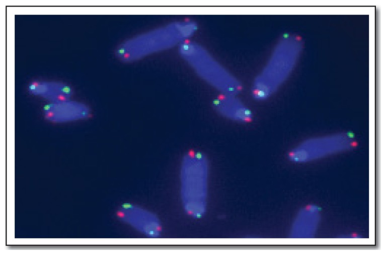
One of the big surprises to emerge from the human genome project was the fact that less than 3% of our DNA encodes our genes. While the purpose—if any—of most noncoding DNA is still being determined, the function of some of this gene-free DNA has long been known to be important in preserving our chromosomes during the cell divisions that occur during the normal wear and tear of age and during wound repair after injury. Each of our chromosomes contains three DNA sequences that are key to maintaining healthy chromosome numbers and structure during our lifetimes. One of these sequences defines the centromere, which helps assure that each chromosome is properly sorted at cell division. The other two sequences occur at each end of the chromosome. These sequences define protective caps called telomeres (See Figure 1).
Figure 1.
Metaphase chromosome (blue) with telomeres highlighted in red or green.
Image courtesy of Susana Gonzalo, Phd
Telomeres Disguise the Ends of Chromosomes
The geneticist Hermann Muller (Nobel Prize in Physiology or Medicine, 1946) noted the peculiarity of chromosome ends in the course of his work on the genetic effects of ionizing radiation in the fruit fly Drosophila melanogaster. He found that X-irradiation caused chromosome breaks that were repaired by fusing the several newly broken ends together, often in abnormal combinations. Strikingly, though, the new chromosome ends created by X-ray-induced chromosome breakage were never repaired by fusion with the naturally occurring ends of chromosomes. From many such studies, Muller concluded that the natural ends of chromosomes were somehow special, and he called these special structures “telomeres,” from the Greek telos, meaning “end” and meros, meaning “part.”1
At around the same time, Barbara McClintock (Nobel Prize in Physiology or Medicine, 1983) noticed a similar property in the chromosomes of corn. In these studies, the new DNA breaks were caused by the transposable elements that she later became famous for discovering. As with X-rays, though, the new chromosome ends at the sites of breaks only rejoined with one another and never with the naturally occurring ends of chromosomes.2.3
Thus, telomeres somehow disguise the ends of chromosomes from each other and from new DNA breaks. All organisms with linear chromosomes, from fungi to plants to animals, have telomeres. The chromosomes of bacteria, which lack telomeres, are circular and so have no ends to protect.
The Biochemistry of Telomeres
When James D. Watson and Francis Crick solved the double helical structure of DNA, they noted that the structure suggested a mechanism for DNA replication. Their prediction was borne out in much subsequent research on DNA replication biochemistry, but the mechanistic details of replication create a problem for linear chromosomes. Because of limitations in DNA polymerase enzymology and the orientation of the DNA strands in the double helix, it is impossible to make complete copies of both DNA strands at each end of linear DNA molecules. Thus, with each cell division, the biochemistry of replication dictates that chromosomes should gradually shorten from each end. This conundrum is called the “end replication problem.” However, the fact that so many living organisms have maintained linear chromosomes testifies to nature’s ability to solve this problem.
With the advent of DNA sequencing technology in the 1970s, it soon became apparent that the DNA sequences at chromosomes ends are distinctive, consisting of tandem repeats of short sequence motifs. The number of repeats and the sequence of the repeated motif differ between organisms, but the basic repetitious structure is found for telomeres of fungi, protozoa and most animals including humans. Since these sequences don’t encode genes, they can be lost without eroding the genetic information carried by the rest of the chromosomal DNA. Additionally, detailed genetic and biochemical studies, primarily using yeast, have shown that the repeated sequences at the end of chromosomes bind to specialized proteins that help conceal the natural ends of chromosomal DNA so that they don’t resemble broken DNA.
This protein “cap” explains why Muller and McClintock never saw telomeres fuse with other DNA breaks. It also explains another biological puzzle. DNA breaks normally cause dividing cells to arrest. This arrest provides a window of opportunity for the cell to repair the break before the onset of the next cell division, which otherwise could result in chromosome fragments being mis-sorted or lost. If repair takes too long, the damaged cells will initiate “apoptosis,” or programmed cell death. The caps on telomeres prevent dividing cells from seeing their own chromosome ends as DNA breaks, permitting normal cell division to proceed.
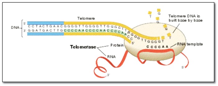
But telomere repeats and their associated caps don’t actually solve the end replication problem. The answer to this proved to be a novel DNA replication biochemistry uncovered by Elizabeth Blackburn, Jack Szostak and Carol Greider, who shared the Nobel Prize in Physiology or Medicine in 2009 (See Figure 2). Using the peculiar biology of a ciliated protozoan, Tetrahymena thermophila, the versatile genetics of the yeast Saccharomyces cerevisiae and the classical toolkit of enzymology, these scientists discovered an enzyme that could add new DNA sequence to chromosome ends, reversing the erosion due to incomplete replication by the standard DNA replication pathway. Remarkably, this enzyme uses RNA, a type of molecule usually made as a copy of DNA sequence, as a blueprint to make new DNA sequences at telomeres. The RNA blueprint is short, but it is copied over and over, creating the characteristic tandem repeat sequences of telomere DNA. In biochemistry, an enzyme name usually has the suffix –ase added to the process it controls, so the telomere-adding enzyme was called “telomerase.”
Figure 2.
Representation of telomerase (being oval) and associated RNA (red ribbon) polymerizing new telomere DNA (yellow).
Adapted from The 2009 Nobel Prize in Physiology or Medicine - Illustrated Presentation. Nobelprize.org. 13 Jul 2012 http://www.nobelprize.org/nobel_prizes/medicine/laureates/2009/illpres.html
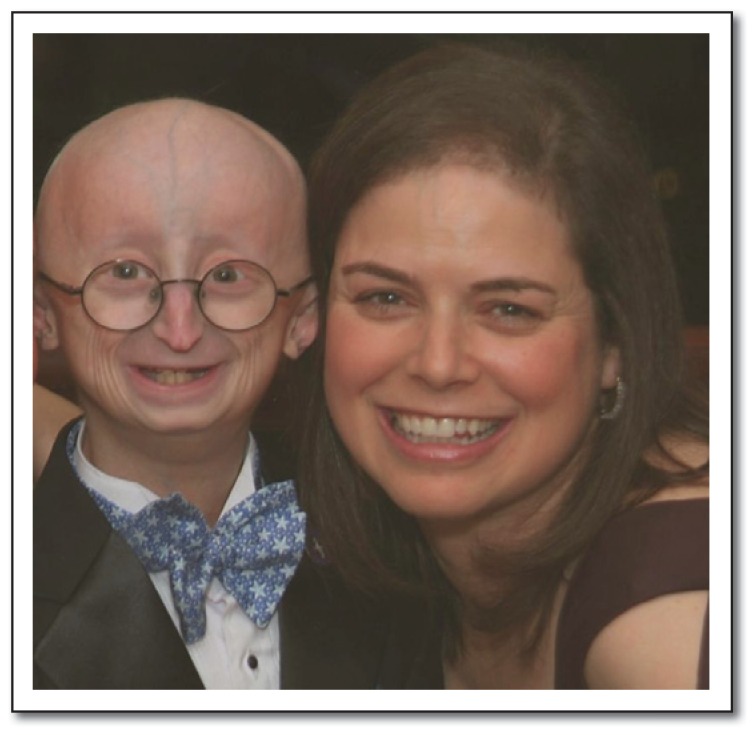
The importance of telomerase in human health is underscored by the clinical presentation of patients with genetic defects in the telomerase enzyme. One example is Dyskeratosis Congenita (DKC), a rare genetic disease associated with dramatically shortened telomeres. DKC mutations may be inherited in X-linked recessive, autosomal dominant and autosomal recessive forms. The autosomal dominant form of this disease is caused by mutations in genes encoding telomerase components including the RNA component of telomerase, or in genes that encode proteins associated with telomerase. Patients with this form of DKC accumulate much less telomerase RNA but remain healthy until young adulthood. Mortality in these patients is the result of progressive bone marrow failure, although there are accompanying defects in skin, hair, nails, epithelia and lung tissue. From these symptoms, it seems probable that the cells directly affected in DKC are stem cells in tissues with a high cell turnover. It should be noted that the clinical presentation of DKC is not that of premature aging syndromes (progerias), which include atherosclerosis and dementia (See Figure 3). Surprisingly, in light of the requirement for stable telomeres in cancer (discussed below), DKC patients show an elevated risk of leukemia and certain other cancers. It may be that telomere instability in these patients predisposes to telomere fusions, with subsequent chromosome breakage and rearrangements leading to oncogenic transformation.
Figure 3.
Leslie B. Gordon, Phd, and her son Sam, who has Hutchison-Gilford Progeria Syndrome. In this photo, Sam is 14 years old.
Telomerase and Aging
The landmark studies of Leonard Hayflick and colleagues established that embryonic cells have a finite potential to divide. Using embryonic fibroblasts, Hayflick found that these cells could only double about 50 times when cultured in vitro before they lost the ability to divide further and underwent growth arrest, also called “replicative senescence.”4 Subsequent studies extended this replicative limit on doubling number to a variety of other cells, a phenomenon termed their “Hayflick limit.” Striking correlations have been found between the natural lifespan of different animals and the Hayflick limit of cultured primary cells from those animals. Intriguingly, cells from patients with progerias have a reduced Hayflick limit.
Taken together, these observations suggest a causal relationship between replicative senescence and organismal aging. This inference should be approached with some caution, however. The correlation can disappear when patient samples are corrected for health status.5 With an estimated 236 cells in an average adult, fifty population doublings (250 cells) is more than enough to provide for even the longest recorded human lifespan. Indeed, most human tissues lack detectable telomerase and the cells of many tissues whose degeneration marks the progression of age, such as the central nervous system and the heart, show little or no evidence of telomere erosion.
Are telomeres limiting in replicative senescence? Here again, the scientific literature is replete with paradoxes. There is a correlation between telomere length and replicative senescence in cultured cells.6 On the other hand, mouse cells have a lower Hayflick limit than human cells and the typical lab mouse lives for only two years, yet laboratory mice have an average telomere length three times longer than that of humans. Mice engineered to lack telomerase activity are viable for up to six generations, clearly demonstrating that a normal mouse does not die from telomere insufficiency. Humans have the shortest telomeres, but the longest life spans, of all the primates. In a survey of over 60 mammalian species, the short-lived species tended to have the longest telomeres.7
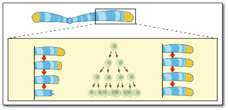
Our germ line cells, the cells that produce sperm and eggs, express the telomerase enzyme. This insures that each generation inherits chromosomes with an adequate length of telomere repeat sequences to support the many cell divisions between fertilization and adulthood (See Figure 4). We are born with an average of ca. 10,000–20,000 nucleotides of telomeric DNA on the ends of our chromosomes, although the exact amount depends on the chromosome and the individual. Our stem cells, the cells that replace the regular loss of cells that turn over during our lifespan, also express telomerase. It is estimated that, without telomerase, our cells lose about 100 nucleotides from each telomere with each cell cycle.
Figure 4.
In the absence of telomerase activity (left), telomeric repeat sequences are progressively lost with each cell division (center). With balancing telomerase activity (right), telomere length is maintained during cell proliferation.
Adapted from The 2009 Nobel Prize in Physiology or Medicine - Illustrated Presentation. Nobelprize.org. 13 Jul 2012 http://www.nobelprize.org/nobel_prizes/medicine/laureates/2009/illpres.html
However, there is evidence that aging is accompanied by the loss of telomeric DNA sequences, suggesting that the level of telomerase activity in somatic stem cells is not sufficient to completely compensate for telomere loss over decades. This possibility was first suggested by the finding that telomeres in human germ line cells are considerably longer than those of somatic cells.8
So if we could induce our somatic cells to express significant amounts of telomerase, would we live longer? While the clinical trials necessary to test this idea in human patients are not yet underway, the evidence based on studies of human cells in culture make this appear possible. Artificially inducing telomerase expression in primary retinal epithelial cells, foreskin fibroblasts, and vascular endothelial cells extends the proliferation of each of these cell types in culture for many cell divisions beyond their Hayflick limit.9 Importantly, these cells also lack markers for malignant transformation, so they are not being turned into cancer cells. Instead, they simply maintain the appearance and behavior of younger cells. If these benefits at the cellular level could be achieved throughout all the cells of our bodies, it would represent rejuvenation beyond that achievable by any cosmetic or surgical intervention.
While correlations have been reported between telomere shortening and age in humans, these correlations often disappear when the epidemiological data are corrected for other variables that also correlate with age. A recent review of the literature suggests that the use of telomere length as a biomarker for aging is premature.10 Longitudinal studies spanning decades of the lives of the same individuals would provide a strong test of the utility of telomere length as an aging biomarker.
At this point, telomere shortening could best be described as a frequent concomitant of aging, much like gray hair or wrinkled skin. Certainly, people with gray hair and wrinkled skin have fewer years of life left, on average, than those with smooth skin and pigmented hair. But nobody would say that gray hair or wrinkled skin cause aging, nor would anyone conclude that we would live longer if we just colored our hair and got a facelift.
Telomeres and Cancer
The defining feature of cancer is uncontrolled cell proliferation. In effect, cancer cells are immortal. In culture, cancer cells escape the Hayflick limit. Unlike most of our somatic cells, 85–90% of cancer cells express telomerase, and compared to normal somatic cells that do express telomerase, cancer cells achieve much higher telomerase expression levels.
Are tumor cell telomeres the Achilles heel of cancer? Telomerase inhibition can induce senescence in certain cancer cells.11 Mice engineered to lack telomerase activity have a lower incidence of cancer than telomerase-replete strains, although they are not cancer-free.12,13 This contrasts with DKC patients, who have a significantly elevated risk for several forms of cancer. On the other hand, mice engineered to over-express telomerase do have a higher incidence of cancer.14 Thus, drugs that target telomerase would seem to hold great promise for cancer therapy. Importantly, since telomerase is dispensable in most cells, there should be little or no toxicity to the patient, in contrast with current cancer chemotherapies. Indeed, telomerase-targeted anti-tumor drugs (imetelstat, a lipidated nucleic acid that targets the catalytic site of telomerase) and immunotherapies (GV1001, GRNVAC1) are in clinical trials.
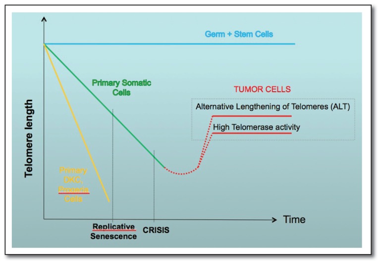
Unfortunately, 10–15% of cancers achieve telomere stability through a telomerase-independent mechanism. This mechanism, first described in yeast cells in which telomerase was genetically inactivated, is termed “Alternative Lengthening of Telomeres” (ALT) (See Figure 5). The ALT mechanism involves the use of the cell’s homologous recombination machinery to copy longer telomere sequences onto chromosomes with shorter telomeres. ALT-dependent tumors would be resistant to telomerase-based therapies. While ALT-dependent tumors are relatively rare, ALT-dependent cells could emerge among tumors cells under strong selective pressure from antitelomerase therapy. There are currently no therapies that target this mechanism.
Figure 5.
Time-dependent change in telomere length in germline and somatic stem cells (blue), primary somatic stem cells (green) and primary cells from DKC and progeria patients (yellow). Tumor cells arising from primary somatic cells (red) stabilize their telomeres either through activation of telomerase or through the ALT pathway.
Graph courtesy of Susana Gonzalo, Phd
Are Telomeres the Foundation of Youth?
The case for telomeres as the “fountain of youth” for cancer cells is strong.15 The alternative lengthening of telomeres (ALT) pathway poses a challenge to telomerase-based anti-tumor therapies, but the evidence that targeting of telomeres of cancer cells should have a high therapeutic index is compelling.
Much less compelling is the evidence that telomere length, per se, is limiting for the human lifespan. This could reflect technical limitations in the published studies. For example, telomere lengths are typically estimated using blood samples. If leukocyte telomeres are not representative of telomere erosion in other tissues, important correlations could be missed. Also, in most studies, comparisons are made between average telomere lengths, while it is likely that the shortest telomeres are physiologically relevant for age-dependent senescence.
Recent work has shown a correlation between chronic severe stress and shorter telomeres, at least in white blood cells.16 However, studies by some of the same authors suggest that telomerase activity is enhanced in stressed patients and in stressed rats. Since enhanced telomerase activity would be expected to extend telomeres, the mechanistic connections between acute stress, enhanced telomerase activity, shorter telomeres and long-term health are still unclear. It is safe to say, however, that minimizing acute stress has long-term health benefits.
The most consistent predictor of longevity in every organism tested is caloric restriction.17 Thus, any model for a rate-limiting effect of telomeres on organismal aging must account for this observation. Correlations pointing to telomere length as a potential driver for organismal longevity are balanced by other studies showing a lack of a relationship, as noted above. The weight of evidence at present supports the view that telomere shortening could magnify the morbidity associated with other stressors and the pathophysiology of certain diseases. By this view, telomere-extending therapies would be expected to have only an incremental effect on prolonging life span. Furthermore, evidence from transgenic mice suggests that telomere-extending therapy could increase the risk of cancer. For the foreseeable future, the best hope for our longevity, as with our wardrobe, is to take good care of what we already have.
Biography
Joel C. Eissenberg, PhD, is a Professor in the Edward A. Doisy Department of Biochemistry and Molecular Biology and Associate Dean for Research at the Saint Louis University School of Medicine.
Contact: eissenjc@slu.edu
Footnotes
Disclsoure
None reported.
References
- 1.Muller HJ. The remaking of chromosomes. Collecting Net. Woods Hole. 1938;13:181–198. [Google Scholar]
- 2.McClintock B. The behavior in successive nuclear divisions of a chromosome broken at meiosis. Proc Natl Acad Sci USA. 1939;25:405–416. doi: 10.1073/pnas.25.8.405. [DOI] [PMC free article] [PubMed] [Google Scholar]
- 3.McClintock B. The stability of broken ends of chromosomes in Zea mays. Genetics. 1941;26:234–282. doi: 10.1093/genetics/26.2.234. [DOI] [PMC free article] [PubMed] [Google Scholar]
- 4.Hayflick L, Moorhead PS. The serial cultivation of human diploid cell strains. Exp Cell Res. 1961;25:585–621. doi: 10.1016/0014-4827(61)90192-6. [DOI] [PubMed] [Google Scholar]
- 5.Christofalo VJ, Allen RG, Pignolo RJ, Martin BG, Beck JC. Relationship between donor age and the replicative livespan of human cells in culture: A reevaluation. Proc Natl Acad Sci USA. 1998;95:10614–10619. doi: 10.1073/pnas.95.18.10614. [DOI] [PMC free article] [PubMed] [Google Scholar]
- 6.Allsopp RC, Vaziri H, Patterson C, Goldstein S, Younglai EV, Futcher AB, Greider CW, Harley CB. Telomere length predicts replicative capacity of human fibroblasts. Proc Natl Acad Sci U S A. 1992;89:10114–10118. doi: 10.1073/pnas.89.21.10114. [DOI] [PMC free article] [PubMed] [Google Scholar]
- 7.Gomes NM, Ryder OA, Houck ML, Charter SJ, Walker W, Forsyth NR, Austad SN, Venditti C, Pagel M, Shay JW, Wright WE. Comparative biology of mammalian telomeres: hypotheses on ancestral states and the roles of telomeres in longevity determination. Aging Cell. 2011;10:761–768. doi: 10.1111/j.1474-9726.2011.00718.x. [DOI] [PMC free article] [PubMed] [Google Scholar]
- 8.Cooke HJ, Smith BA. Variability at the telomeres of the human X/Y pseudoautosomal region. Cold Spring Harb Symp Quant Biol. 1986;51(Pt 1):213–219. doi: 10.1101/sqb.1986.051.01.026. [DOI] [PubMed] [Google Scholar]
- 9.Bodnar AG, Ouellette M, Frolkis M, Holt SE, Chiu C, Morin GB, Harley CB, Shay JW, Lichtsteiner S, Wright WE. Extension of life-span by introduction of telomerase into normal human cells. Science. 1998;279:349–352. doi: 10.1126/science.279.5349.349. [DOI] [PubMed] [Google Scholar]
- 10.Mather KA, Jorm AF, Parslow RA, Christensen H. Is telomere length a biomarker of aging? A review. J Gerontol: Biol Sci. 2009;66A:202–213. doi: 10.1093/gerona/glq180. [DOI] [PubMed] [Google Scholar]
- 11.Shammas MA, Simmons CG, Corey DR, Shmookler Reis RJ. Telomerase inhibition by peptide nucleic acids reverses ‘immortality’ of transformed human cells. Oncogene. 1999;18(46):6191–6200. doi: 10.1038/sj.onc.1203069. [DOI] [PubMed] [Google Scholar]
- 12.Gonzalez-Suarez E, Samper E, Flores JM, Blasco MA. Telomerase-deficient mice with short telomeres are resistant to skin tumorigenesis. Nat Genet. 2000;26:114–117. doi: 10.1038/79089. [DOI] [PubMed] [Google Scholar]
- 13.Rudolph KL, Millard M, Bosenberg MW, DePinho RA. Telomere dysfunction and evolution of intestinal carcinoma in mice and humans. Nat Genet. 2001;28(2):155–159. doi: 10.1038/88871. [DOI] [PubMed] [Google Scholar]
- 14.Artandi SE, Alson S, Tietze MK, Sharpless NE, Ye S, Greenberg RA, Castrillon DH, Horner JW, Weiler SR, Carrasco RD, DePinho RA. Constitutive telomerase expression promotes mammary carcinomas in aging mice. Proc Natl Acad Sci U S A. 2002;99:8191–8196. doi: 10.1073/pnas.112515399. [DOI] [PMC free article] [PubMed] [Google Scholar]
- 15.Artandi SE, DePinho RA. Telomeres and telomerase in cancer. Carcinogenesis. 2010;31:9–18. doi: 10.1093/carcin/bgp268. [DOI] [PMC free article] [PubMed] [Google Scholar]
- 16.Blackburn EH, Epel ES. Telomeres and adversity: Too toxic to ignore. Nature. 2012;490:169–171. doi: 10.1038/490169a. [DOI] [PubMed] [Google Scholar]
- 17.Everitt AV, Rattan SIS, Couteur DG, de Cabo R, editors. Calorie Restriction, Aging and Longevity. Springer; Dordrecht: 2010. p. 500. [Google Scholar]


