Abstract
Background
In order to understand the role of dengue virus (DENV) specific T cell responses that associate with protection, we studied their frequency and phenotype in relation to clinical disease severity and resolution of viraemia in a large cohort of patients with varying severity of acute dengue infection.
Methodology/Principal findings
Using ex vivo IFNγ ELISpot assays we determined the frequency of dengue viral peptide (DENV)-NS3, NS1 and NS5 responsive T cells in 74 adult patients with acute dengue infection and examined the association of responsive T cell frequency with the extent of viraemia and clinical disease severity. We found that total DENV-specific and DENV-NS3-specific T cell responses, were higher in patients with dengue fever (DF), when compared to those with dengue haemorrhagic fever (DHF). In addition, those with DF had significantly higher (p = 0.02) DENV-specific T cell responses on day 4 of infection compared to those who subsequently developed DHF. DENV peptide specific T cell responses inversely correlated with the degree of viraemia, which was most significant for DENV-NS3 specific T cell responses (Spearman’s r = -0.47, p = 0.0003). The frequency of T cell responses to NS1, NS5 and pooled DENV peptides, correlated with the degree of thrombocytopenia but had no association with levels of liver transaminases. In contrast, total DENV-IgG inversely correlated with the degree of thrombocytopenia and levels of liver transaminases.
Conclusions/Significance
Early appearance of DENV-specific T cell IFNγ responses before the onset of plasma leakage, appears to associate with milder clinical disease and resolution of viraemia, suggesting a protective role in acute dengue infection.
Author summary
In order to understand the role of dengue virus (DENV) specific T cell responses in protection against infection, we studied T cell cytokine production in relation to clinical disease severity and resolution of viraemia in a large cohort of patients with varying severity of acute dengue infection. We found that DENV-specific T cell responses were higher in patients with dengue fever, when compared to those with dengue haemorrhagic fever. In addition, early appearance of DENV-specific T cell responses was significantly associated with milder clinical disease (p = 0.02). DENV peptide specific T cell responses inversely correlated with the degree of viraemia, which was most significant for DENV-NS3 specific T cell responses (Spearman’s r = -0.47, p = 0.0003). The frequency of NS1, NS5 and pooled DENV peptides, correlated with the degree of thrombocytopenia but had no association with liver transaminases. Our data suggest that early appearance of DENV-specific T cell IFNγ responses appear to associate with milder clinical disease and resolution of viraemia, suggesting a protective role in acute dengue infection.
Introduction
Dengue virus is the cause of the most common mosquito-borne viral infection worldwide, indeed over half of the global population live in areas where there is intense dengue transmission putting them at risk of dengue infection [1]. Dengue virus causes 390 million infections annually, of which nearly a quarter are clinically apparent causing a spectrum of disease phenotypes ranging from mild dengue fever (DF) to dengue hemorrhagic fever (DHF). DHF is defined by a transient increase in vascular permeability resulting in plasma leakage, with high fever, bleeding, thrombocytopenia and haemoconcentration, which can lead to shock (dengue shock syndrome (DSS))[2]. It is however not fully understood why some people develop more severe forms of the disease, with patient history, immunity, age, viral serotype, sub-strain and epidemiological factors all postulated to play a role [3]. It was highlighted during a recent summit to identify correlates of protection for dengue, that dengue virus (DENV) specific T cell immunity should be studied in more detail, in order to develop safe and effective dengue vaccines [4].
Although a dengue vaccine (Denvaxia) is now licensed in several countries, the efficacy is low in dengue seronegative individuals and provides only partial protection against DENV2 [5]. Although it is now generally believed that DENV specific T cells are protective, it is important that dengue vaccines should not induce “harmful” T cell immunity [4, 6–8]. Indeed, a significant hurdle in developing an efficacious dengue vaccine has been our limited understanding of the protective immune response in acute dengue infection and the added complexity of the presence of four DENV serotypes that are highly homologous. Although the evidence for T cells playing a possible protective role in DENV infections is emerging, there is still conflicting evidence as to the role of antigen-specific T cells during dengue infection is reported in the literature.
T cell responses to DENV are predominantly directed towards the nonstructural proteins (NS), with the majority of the CD8+ T cell responses directed towards NS3 followed by NS5 and CD4+ T cell responses to envelope, PrM and NS1 proteins [9–11]. It was believed that highly cross-reactive T cells specific to DENV-NS3, and other proteins, associate with severe clinical disease (DHF), and it was thought that these cells contribute to DHF by inducing a ‘cytokine storm’[12–15]. It is hypothesized in the ‘original antigenic sin’ theory that T cell responses against the initial DENV serotype of primary infection persist and dominate during subsequent infections; and that these T cells are suboptimal in inducing robust antiviral responses upon re-challenge [13, 14, 16]. However, it has been shown that DENV-NS3 specific T cell responses were at very low frequency during acute disease, and only detected in the convalescent phase pointing away from a role in vascular leak [14, 16, 17]. Recently it was observed that DENV-specific T cells are found in large numbers in the skin during acute dengue infection, and it is speculated that highly cross-reactive, pathogenic skin T cells could be contributing to DHF, despite being absent or present at low frequencies in the peripheral blood [8, 18]. As the frequency of skin resident DENV-specific T cells was investigated in a small patient cohort, it is not yet clear whether the frequency of the skin T cells associated with clinical disease severity.
Conversely some studies in both humans and mouse models have shown that DENV-specific T cells in the blood are likely to be protective [19–23]. It was shown that in individuals who were naturally infected with DENV, polyfunctional CD8+ T cells responses of higher magnitude and breadth were seen for HLA alleles associated with protection [21]. Similar findings were seen with DENV-specific CD4+ T cell responses [23]. Our previous studies have also shown that the magnitude of IFNγ-producing DENV NS3-specific memory T cell responses was similar in those who had varying severity of recovered past dengue infection, suggesting that the magnitude of the memory T cell response does not correlate with clinical disease severity[22]. While many studies have been carried out to elucidate the functionality of T cell responses in dengue, these have been limited to studying T cells specific for particular HLA types by using tetramers/pentamers [16, 18], or to investigating T cell responses in individuals with unknown severity of dengue.
To aid the generation of an effective vaccine it will be important to understand the role, phenotype and frequency of dengue-specific T cell responses in relation to clinical disease severity and clearance of viraemia [6, 7]. Therefore, here we investigate T cell responses to immunodominant DENV NS proteins in patients with DHF and DF, and analyse the association of such responses with resolution of viraemia.
Methods
Ethics statement
The study was approved by the Ethical Review Committee of The University of Sri Jayewardenepura. All patients were adults and recruited post written consent.
Recruitment of patients for analysis of the functionality of T cell responses
We recruited 74 adult patients with acute dengue infection from the National Infectious Diseases Institute, between day 4–8 of illness, following informed written consent. The patients were followed up throughout their hospital stay and all clinical features were recorded several times each day, from time of admission to discharge. Ultra sound scans were performed to determine the presence of fluid leakage in pleural and peritoneal cavities. Full blood counts, and liver transaminase measurements were performed serially through the illness. Clinical disease severity was classified according to the 2011 WHO dengue diagnostic criteria [24]. Accordingly, patients with ultrasound scan evidence of plasma leakage (those who had pleural effusions or ascites) were classified as having DHF. Those who did not develop any clinical or laboratory features of plasma leakage throughout their hospital stay, were diagnosed as having DF. Shock was defined as having cold clammy skin, along with a narrowing of pulse pressure of ≤ 20 mmHg. Based on this classification, 45 patients had DHF and 29 patients had dengue fever (DF) of the 74 patients recruited for the study.
Serology
Acute dengue infection was confirmed in serum samples using a PCR (see below) and dengue antibody detection. Dengue antibody assays were completed using a commercial capture-IgM and IgG ELISA (Panbio, Brisbane, Australia) [25, 26]. Based on the WHO criteria, those who had an IgM: IgG ratio of >1.2 were considered to have a primary dengue infection, while patients with IgM: IgG ratios <1.2 were categorized under secondary dengue infection [27]. The DENV-specific IgM and IgG ELISA was also used to semi-quantitatively determine the DENV-specific IgM and IgG titres, which were expressed in PanBio units.
Qualitative and quantitative assessment of viral loads
DENV were serotyped and viral titres quantified as previously described [28]. RNA was extracted from the serum samples using QIAamp Viral RNA Mini Kit (Qiagen, USA) according to the manufacturer’s protocol. Multiplex quantitative real-time PCR was performed as previously described using the CDC real time PCR assay for detection of the dengue virus [29], and modified to quantify the DENV. Oligonucleotide primers and a dual labeled probe for DENV 1,2,3,4 serotypes were used (Life technologies, India) based on published sequences [29]. In order to quantify viruses, standard curves of DENV serotypes were generated as previously described in Fernando, S. et.al [28].
Peptides
The peptide arrays spanning DENV NS1 (DENV-1 Singapore/S275/1990, NS1 protein NR-2751), NS3 (DENV-3, Philippines/H87/1956, NS3 protein, NR-2754) and NS5 proteins (DENV-2, New Guinea C (NGC), NS5 protein, NR-2746) were obtained from the NIH Biodefense and Emerging Infections Research Resource Repository, NIAID, NIH. The DENV NS3 peptide array consisted of 105, 14–17 mers peptides, NS1 and NS5 proteins were comprised of 60 and 156 peptides respectively. The peptides were reconstituted as previously described [30]. NS1, NS3 and NS5 peptides were pooled separately to represent the DENV- NS1, NS3 and NS5 proteins. In addition, total NS1, NS3 and NS5 peptides were combined to represent a ‘DENV-all’ pool of peptides.
Ex vivo ELISpot assay
Ex vivo IFNγ ELISpot assays were carried out as previously discussed using freshly isolated peripheral blood mononuclear cells (PBMC) obtained from 74 patients [22]. Fresh PBMCs were used due to concerns of possible reduction/alterations in IFNγ production by cryopreserved PBMCs [31]. DENV-NS3, NS1, NS5 and the combined DENV-ALL peptides were added at a final concentration of 10 μM and incubated overnight as previously described [16, 32]. All peptides were tested in duplicate. PHA was included as a positive control of cytokine stimulation and media alone was applied to the PBMCs as a negative control. The spots were enumerated using an automated ELISpot reader (AID Germany). Background (PBMCs plus media alone) was subtracted and data expressed as number of spot-forming units (SFU) per 106 PBMCs.
Quantitative cytokine assays
Quantitative ELISA for TNFα (Biolegend USA) and IL-2 (Mabtech, Sweden) were performed on ELISpot culture supernatants according to the manufacturer’s instructions.
Statistical analysis
PRISM version 6 was used for statistical analysis. As the data were not normally distributed, differences in means were compared using the Mann-Whitney U test (two tailed). Spearman rank order correlation coefficient was used to evaluate the correlation between variables including the association between DENV-specific T cell responses and platelet counts, degree of viraemia and liver transaminases.
Results
Patient clinical and laboratory features
To investigate the role of T cells in the progression of dengue infection we stratified patients based on disease severity. The clinical and laboratory features of the 74 patients recruited to the study are shown in Table 1. There was no statistically significant difference in the age of the individuals who had DF (median 29, IQR 29 to 42 years), when compared to those who had DHF (median 33, IQR 33 to 39 years). Of those who had DF, 18 (62.1%) were males and in those who had DHF 31 (68.9%) were males. Of these 74 patients, 45 had DHF and 29 had DF, and all 45 patients with DHF had ascites with 10 of them also experiencing pleural effusions. None of the patients developed shock and only one person progressed to bleeding manifestations (Table 1). The median duration of illness when recruited to the study was similar for patients with DF (median 5, IQR 5 to 6 days) and DHF (median 5, IQR 4 to 6 days).
Table 1. Clinical and laboratory characteristics of patients with DHF and DF.
| Clinical findings | DHF (n = 45) | DF (n = 29) |
|---|---|---|
| Vomiting | 15 (33.33%) | 8 (27.59%) |
| Abdominal pain | 25 (55.56%) | 7 (24.14%) |
| Hepatomegaly | 10 (22.22%) | 2 (6.9%) |
| Bleeding manifestations | 1 (2.22%) | 0 |
| Pleural effusion | 10 (22.22%) | 0 |
| Ascites | 45 (100%) | 0 |
| Lowest platelet count | ||
| <20,000 cells/mm3 | 25 (55.56%) | 1 (3.45%) |
| 20,000 to 50,000 | 13 (28.89%) | 10 (34.48%) |
| 50,000–100,000 | 6 (13.33%) | 12(41.38%) |
| >100,000 | 1 (2.22%) | 6 (20.69%) |
| Lowest Lymphocyte count | ||
| <750 | 20 (44.44%) | 7 (24.14%) |
| 750–1500 | 20 (44.44%) | 20(68.97%) |
| >1500 | 5 (11.11%) | 2 (6.90%) |
| Infecting serotype | ||
| DENV1 | 14 (31.1) | 16 (55.2) |
| DENV2 | 15 (33.3) | 4 (13.8) |
| DENV3 | 3 (6.7) | 1 (3.4) |
| DENV4 | 2 (4.4) | 0 (0) |
| Aviraemia | 11 (24.4) | 8 (27.5) |
| Highest AST (IU/L) | ||
| Median (IQR) | 133.6 (86 to 366.1) | Median 72.5, IQR 55 to 183 |
| Highest ALT (IU/L) | ||
| Median (IQR) | 172 (81 to 291) | 90 (30.3 to 96) |
Ex vivo IFNγ responses in patients with acute dengue infection
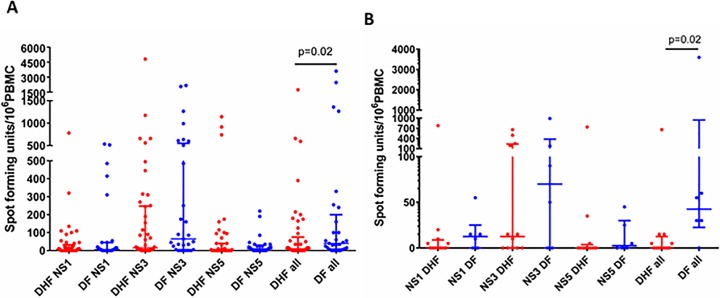
To evaluate the role of T cell derived cytokine in the immunopathology or regulation of acute dengue infection, we stimulated PBMCs isolated from patients with either DF or DHF with peptides constituting DENV-derived non-structural protein (NS) and assessed cytokine production by ELISPOT. We stimulated the patient PBMCs with different pools of overlapping peptides making up either full length NS1, NS3 or NS5 protein, or a pool of total NS1, 3 and 5 peptides (DENV-all). NS3 and NS5 were selected for investigation as CD8+ T cell responses have been shown to be directed to these proteins and CD4+ T cells have been shown to target structural proteins and NS1 as the main non-structural protein[11, 21]. This combination of NS proteins from the particular DENV strains has previously been used to study DENV specific T cell responses [33]. We used this ex vivo ELISPOT method to model antigen presentation of dengue-derived peptides to antigen-specific T cells in vitro and assessed T cell activation by IFNγ production, as a representative cytokine produced by T cells during dengue infection. T cell responses to the pooled DENV peptides (DENV-ALL) (p = 0.02) were higher in PBMCs derived from patients with DF than DHF patients and the NS3-specific responses showed a trend to be higher in those with DF than DHF (Fig 1A). T cell responses to DENV-NS1 peptides were similar in patients with DF and DHF. 26/45 patients with DHF and 12/29 patients with DF, had zero responses to NS5. We did not detect TNFα in the ex vivo ELISpot culture supernatants, which is in contrast to studies performed by others on T cell clones that implied TNFα producing DENV-specific T cells contribute to disease pathogenesis [15]. We also did not detect significant quantities of IL-2. The T cell responses to the overlapping peptides were not different in males compared to females, and none of the peptide-specific responses correlated with the age of the individuals.
Fig 1. Ex vivo ELISpot responses to DENV peptides in patients with DHF and DF.
(A) Ex vivo IFNγ ELISpot responses to DENV NS1, NS3, NS5 and combined DENV overlapping peptides in patients with DF (n = 29) and DHF (n = 45). (B) Ex vivo IFNγ ELISpot responses to DENV peptides in patients who were recruited on day 4 since onset of illness with DF (n = 6) and DHF (n = 12). Error bars represent the median and the interquartile range. *P<0.05.
To assess if detection of DENV specific T cell responses before the onset of the critical phase (vascular leakage phase), was associated with a reduced likelihood of developing leakage, we isolated the data from DF and DHF patients recruited on day 4 post the onset of illness and analysed IFNγ production by peptide stimulated PBMCs. None of the patients had evidence of vascular leakage on day 4 of illness and those who developed leakage (patients with DHF), did so on day 5 or 6. DF patients (those who did not develop any plasma leakage throughout their illness) had a significantly higher IFNγ secretion response (p = 0.02) to the DENV-all peptide pool (median 42.5, IQR = 22.5 to 945 SFU/106 PBMCs), when compared to DHF patients (median 0, IQR = 0 to 12.5 SFU/106 PBMCs) (Fig 1B). As such, significantly higher DENV-specific T cell responses were seen in those who did not develop fluid leakage, and those who had lower DENV-specific T cell responses proceeded to develop fluid leakage (DHF). Responses to DENV-NS3, NS1 and NS5 also appeared higher in patients with DF at this time point, although this did not reach statistical significance (Fig 1B).
To further assess the time-course of the response we obtained a second blood sample from eight patients within our cohort two days after collection of the first sample. T cell responses to DENV-ALL and DENV-NS3 peptides increased from the first sample (day 4) to the second (day 6), but it was not statistically significant (p>0.05) (S1 Fig)
Laboratory parameters and DENV-specific T cell responses
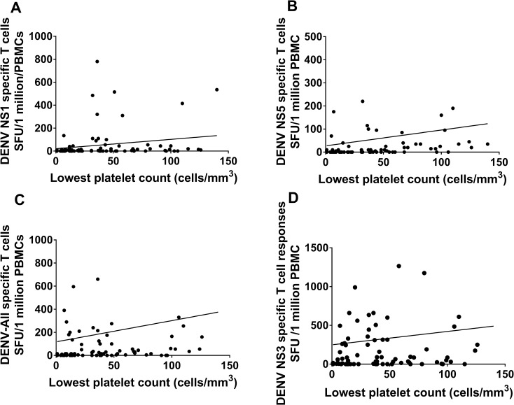
Thrombocytopenia is associated with clinical disease severity and higher degrees of thrombocytopenia are seen in those with DHF compared to those with DF[24]. We found that DENV peptide specific T cell responses correlated with the degree of thrombocytopenia. While this correlation with T cell responses and platelet counts was significant for DENV-NS1 (Spearmans r = 0.26, p = 0.01) (Fig 2A), NS5 (Spearmans r = 0.4, p = 0.0002) (Fig 2B) and DENV-All (Spearmans r = 0.31, p = 0.005) (Fig 2C), it was not significant for NS3 (Spearmans r = 0.18, p = 0.09) (Fig 2D). No association was seen with DENV-peptide specific T cell responses and aspartate transaminase (AST) and alanine transaminase (ALT) (S2 Fig), which are indicators of liver dysfunction [28, 34].
Fig 2. Relationship between laboratory parameters and DENV specific T cells in patients with acute dengue.
Platelet counts were correlated with ex vivo IFNγ ELISpot responses to DENV NS1 (Spearmans r = 0.26, p = 0.01) (A), NS5 (Spearmans r = 0.4, p = 0.0002) (B) and the overall DENV (Spearmans r = 0.31, p = 0.005) (C) and NS3 (Spearmans r = 0.18, p = 0.09) (D) overlapping peptides in patients (n = 74).
The relationship between DENV serotype and T cell responses
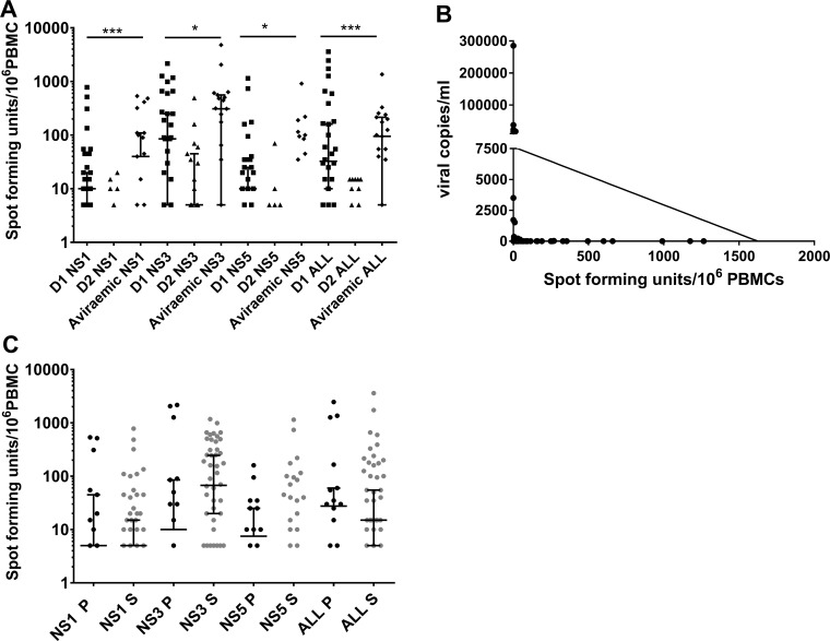
While some studies report that certain DENV serotypes associate with DHF [35, 36], others have shown that the risk of DHF is similar regardless of serotype [37]. Therefore, we proceeded to determine whether there were differences in the T cell responses to DENV-proteins based on the viral serotype that the patients were infected with. Within our cohort 30 (40.5%) patients were infected with DENV1, 19 (25.7%) with DENV2, 4 (5.4%) with DENV-3 and 2 (2.7%) with DENV-4 (Table 1). The serotype could not be determined in 19 (25.7%) patients, as they were not viraemic at the time of recruitment. DHF developed in 14/30 (46.7%) of the patients infected with DENV-1 and 15/19 (78.9%) of those infected with the DENV-2 and in 11/19 (57.9%) who were aviraemic at the time of recruitment (Fig 3A). Thus, it appeared that DENV-2 infection was more likely to lead to development of DHF (odds ratio 3.3, 95% CI 0.93 to 12.1), however the association was not statistically significant (p = 0.08) in this cohort. Aviraemic individuals displayed significantly higher IFNγ T cell responses to NS1 (p = 0.002), NS3 (p = 0.02), NS5 (p = 0.02) and DENV-ALL pooled peptides (p = 0.0004) when compared to those who were viraemic at the time of recruitment. In addition, those who were infected with the DENV-2 serotype, with a trend towards increased DHF susceptibility, had significantly lower responses to NS1 (p = 0.002), NS3 (p = 0.04), NS5 (p = 0.003) and DENV-All (p = 0.0003) peptides when compared to those who were infected with DENV-1.
Fig 3. Relationship between DENV serotype, clinical disease severity, viraemia and T cell responses.
A: Ex vivo ELISpot responses to DENV NS1, NS3, NS5 and combined DENV-ALL overlapping peptides in patients who were infected with DENV-1 (n = 30), DENV-2 (n = 19) or who were aviraemic (n = 19). Error bars represent the median and the interquartile range. **P<0.05, **P<0.01, ***P<0.001. (B) Correlation between DENV NS3-specific T cell responses and degree of viraemia (Spearman’s r = -0.47, p = 0.0003). (C) Ex vivo ELISpot responses to DENV NS1, NS3, NS5 and the overall DENV overlapping peptides in patients who had primary dengue (P) (n = 19) and secondary dengue infection (S) (n = 48).
As the PBMCs of patients infected with different DENV serotypes, were stimulated with the non-structural proteins from different virus serotypes (NS1-DENV1, NS3-DEN3 and NS5-DEN2), we sought to assess the sequence identity of these proteins between serotypes. Multiple alignment of the NS5 protein sequences of DENV2 (58 sequences) and DENV3 (28 sequences) was performed using virus variation resource [38] and analysed using Clustal omega and showed a sequence identity of > 72.1% between the NS5 proteins of these viral serotypes [39]. Multiple alignment of the NS3 protein of DENV2 (61 sequences) and DENV3 (28 sequences) showed a sequence identity of > 72.02%. Multiple alignment of the NS1 protein of DENV2 (62 sequences) and DENV3 (28 sequences) showed a sequence identity of >65.11%. The homology between DENV1 and DEN2 NS5 was >71.83% (comparison of 102 DENV1 sequences and 58 DENV2 sequences) [38], while the homology between DENV1 and DENV3 NS3 was >76.9% (comparison of 102 DENV1 sequences and 28 DENV3 sequences) [38]. Therefore, the T cell responses to different DENV serotypes is unlikely to be profoundly influenced by using non-structural protein sequences of different DENV serotypes. However, since there was some concern that stimulating using different serotypes would influence the results, we carried out further analysis. Since the most immunodominant protein was NS3, which was from DENV3, we also compared NS3 specific T cell responses in patients with DENV1 (n = 30) and DENV2 (n = 19). Although we did not find any significant difference in the T cell responses to NS3 based on infecting DENV serotype (p = 0.06), the responses to DENV3 NS3, appeared higher in patients with an acute DENV1 infection (median 85, IQR 5 to 435 SFU/million PBMBs) compared to those with an acute DENV2 infection (median 10, IQR 0 to 66.25 SFU/million PBMBs) (S3A Fig).
We then went on to compare responses to NS5 peptides, which were derived from DENV2, in patients with an acute DENV1 and DENV2 infection. Again, those with an acute DENV2 infection had significantly lower responses to the NS5 (p<0.0001) compared to patients with DENV1 (S3B Fig). We believe this difference is due to 15/19 (78.9%) patients with DENV2 having DHF (and thus poor T cell responses), whereas only 14/30 (46.7%) of those with DENV1 had DHF. We carried out a similar comparison within the group of patients with acute DENV1 for NS1 (which was from a DENV1 strain) and again did not see any difference within the DENV1 group in those with DF and DHF (S3C Fig), which was compatible with responses of the overall results.
Viraemia and DENV specific T cell responses
DHF patients have been shown to have higher viral loads, exhibit prolonged viraemia [40, 41] and persistent DENV-NS1 antigenaemia [42, 43]. As such we attempted to elucidate a correlation between T cell cytokine responses and viremia. DENV specific T cell responses to NS1, NS3 and NS5 peptides in addition to the pooled peptides (DENV-ALL) inversely correlated with the degree of viraemia, which was most significant for DENV-NS3 specific T cell responses (Spearman’s r = -0.47, p = 0.0003) (Fig 3B and S4 Fig). The viral loads significantly inversely correlated with the platelet counts (Spearmans r = -0.34, p = 0.01), with the platelet counts being lowest in individuals with the highest viral loads.
It is thought that a second dengue virus infection with a different viral serotype is a risk factor for developing DHF[44]. To determine the effect of secondary infection on the resulting T cell response, we characterized patient infection history and assayed patient blood for the presence of dengue specific IgM and IgG. Primary infection was defined by DENV- specific IgM:IgG >1.2 [24]. Accordingly, 19 (25.7%) patients were classified as experiencing a primary dengue infection and 48 (64.9%) were defined as secondary dengue infection. The antibody results were inconclusive for 7 (9.4%) patients. Our results showed no significant difference in DENV specific T cell responses between primary and secondary dengue infection patient groups (p>0.05) for any of the DENV peptide pools (Fig 3C).
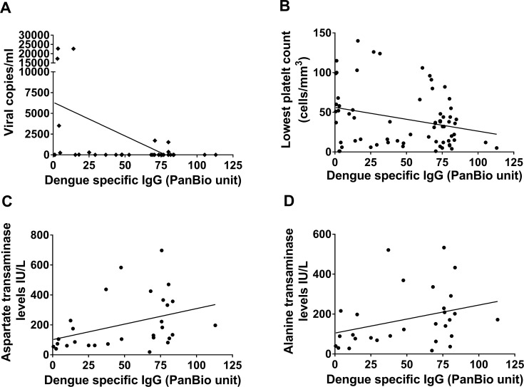
We semi-quantitatively determined the DENV-specific IgM and IgG antibody titres in all patients with DF and DHF, and we found that neither the DENV-IgM nor IgG antibody titres correlated with T cell responses to DENV-NS1, NS5 and NS3. However, the DENV-specific IgG antibody titres inversely correlated with viral loads in those with DHF (Spearman’s r = -0.37, p = 0.03) (Fig 4A), but not in those with DF (Spearmans r = -0.25, p = 0.16). In analysis of the IgG antibody titres of all patients (n = 74) they too inversely correlated with the degree of thrombocytopenia (Spearmans r = -0.29, p = 0.009) (Fig 4B). DENV-specific IgG also correlated with the highest aspartate transaminase (AST) (Spearmans r = 0.51, p = 0.004) (Fig 4C) and alanine transaminase (ALT) levels (Spearmans r = 0.4, p = 0.03) in all patients with acute dengue infection (Fig 4D).
Fig 4. DENV-specific IgG responses and laboratory parameters of clinical disease severity in patients with acute dengue.
Analysis of the correlation between DENV-specific IgG responses and (A) degree of viraemia (Spearmans r = -0.25, p = 0.16) in patients with DHF, (B) degree of thrombocytopenia (Spearmans r = -0.29, p = 0.009), (C) aspartate transaminase (AST) levels (Spearmans r = 0.51, p = 0.004) and (D) alanine transaminase (ALT) levels (Spearmans r = 0.51, p = 0.004) in all patients (n = 74) with acute dengue infection.
Discussion
In this study we set out to investigate the role of T cells in dengue immunity and found that DENV-specific T cells are present at low frequency during acute infection, consistent with previous reports published by us and by others [16, 17, 45]. IFNγ production was significantly higher in patients with DF as opposed to DHF, especially during early infection. Those who had lower DENV-NS3 specific T cell responses on day four since the onset of illness (before development of fluid leakage), were significantly more likely to subsequently develop vascular leakage of DHF. In addition, the frequency of pooled DENV-peptide specific, in particular DENV-NS3 specific, T cell responses was associated with resolution of viraemia. Aviraemic patients had significantly higher DENV- specific T cell responses when compared to those who were viraemic. However, overall the majority of patients with both DF and DHF, had a very low magnitude of DENV specific T cell responses. T cell IFNγ responses to DENV NS1, NS5 and pooled DENV (NS1, NS3 and NS5) peptides inversely correlated with the degree of thrombocytopenia, but we did not show any relationship with liver transaminases (AST and ALT levels). Both the degree of thrombocytopenia and a rise in both AST and ALT, have been shown to associate with dengue severity [24, 28]. Therefore, our data show that the early appearance (on day 4 of illness before any patient developed plasma leakage) of DENV-NS3 specific T cell responses is associated with milder disease, which is compatible with recent studies regarding the role of T cells in DENV infection [18, 21–23]. This suggests that DENV-peptide specific T cells are possibly have a protective role against developing severe forms of dengue infection.
Although Appana et al also evaluated ex vivo IFNγ to selected peptides of structural and non-structural DENV proteins by ELISpot assays, they did not find any differences in the frequency of DENV-specific T cell responses in patients with DF when compared to those with DHF [46]. However, only peptides that were predicted to bind to certain major HLA alleles were included in the authors’ peptide pools used in the ELISpot assays [46], whereas here we utilised peptides spanning the entire length of DENV NS1, NS3 and NS5, proteins. This difference in experimental approach may have affected the cytokine production profile of the responding T cells in the different disease states. In addition, as the viability and function of T cells have been shown to be affected in those with acute dengue infection [31], we used freshly isolated PBMCs in all our experiments to limit extraneous cellular stress in contrast to previous studies [15, 21, 46, 47].
In this study, some of the non-structural peptides used were of a different DENV serotype than that causing the acute infection as we used fresh PBMCs in our assays before the infecting serotype was known. As many previous studies on T cell responses have used peptides of unmatched DENV serotypes to assess T cell responses[33, 46] and also as it was shown that the breadth or the magnitude of DENV specific T cell responses, were similar even when peptides of unmatched serotypes were used [9], we too proceeded with the same approach. However, there is a possibility that using unmatched peptides could affect the magnitude of the T cell responses. Subsequent subanalysis of T cell responses to DENV1-NS1 and DENV3-NS3 of patients with an acute DENV1 and DENV2 infection, carried out by us also did not show any difference. However, analysis of T cell responses to DENV2 derived NS5 overlapping peptides, showed that patients with an acute DENV2 infection (matched peptides) had significantly lower responses than patients with DENV1 infection. We believe this difference is possibly due to 78.9% patients with DENV2 having DHF (and thus poor T cell responses), whereas only 46.7% of those with DENV1 had DHF.
As with most viral infections, the severity of the illness and T cell responses to the DENV has also been shown to depend on the HLA type of the individual [21, 23, 48]. However, we did not assess the T cell responses in relation to their HLA type as the primary aim of this study was to assess the overall DENV specific T cell responses based on clinical disease severity and viraemia, irrespective of their HLA types in a large cohort, with relevance to pathogenesis and vaccine design. As the HLA type is known to influence the magnitude of the T cell response, there is a possibility that the differences in the magnitude of T cell responses to different DENV overlapping peptides, could also be due to the differences in their HLA types, which were not assessed here.
In general, more severe forms of dengue infection are observed during a secondary heterologous dengue infection [4], which gave rise to the hypothesis that cross reactive T cells responding to the primary infecting DENV serotype are suboptimal in clearing the secondary virus, and lead to development of more severe disease [14, 16]. In these studies, it was shown that a tetramer of different viral specificity to the current infecting DENV serotype, sometimes had a higher affinity to the DENV specific T cells [16]. In our study, we did not observe any difference in IFNγ production in overall ex vivo ELISpot assays from PBMCs derived from patients with primary or secondary dengue infection; however, we did not examine variant peptide-specific responses. The broad differences we observed in DENV-specific T cell responses correlated only with clinical disease severity. Interestingly, the DENV-specific IgG levels, which were measured semi-quantitatively, inversely correlated with the degree of thrombocytopenia and also AST and ALT levels, which are known to associate with liver damage. DENV-specific IgG levels are known to be significantly higher in patients with secondary dengue, compared to primary dengue, indeed it is one of the criteria for definition of a secondary dengue infection. However, as we assessed the relationship of total IgG with these laboratory parameters and did not assess the presence of neutralizing antibodies, which are more likely to be protective, this needs to be further evaluated.
In summary, we found that DENV-specific T cell IFNγ responses, were associated with milder clinical disease severity and resolution of viraemia, suggesting a protective role for peptide specific T cells early in acute dengue infection.
Supporting information
(TIF)
Association of DENV specific NS1, NS3, NS5 and All overlapping peptide responses in patients with acute dengue (n = 74) with the highest recorded aspartate transaminase level (NS1 Spearnman’s r = 0.15, p = 0.4; NS3 Spearman’s r = 0.34, p = 0.05; NS5 Spearman’s r = 0.16, p = 0.37; All Spearman’s r = 0.33, p = 0.06) (A) and alanine transaminase level (NS1 Spearman’s r = 12, p = 0.5; NS3 Spearman’s r = 0.25, p = 0.15; NS5 Spearman’s r = 0.19, p = 0.29; All Spearman’s r = 0.31, p = 0.08) (B).
(TIF)
(A) IFNγ ELISpot responses were assessed in patients with acute DENV1 (n = 30) and acute DENV2 (n = 19) to NS3 overlapping peptides derived from DENV3
(B) IFNγ ELISpot responses were assessed in patients with acute DENV1 (n = 30) and acute DENV2 (n = 19) to NS5 overlapping peptides derived from DENV2
(C) IFNγ ELISpot responses were assessed in patients with acute DENV1 resulting in DF (n = 16) or DHF (n = 14) for NS1 overlapping peptides derived from DENV1.
(TIF)
(A) Correlation between DENV All-Specific T cell responses and degree of viraemia (Spearman’s r = -0.38, p = 0.004).
(B) Correlation between DENV NS5-Specific T cell responses and degree of viraemia (Spearman’s r = -0.28, p = 0.04).
(C) Correlation between DENV NS1-Specific T cell responses and degree of viraemia (Spearman’s r = -0.31, p = 0.02).
(TIF)
Data Availability
All relevant data are within the paper and its Supporting Information files.
Funding Statement
Funding was provided by the Centre for Dengue Research, University of Sri Jayewardenapura, National Science Foundation, Sri Lanka (NSF/RG/2016/HS/05) and by the Medical Research Council (UK). GSO receives support from the National Institute for Health Research (NIHR) Oxford Biomedical Research Centre (BRC). The funders had no role in study design, data collection and analysis, decision to publish, or preparation of the manuscript.
References
- 1.Guzman MG, Gubler DJ, Izquierdo A, Martinez E, Halstead SB. Dengue infection. Nat Rev Dis Primers. 2016;2:16055 10.1038/nrdp.2016.55 . [DOI] [PubMed] [Google Scholar]
- 2.Bhatt S, Gething PW, Brady OJ, Messina JP, Farlow AW, Moyes CL, et al. The global distribution and burden of dengue. Nature. 2013;496(7446):504–7. Epub 2013/04/09. 10.1038/nature12060 ; PubMed Central PMCID: PMC3651993. [DOI] [PMC free article] [PubMed] [Google Scholar]
- 3.Malavige GN, Ogg GS. T cell responses in dengue viral infections. J Clin Virol. 2013;58(4):605–11. Epub 2013/11/14. 10.1016/j.jcv.2013.10.023 . [DOI] [PubMed] [Google Scholar]
- 4.Katzelnick LC, Harris E, Participants in the Summit on Dengue Immune Correlates of P. Immune correlates of protection for dengue: State of the art and research agenda. Vaccine. 2017;35(36):4659–69. 10.1016/j.vaccine.2017.07.045 ; PubMed Central PMCID: PMCPMC5924688. [DOI] [PMC free article] [PubMed] [Google Scholar]
- 5.Halstead SB, Russell PK. Protective and immunological behavior of chimeric yellow fever dengue vaccine. Vaccine. 2016;34(14):1643–7. 10.1016/j.vaccine.2016.02.004 . [DOI] [PubMed] [Google Scholar]
- 6.Simmons CP, McPherson K, Van Vinh Chau N, Hoai Tam DT, Young P, Mackenzie J, et al. Recent advances in dengue pathogenesis and clinical management. Vaccine. 2015;33(50):7061–8. 10.1016/j.vaccine.2015.09.103 . [DOI] [PubMed] [Google Scholar]
- 7.Rivino L. T cell immunity to dengue virus and implications for vaccine design. Expert review of vaccines. 2016;15(4):443–53. 10.1586/14760584.2016.1116948 . [DOI] [PubMed] [Google Scholar]
- 8.Screaton G, Mongkolsapaya J, Yacoub S, Roberts C. New insights into the immunopathology and control of dengue virus infection. Nature reviews Immunology. 2015;15(12):745–59. 10.1038/nri3916 . [DOI] [PubMed] [Google Scholar]
- 9.Simmons CP, Dong T, Chau NV, Dung NT, Chau TN, Thao le TT, et al. Early T-cell responses to dengue virus epitopes in Vietnamese adults with secondary dengue virus infections. Journal of virology. 2005;79(9):5665–75. 10.1128/JVI.79.9.5665-5675.2005 . [DOI] [PMC free article] [PubMed] [Google Scholar]
- 10.Duangchinda T, Dejnirattisai W, Vasanawathana S, Limpitikul W, Tangthawornchaikul N, Malasit P, et al. Immunodominant T-cell responses to dengue virus NS3 are associated with DHF. Proceedings of the National Academy of Sciences of the United States of America. 2010;107(39):16922–7. 10.1073/pnas.1010867107 ; PubMed Central PMCID: PMCPMC2947904. [DOI] [PMC free article] [PubMed] [Google Scholar]
- 11.Rivino L, Kumaran EA, Jovanovic V, Nadua K, Teo EW, Pang SW, et al. Differential targeting of viral components by CD4+ versus CD8+ T lymphocytes in dengue virus infection. Journal of virology. 2013;87(5):2693–706. Epub 2012/12/21. 10.1128/JVI.02675-12 ; PubMed Central PMCID: PMC3571409. [DOI] [PMC free article] [PubMed] [Google Scholar]
- 12.Duangchinda T, Dejnirattisai W, Vasanawathana S, Limpitikul W, Tangthawornchaikul N, Malasit P, et al. Immunodominant T-cell responses to dengue virus NS3 are associated with DHF. Proceedings of the National Academy of Sciences of the United States of America. 2011;107(39):16922–7. 10.1073/pnas.1010867107 . [DOI] [PMC free article] [PubMed] [Google Scholar]
- 13.Midgley CM, Bajwa-Joseph M, Vasanawathana S, Limpitikul W, Wills B, Flanagan A, et al. An in-depth analysis of original antigenic sin in dengue virus infection. Journal of virology. 2011;85(1):410–21. Epub 2010/10/29. 10.1128/JVI.01826-10 ; PubMed Central PMCID: PMC3014204. [DOI] [PMC free article] [PubMed] [Google Scholar]
- 14.Mongkolsapaya J, Dejnirattisai W, Xu XN, Vasanawathana S, Tangthawornchaikul N, Chairunsri A, et al. Original antigenic sin and apoptosis in the pathogenesis of dengue hemorrhagic fever. Nature medicine. 2003;9(7):921–7. 10.1038/nm887 . [DOI] [PubMed] [Google Scholar]
- 15.Dong T, Moran E, Vinh Chau N, Simmons C, Luhn K, Peng Y, et al. High pro-inflammatory cytokine secretion and loss of high avidity cross-reactive cytotoxic T-cells during the course of secondary dengue virus infection. PloS one. 2007;2(12):e1192 10.1371/journal.pone.0001192 . [DOI] [PMC free article] [PubMed] [Google Scholar]
- 16.Mongkolsapaya J, Duangchinda T, Dejnirattisai W, Vasanawathana S, Avirutnan P, Jairungsri A, et al. T cell responses in dengue hemorrhagic fever: are cross-reactive T cells suboptimal? J Immunol. 2006;176(6):3821–9. . [DOI] [PubMed] [Google Scholar]
- 17.Malavige GN, Jeewandara C, Alles KM, Salimi M, Gomes L, Kamaladasa A, et al. Suppression of virus specific immune responses by IL-10 in acute dengue infection. PLoS neglected tropical diseases. 2013;7(9):e2409 Epub 2013/09/17. 10.1371/journal.pntd.0002409 ; PubMed Central PMCID: PMC3764236. [DOI] [PMC free article] [PubMed] [Google Scholar]
- 18.Rivino L, Kumaran EA, Thein TL, Too CT, Gan VC, Hanson BJ, et al. Virus-specific T lymphocytes home to the skin during natural dengue infection. Science translational medicine. 2015;7(278):278ra35 10.1126/scitranslmed.aaa0526 . [DOI] [PubMed] [Google Scholar]
- 19.Amorim JH, dos Santos Alves RP, Bizerra R, Araujo Pereira S, Ramos Pereira L, Nascimento Fabris DL, et al. Antibodies are not required to a protective immune response against dengue virus elicited in a mouse encephalitis model. Virology. 2016;487:41–9. 10.1016/j.virol.2015.10.006 . [DOI] [PubMed] [Google Scholar]
- 20.Yauch LE, Zellweger RM, Kotturi MF, Qutubuddin A, Sidney J, Peters B, et al. A protective role for dengue virus-specific CD8+ T cells. J Immunol. 2009;182(8):4865–73. 10.4049/jimmunol.0801974 . [DOI] [PMC free article] [PubMed] [Google Scholar]
- 21.Weiskopf D, Angelo MA, de Azeredo EL, Sidney J, Greenbaum JA, Fernando AN, et al. Comprehensive analysis of dengue virus-specific responses supports an HLA-linked protective role for CD8+ T cells. Proceedings of the National Academy of Sciences of the United States of America. 2013;110(22):E2046–53. Epub 2013/04/13. 10.1073/pnas.1305227110 ; PubMed Central PMCID: PMC3670335. [DOI] [PMC free article] [PubMed] [Google Scholar]
- 22.Jeewandara C, Adikari TN, Gomes L, Fernando S, Fernando RH, Perera MK, et al. Functionality of dengue virus specific memory T cell responses in individuals who were hospitalized or who had mild or subclinical dengue infection. PLoS neglected tropical diseases. 2015;9(4):e0003673 Epub 2015/04/16. 10.1371/journal.pntd.0003673 ; PubMed Central PMCID: PMC4395258. [DOI] [PMC free article] [PubMed] [Google Scholar]
- 23.Weiskopf D, Bangs DJ, Sidney J, Kolla RV, De Silva AD, de Silva AM, et al. Dengue virus infection elicits highly polarized CX3CR1+ cytotoxic CD4+ T cells associated with protective immunity. Proceedings of the National Academy of Sciences of the United States of America. 2015;112(31):E4256–63. 10.1073/pnas.1505956112 . [DOI] [PMC free article] [PubMed] [Google Scholar]
- 24.WHO, editor. Comprehensive guidelines for prevention and control of dengue fever and dengue haemorrhagic fever SEARO, New Delhi, India: World Health Organization; 2011. [Google Scholar]
- 25.Vaughn DW, Nisalak A, Solomon T, Kalayanarooj S, Nguyen MD, Kneen R, et al. Rapid serologic diagnosis of dengue virus infection using a commercial capture ELISA that distinguishes primary and secondary infections. The American journal of tropical medicine and hygiene. 1999;60(4):693–8. . [DOI] [PubMed] [Google Scholar]
- 26.Sang CT, Cuzzubbo AJ, Devine PL. Evaluation of a commercial capture enzyme-linked immunosorbent assay for detection of immunoglobulin M and G antibodies produced during dengue infection. Clinical and diagnostic laboratory immunology. 1998;5(1):7–10. Epub 1998/02/10. ; PubMed Central PMCID: PMC121382. [DOI] [PMC free article] [PubMed] [Google Scholar]
- 27.Dengue guidelines for diagnosis, prevention and control, (2009).
- 28.Fernando S, Wijewickrama A, Gomes L, Punchihewa CT, Madusanka SD, Dissanayake H, et al. Patterns and causes of liver involvement in acute dengue infection. BMC infectious diseases. 2016;16:319 10.1186/s12879-016-1656-2 ; PubMed Central PMCID: PMCPMC4938910. [DOI] [PMC free article] [PubMed] [Google Scholar]
- 29.Santiago GA, Vergne E, Quiles Y, Cosme J, Vazquez J, Medina JF, et al. Analytical and clinical performance of the CDC real time RT-PCR assay for detection and typing of dengue virus. PLoS neglected tropical diseases. 2013;7(7):e2311 10.1371/journal.pntd.0002311 ; PubMed Central PMCID: PMCPMC3708876. [DOI] [PMC free article] [PubMed] [Google Scholar]
- 30.Malavige GN, McGowan S, Atukorale V, Salimi M, Peelawatta M, Fernando N, et al. Identification of serotype-specific T cell responses to highly conserved regions of the dengue viruses. Clinical and experimental immunology. 2012;168(2):215–23. 10.1111/j.1365-2249.2012.04566.x . [DOI] [PMC free article] [PubMed] [Google Scholar]
- 31.Perdomo-Celis F, Salgado DM, Castaneda DM, Narvaez CF. Viability and functionality of Cryopreserved Peripheral Blood Mononuclear Cells in pediatric dengue. Clin Vaccine Immunol. 2016. 10.1128/CVI.00038-16 ; PubMed Central PMCID: PMCPMC4860468. [DOI] [PMC free article] [PubMed] [Google Scholar]
- 32.Malavige GN, Jones L, Kamaladasa SD, Wijewickrama A, Seneviratne SL, Black AP, et al. Viral load, clinical disease severity and cellular immune responses in primary varicella zoster virus infection in Sri Lanka. PloS one. 2008;3(11):e3789 10.1371/journal.pone.0003789 . [DOI] [PMC free article] [PubMed] [Google Scholar]
- 33.Chu H, George SL, Stinchcomb DT, Osorio JE, Partidos CD. CD8+ T-cell Responses in Flavivirus-Naive Individuals Following Immunization with a Live-Attenuated Tetravalent Dengue Vaccine Candidate. The Journal of infectious diseases. 2015;212(10):1618–28. 10.1093/infdis/jiv258 ; PubMed Central PMCID: PMCPMC4621245. [DOI] [PMC free article] [PubMed] [Google Scholar]
- 34.Huy NT, Van Giang T, Thuy DH, Kikuchi M, Hien TT, Zamora J, et al. Factors associated with dengue shock syndrome: a systematic review and meta-analysis. PLoS neglected tropical diseases. 2013;7(9):e2412 Epub 2013/10/03. 10.1371/journal.pntd.0002412 ; PubMed Central PMCID: PMC3784477. [DOI] [PMC free article] [PubMed] [Google Scholar]
- 35.Fried JR, Gibbons RV, Kalayanarooj S, Thomas SJ, Srikiatkhachorn A, Yoon IK, et al. Serotype-specific differences in the risk of dengue hemorrhagic fever: an analysis of data collected in Bangkok, Thailand from 1994 to 2006. PLoS neglected tropical diseases. 2010;4(3):e617 10.1371/journal.pntd.0000617 ; PubMed Central PMCID: PMCPMC2830471. [DOI] [PMC free article] [PubMed] [Google Scholar]
- 36.Yung CF, Lee KS, Thein TL, Tan LK, Gan VC, Wong JG, et al. Dengue serotype-specific differences in clinical manifestation, laboratory parameters and risk of severe disease in adults, singapore. The American journal of tropical medicine and hygiene. 2015;92(5):999–1005. 10.4269/ajtmh.14-0628 ; PubMed Central PMCID: PMCPMC4426593. [DOI] [PMC free article] [PubMed] [Google Scholar]
- 37.Fox A, Le NM, Simmons CP, Wolbers M, Wertheim HF, Pham TK, et al. Immunological and viral determinants of dengue severity in hospitalized adults in Ha Noi, Viet Nam. PLoS neglected tropical diseases. 2011;5(3):e967 Epub 2011/03/11. 10.1371/journal.pntd.0000967 ; PubMed Central PMCID: PMC3046970. [DOI] [PMC free article] [PubMed] [Google Scholar]
- 38.Brister JR, Bao Y, Zhdanov SA, Ostapchuck Y, Chetvernin V, Kiryutin B, et al. Virus Variation Resource—recent updates and future directions. Nucleic Acids Res. 2014;42(Database issue): D660–5. 10.1093/nar/gkt1268 ; PubMed Central PMCID: PMCPMC3965055. [DOI] [PMC free article] [PubMed] [Google Scholar]
- 39.Sievers F, Wilm A, Dineen D, Gibson TJ, Karplus K, Li W, et al. Fast, scalable generation of high-quality protein multiple sequence alignments using Clustal Omega. Mol Syst Biol. 2011;7:539 10.1038/msb.2011.75 ; PubMed Central PMCID: PMCPMC3261699. [DOI] [PMC free article] [PubMed] [Google Scholar]
- 40.Guilarde AO, Turchi MD, Siqueira JB Jr., Feres VC, Rocha B, Levi JE, et al. Dengue and dengue hemorrhagic fever among adults: clinical outcomes related to viremia, serotypes, and antibody response. The Journal of infectious diseases. 2008;197(6):817–24. 10.1086/528805 . [DOI] [PubMed] [Google Scholar]
- 41.Wang WK, Chen HL, Yang CF, Hsieh SC, Juan CC, Chang SM, et al. Slower rates of clearance of viral load and virus-containing immune complexes in patients with dengue hemorrhagic fever. Clin Infect Dis. 2006;43(8):1023–30. 10.1086/507635 . [DOI] [PubMed] [Google Scholar]
- 42.Paranavitane SA, Gomes L, Kamaladasa A, Adikari TN, Wickramasinghe N, Jeewandara C, et al. Dengue NS1 antigen as a marker of severe clinical disease. BMC infectious diseases. 2014;14(1):570 Epub 2014/11/05. 10.1186/s12879-014-0570-8 ; PubMed Central PMCID: PMC4222370. [DOI] [PMC free article] [PubMed] [Google Scholar]
- 43.Adikari TN, Gomes L, Wickramasinghe N, Salimi M, Wijesiriwardana N, Kamaladasa A, et al. Dengue NS1 antigen contributes to disease severity by inducing interleukin (IL)-10 by monocytes. Clinical and experimental immunology. 2016;184(1):90–100. 10.1111/cei.12747 ; PubMed Central PMCID: PMCPMC4778103. [DOI] [PMC free article] [PubMed] [Google Scholar]
- 44.Guzman MG, Alvarez M, Halstead SB. Secondary infection as a risk factor for dengue hemorrhagic fever/dengue shock syndrome: an historical perspective and role of antibody-dependent enhancement of infection. Archives of virology. 2013;158(7):1445–59. Epub 2013/03/09. 10.1007/s00705-013-1645-3 . [DOI] [PubMed] [Google Scholar]
- 45.Chandele A, Sewatanon J, Gunisetty S, Singla M, Onlamoon N, Akondy RS, et al. Characterization of human CD8 T cell responses in dengue virus infected patients from India. Journal of virology. 2016. 10.1128/JVI.01424-16 . [DOI] [PMC free article] [PubMed] [Google Scholar]
- 46.Appanna R, Huat TL, See LL, Tan PL, Vadivelu J, Devi S. Cross-reactive T-cell responses to the nonstructural regions of dengue viruses among dengue fever and dengue hemorrhagic fever patients in Malaysia. Clin Vaccine Immunol. 2007;14(8):969–77. 10.1128/CVI.00069-07 . [DOI] [PMC free article] [PubMed] [Google Scholar]
- 47.Luhn K, Simmons CP, Moran E, Dung NT, Chau TN, Quyen NT, et al. Increased frequencies of CD4+ CD25(high) regulatory T cells in acute dengue infection. The Journal of experimental medicine. 2007;204(5):979–85. 10.1084/jem.20061381 . [DOI] [PMC free article] [PubMed] [Google Scholar]
- 48.Malavige GN, Rostron T., Rohanachandra L.T., Jayaratne S.D., Fernando N., De Silva A.D., Liyanage M., Ogg G.S. HLA class I and class II associations in dengue viral infections in a Sri Lankan population. PloS one. 2011. [DOI] [PMC free article] [PubMed] [Google Scholar]
Associated Data
This section collects any data citations, data availability statements, or supplementary materials included in this article.
Supplementary Materials
(TIF)
Association of DENV specific NS1, NS3, NS5 and All overlapping peptide responses in patients with acute dengue (n = 74) with the highest recorded aspartate transaminase level (NS1 Spearnman’s r = 0.15, p = 0.4; NS3 Spearman’s r = 0.34, p = 0.05; NS5 Spearman’s r = 0.16, p = 0.37; All Spearman’s r = 0.33, p = 0.06) (A) and alanine transaminase level (NS1 Spearman’s r = 12, p = 0.5; NS3 Spearman’s r = 0.25, p = 0.15; NS5 Spearman’s r = 0.19, p = 0.29; All Spearman’s r = 0.31, p = 0.08) (B).
(TIF)
(A) IFNγ ELISpot responses were assessed in patients with acute DENV1 (n = 30) and acute DENV2 (n = 19) to NS3 overlapping peptides derived from DENV3
(B) IFNγ ELISpot responses were assessed in patients with acute DENV1 (n = 30) and acute DENV2 (n = 19) to NS5 overlapping peptides derived from DENV2
(C) IFNγ ELISpot responses were assessed in patients with acute DENV1 resulting in DF (n = 16) or DHF (n = 14) for NS1 overlapping peptides derived from DENV1.
(TIF)
(A) Correlation between DENV All-Specific T cell responses and degree of viraemia (Spearman’s r = -0.38, p = 0.004).
(B) Correlation between DENV NS5-Specific T cell responses and degree of viraemia (Spearman’s r = -0.28, p = 0.04).
(C) Correlation between DENV NS1-Specific T cell responses and degree of viraemia (Spearman’s r = -0.31, p = 0.02).
(TIF)
Data Availability Statement
All relevant data are within the paper and its Supporting Information files.


