Abstract
Background:
With the aging of the population and the use of video terminals, the incidence of Conjunctivochalasis is getting higher, and related research is increasing. So our research aimed to use visualization software to display the research trends of Conjunctivochalasis.
Methods:
Retrieved the document (from 1986 to 2017) of conjunctivochalasis in the web of science core collection, analyzed by Citespace V.
Results:
The main language is English. Article is the key type of document. The average annual number of publications in the time period from 2008 to 2017 was 11.6, which was significantly higher than the period from 1994 to 2007, indicating that the total number of publications has been continuously developed. The law of frequency quoted showed an upward trend yearly. Furthermore, we can find out that Japan, USA, and People's Republic of China were the most productive countries, Kyoto Prefectural University of Medicine was the most prolific institution, Shanghai Jiaotong University is a key institution. The average IF of journals was 3.0508. Cornea and Canadian Journal of Ophthalmology are core journals. Tseng SCG is the most active scholar. All cited author contributed to 5 classifications. Di PMA paper is a classic literature. Huang YK paper can be regarded as the frontier document. All cited-reference dedicated to 7 categories. Conjunctivochalasis is the hot topic, related to observe indicators, risk factors, treatment, graded diagnosis of conjunctivochalasis, etc. In addition, fibroblast was research hotspot. At length, the cluster map of keyword was divided into 7 categories.
Conclusion:
This research will help relevant clinicians and researchers to accurately and quickly grasp the research trends in the field, and continue to conduct new research on the basis.
Keywords: bibliometric analysis, citespace, conjunctivochalasis, research trends, web of science
1. Introduction
Conjunctivochalasis (CCh) is the joint age-related clinical eye disease caused by a loose, non-edema bulbar conjunctiva that accumulates between the eyeball and the lower eyelid margin. Wrinkles formed by the loose conjunctiva affect the stability of tears and tear film, leading to patients with dry eyes, tears, burning sensation, foreign body sensation, and other uncomfortable symptoms. In severe cases, it can induce subconjunctival hemorrhage. Ultimately, the patient's vision and quality of life are cut due to this illness. Epidemiological surveys have reported that the incidence of CCh in Chinese population aged over 60 ranges from 13.92%[1] to 44.08%,[2] and the incidence increased with age. With the aging of the population, CCh will be a prominent health problem.
Bibliometrics takes quantitative and qualitative analysis, adopts mathematical and statistical methods, and aimed at the distribution of citation counts and the publication of journals and articles as time goes on.[3] Bibliometrics has been widely applied to various fields including medicine, ecology, geology, etc.,[4–6] and has also been employed in the ophthalmic field. For example, in the study of dry eye, Boudry et al[7] used the VOSviewer software (Leiden, The Netherlands) to execute a bibliometric analysis of dry eye disease on Web of science database, ultimately got the research trends of dry eye, and Schargus et al[8] have selected studies published in the Institute of Scientific Information database (1900–2016) on dry eye research, offers scholars with a exhaustive analysis on the most cited dry eye literatures, Ramin et al[9] ran HistCite software (Eugene Garfield, Institute for Scientific Information) to analyze 20 years of glaucoma research, Caglar et al[10] resorted to the correlation analysis in routine statistics to find out the rule of diabetic retinopathy paper from 1980 to 2014. In addition, Lin et al[11] and Wen et al[12] PF cataract and Xu et al[13] ametropia bibliometrics studies were also included, these were the specific applications of bibliometrics in ophthalmological diseases. However, using the visualization software Citespace V to do relevant research is rare, so in this paper we turn our attention to the use of this software to analyze the research trends of conjunctivochalasis, the software were invented by professor Chen et al[14] in 2004. This research utilized Citespace V to analyze conjunctivochalasis papers retrieved on the web of science database and forms a retrospective and current network on CCh, which helped us grasp the research trends of conjunctivochalasis over 30 years.
2. Methodology
2.1. Data acquisitions
The literature database is derived from the web of science database. The key topic used for retrieval is “Conjunctivochalasis,” the database was retrieved on June 17, 2018, and the period of publication is between 1986 and 2017, the database selection setting was “web of science core collection” (WoSCC), other conditions didn’t require, then obtained 156 literatures, after the software remove duplicates operation, 154 articles were remained. And because the data in this article were downloaded from a public database, it did not involve ethical approval issues.
2.2. Statistical methods
The analysis of frequency quoted and the total number of publications based on the create citation report module in the web of science core collection database. Citespace was used to conduct research collaboration network analysis, co-citation analysis of documents, and co-occurrence analysis of topics and domains. Finally, formed document cocitation Network/Author cocitation Network/Journal cocitation Network/Co-occuring Author keywords and keywords plus/Coauthorship Network/Network of Coauthors’ institutions/Network of Coauthors’ countries, then, the results were presented in the form of a visual network, and these results relied on “removal of duplicate data.”
2.2.1. The elementary parameter settings of software (Citespace V):
Time slicing (from 1986 to 2017, years per slice: 6), term source (title, abstract, author keywords, keywords plus), node type (check one at a time), selection criteria (Top N, select top 50 levels of most cited or occurred items from each slice), visualization (cluster view-static, show merged network). The retrieval and verification work of the database was completed jointly by YZ and LH to guarantee the accuracy of the research results.
3. Results
3.1. Publication outputs
From 1994 to 2007, the total number of publications about conjunctivochalasis per year was single digits (Fig. 1); however, from 2008 to 2017, the average annual number of publications was 11.6, which was significantly higher than the previous period (from 1994 to 2007: 3.33).
Figure 1.
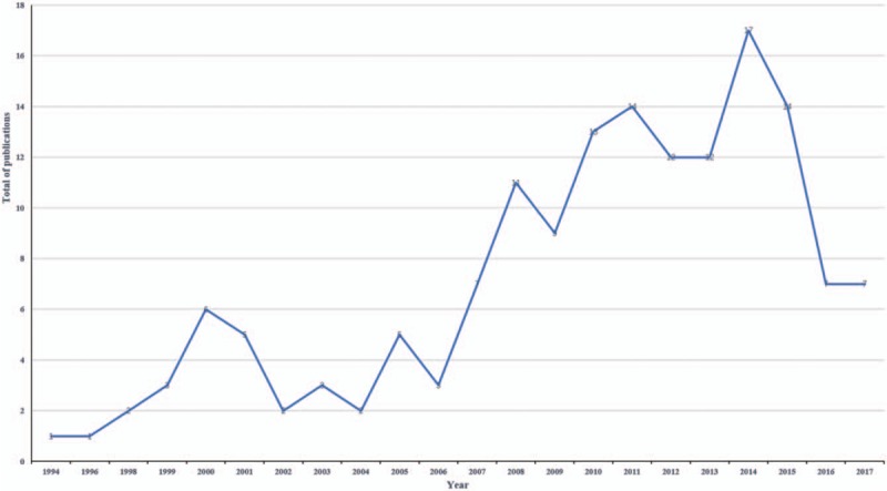
Total number of publications on conjunctivochalasis. The abscissa in the figure represents the year and the ordinate represents the total number of publications.
The number of frequencies quoted (Fig. 2) in the literature showed a slight downward trend in 2006, but the overall trend maintained a steady increase from “1” in 1998 to “302” in 2017.
Figure 2.

Frequency quoted on conjunctivochalasis. The abscissa in the figure represents the year and the ordinate represents the frequency quoted.
3.2. Document types and languages of conjunctivochalasis
The distribution of document types and research languages was given in Table 1. The research results showed that English was the main research language, accounting for 96.795% of languages; and the article was the main type of document, accounting for 78.846% of the study.
Table 1.
Distribution of the document types and languages in the study of the conjunctivochalasis from 1986 to 2017.

3.3. Distribution of countries and institutions
The country map was gained by the aid of installing the pruning (pruning based on Pathfinder) parameter. After running the software, 15 nodes and 15 links have been consisted in the network, and the density value was 0.1429 (Fig. 3). Prolific 10 countries of conjunctivochalasis were presented in Table 2. And the publications about conjunctivochalasis were dedicated by 31 countries. In the map of countries, Japan had the largest number of publications (39), followed by USA (38), People's Republic of China (24), Germany (8), and Wales (7). Moreover, the centrality which ranked the first was USA (0.44).
Figure 3.
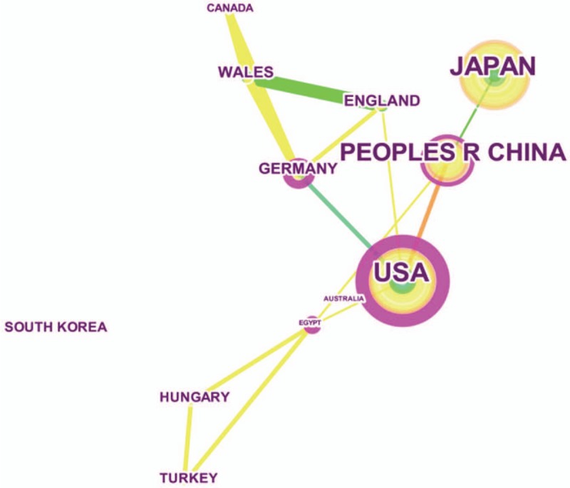
Network of countries on conjunctivochalasis. The purple node in the middle of the annual ring represents the impact and the importance of a country. The larger the node and the more purple it appears, the greater is the importance of the country. The connection line between 2 nodes indicates cocited relations and cocitation intensity. The higher the frequency of the cocitation, the thicker the connection line is and the closer the nodes are.
Table 2.
The top 10 countries, institutions, and authors contributed to publications on the conjunctivochalasis from 1986 to 2017.

The institution map was gained by aid of installing the pruning (choose “Pathfinder”) parameter. After running the software, 38 nodes and 35 links have been consisted in the network, and the density value was 0.0498 (Fig. 4). Table 2 showed the productivity of 10 countries, authors, and institutions on Conjunctivochalasis. The publications of conjunctivochalasis were dedicated by 189 institutions. In the network of coauthors’ institutions, Kyoto Prefectural University of Medicine had the largest productive publications (14), followed by University of Miami (11), Ocular Surface Centre (10), Tokyo Women's Medical University (8), and University of Tokyo (7). What's more, the centrality (Table 3) which arranged the principal was Shanghai Jiao Tong University (0.04).
Figure 4.
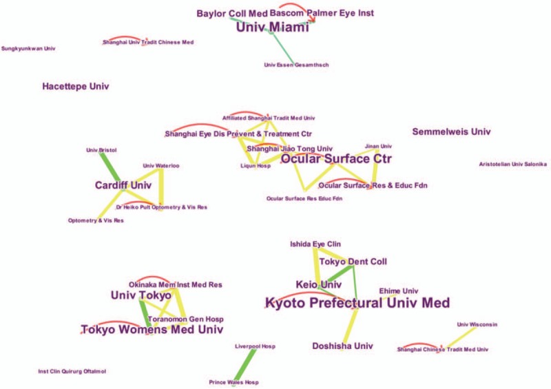
Network of coauthors’ institutions on conjunctivochalasis. The purple node in the middle of the annual ring represents the impact and the importance of an institution. The larger the node and the more purple it appears, the greater is the importance of the institution. The connection line between 2 nodes indicates cocited relations and cocitation intensity. The higher the frequency of the cocitation, the thicker the connection line is and the closer the nodes are. Abbreviation: Ctr = centre, Univ = University, Hosp = hospital, Inst = Institution, etc. These are the abbreviation of the institution. The arrow represents the parameter setting of the guiding line alpha in transparency, the longer the arrow, the greater the citation counts.
Table 3.
The top 10 countries, institutions and authors for the importance index (centrality value) of the conjunctivochalasis literature from 1986 to 2017.

3.4. Distribution of journals and cited journals
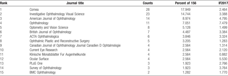
Table 4 showed the top 15 journals associated with conjunctivochalasis. In the aggregate, 39 academic journals have published papers on conjunctivochalasis. The average impact factor for these journals is 3.0508. The journal with the largest number of papers published on conjunctivochalasis was cornea (IF 2017, 2.464; 28 publications, 17.949%), followed by Investigative Ophthalmology Visual Science (IF 2017, 3.388; 23 publications, 14.744%), American Journal of Ophthalmology (IF 2017, 4.795, 14 publications, 8.974%), Ophthalmology (IF 2017, 7.479; 11 publications, 7.051%), Optometry and Vision Science (IF 2017, 1.499; 8 publications, 5.128%).
Table 4.
The top 15 journals contributed to publications on the conjunctivochalasis.
The cited journal map was obtained by the aid of installing the pruning (pruning based on Pathfinder/Pruning the merged network) parameter. After executing the software, 105 nodes and 147 links have been contained in the network, and the density value was 0.0269 (Fig. 5). As can be observed in Table 5, cited journal which ranked in the first was also Cornea (113), followed by AM J Ophthalmol (113), Brit J Ophthalmol (101), Invest Ophth Vis Sci (97), Surv Ophthalmol (91). From the view of centrality (Table 6), Can J Ophthalmol (0.67) was the first, followed by Deut Med Wochenschr (0.64), B Spec Med CHI (0.63), Klinische Monatsblatter Fur Augenheilkunde (0.61), Eye & Contact Lens-Science (0.60).
Figure 5.
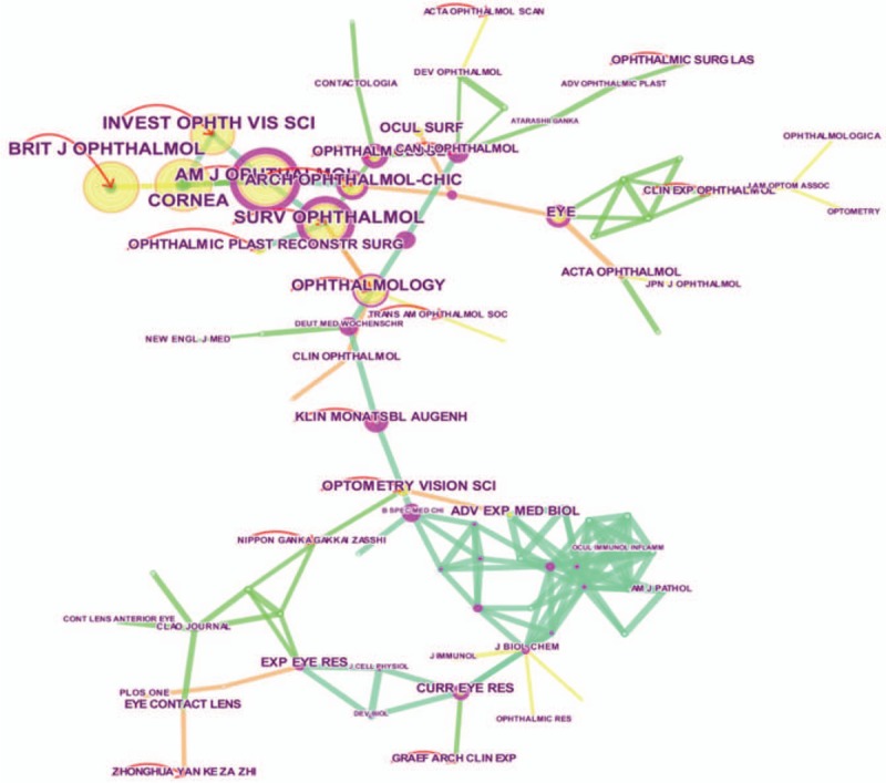
Journal cocitation network on conjunctivochalasis. The purple node in the middle of the annual ring represents the impact and the importance of cited journal. The larger the node and the more purple it appears, the greater is the importance of the cited journal. The connection line between 2 nodes indicates cocited relations and cocitation intensity. The higher the frequency of the cocitation, the thicker the connection line is and the closer the nodes are. The arrow represents the parameter setting of the guiding line alpha in transparency, the longer the arrow, the greater the citation counts.
Table 5.
The top 10 cited journal, cited author, and cited reference contributed to publications on the conjunctivochalasis from 1986 to 2017.

Table 6.
The top 10 cited journal, cited author, and cited reference for the importance index (centrality value) of the conjunctivochalasis literature from 1986 to 2017.

3.5. Distribution of author and cited author
The coauthorship map was gotten by aid of assembling the pruning (Pruning based on Pathfinder/Pruning the merged network) parameter. After running the software, 89 nodes and 145 links have been contained in the map, and the density value was 0.0097 (Fig. 6). Four hundred fourty eight authors contributed to the publications of conjunctivochalasis, for authors who had the most productive (Table 2) were Tseng SCG (19), Yokoi N (13), Kinoshita S (13), Meller D (10), Mimura T (9). Besides, the top ranked item by centrality (Table 3) is also Tseng SCG (0.07).
Figure 6.
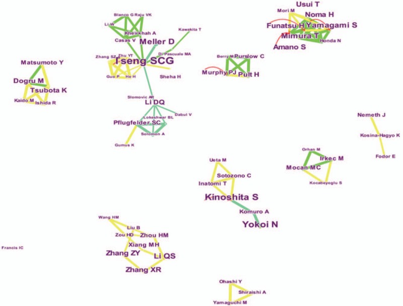
Coauthorship network on conjunctivochalasis. The connection line between two nodes indicates cocited relations and cocitation intensity. The higher the frequency of the cocitation, the thicker the connection line is and the closer the nodes are. The arrow represents the parameter setting of the guiding line alpha in transparency, the longer the arrow, the greater the citation counts.
The cited author map was gained by the aid of installing the pruning (Pruning based on Pathfinder/Pruning the merged network) settings parameter. After running the software, 138 nodes and 273 links have been embodied in the map, and the density value was 0.0289. Figure 7 showed the author cocitation network on conjunctivochalasis, and Fig. 8 was the cluster map of the cited author based on label clusters with title terms. The distribution of cited author showed in Table 5, Meller D was the first (95 counts), followed by Di PMA (55), Yokoi N (55), Hughes WL (45), Liu D (43). Furthermore, from the angle of centrality (Table 6), respectively, the top 5 were Tseng SCG (0.07), Li DQ (0.04), Kinoshita S (0.01), Zhang ZY (0.01), Pflugfelder SC (0.01).
Figure 7.
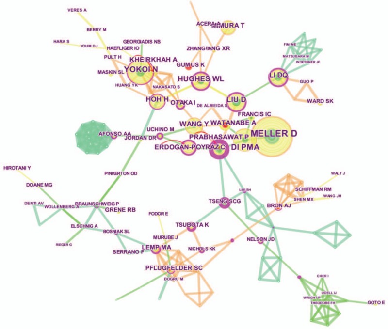
Author Cocitation network on conjunctivochalasis. Burst terms are shown in light red text. Red circles indicate articles with citation bursts, that is, rapid increases of citation counts. The purple node in the middle of the annual ring represents the impact and the importance of cited author. The larger the node and the more purple it appears, the greater is the importance of the cited author. The arrow represents the parameter setting of the guiding line alpha in transparency, the longer the arrow, the greater the citation counts.
Figure 8.
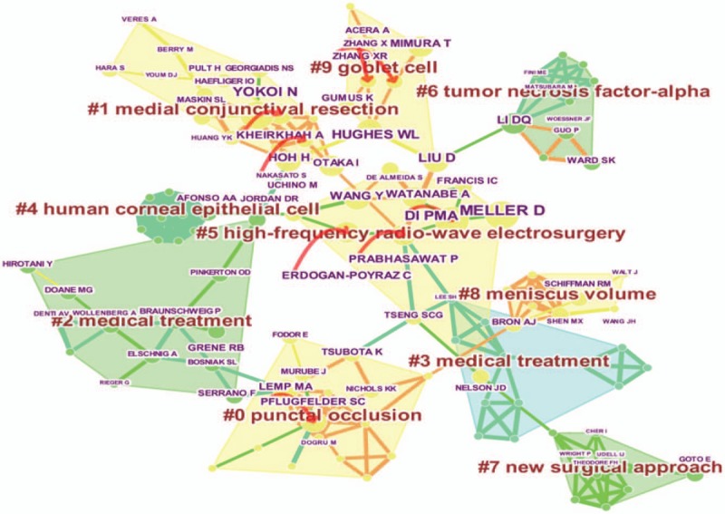
Cluster map of cited author based on label clusters with title terms. Nodes of different colors represent different clusters, the combination of symbols and numbers represents the cited authors’ study of similar categories. The arrow represents the parameter setting of the guiding line alpha in transparency, the longer the arrow, the greater the citation counts.
3.6. Distribution of cited reference
The cited-reference map was gained by the aid of installing the pruning (Pruning based on Pathfinder/Pruning the merged network) parameter. Upon running software, 150 nodes and 249 links have been comprised in the map, and the density value was 0.0223. And Fig. 9 was the cluster map of cited reference based on label clusters with title terms. Furthermore, counts and centrality values of references were shown in Tables 5 and 6. The largest citation counts of reference was Di PMA (35) paper which published his output named Clinical characteristics of Conjunctivochalasis with or without aqueous tear deficiency in 2004, and the 2 largest Centrality on Conjunctivochalasis were Di PMA (0.33) and Kheirkhah A (0.33) literature which published their paper named Amniotic Membrane Transplantation with Fibrin Glue for Conjunctivochalasis in 2007.
Figure 9.
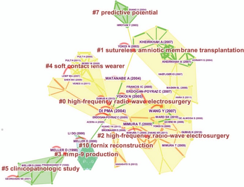
Cluster map of cited reference based on label clusters with title terms. Nodes of different colors represent different clusters, the combination of symbols and numbers represents the cited references’ similarities. The arrow represents the parameter setting of the guiding line alpha in transparency, the longer the arrow, the greater the citation counts.
3.7. Distribution of keywords
Keywords were extracted from 154 records. Then, co-occurring author keywords and keywords plus map were obtained by the aid of installing the pruning (Pruning based on Pathfinder/Pruning the merged network) parameter. When software running, the reference network was created in 103 nodes and 187 links, and the density value was 0.0356 (Fig. 10). Table 7 reflected the top 10 keywords in terms of citation counts and centrality on conjunctivochalasis. In addition to conjunctivochalasis, it was ocular surface (count: 26) who ranked the top, and the highest centrality of keyword was “risk factor” (0.46).
Figure 10.
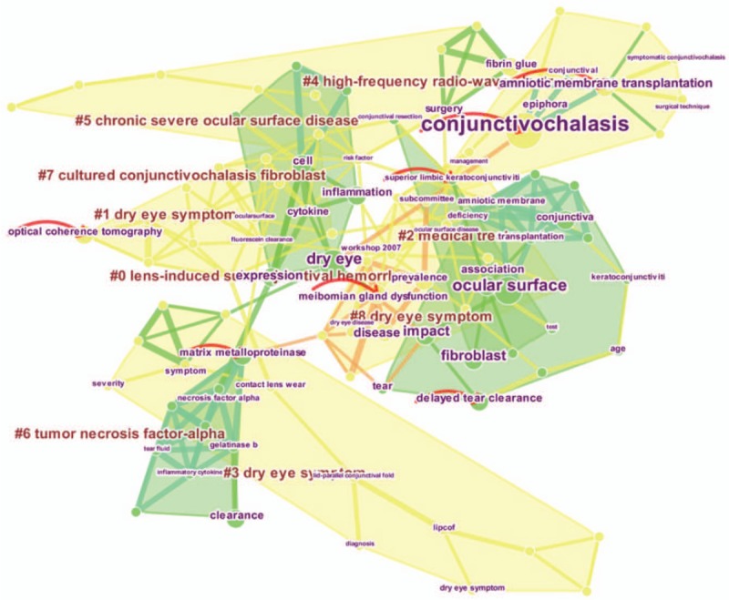
Cluster map of keyword based on label clusters with title terms. Nodes of different colors represent different clusters, the combination of symbols and numbers represents the keywords’ similarities. The arrow represents the parameter setting of the guiding line alpha in transparency, the longer the arrow, the greater the citation counts.
Table 7.
The top 10 keywords in terms of citation counts and centrality on the conjunctivochalasis.

4. Discussion
In our research, the analysis software we used was Citespace V, and there were few bibliometric studies that utilize this software for ophthalmologic diseases by reading the literature, the ones most closely related to our research methods are Liang et al[29] and Miao et al[30] study, the similarities were the intersection of software and research ideas used, but the study of Liang et al[29] did not involve the research of cluster analysis charts. In contrast, the study of Miao is more similar, but the research directions of the 2 are very different. Evidently, there is also bibliometric analysis using this software, but completely different from ours. Such as the study of Zhang et al[31] and Biglu et al,[32] Zhang et al[31] study is only a bibliometric study of authors and research institutions. Furthermore, the study of Biglu et al[32] is just for breast cancer publication network of coauthorship and coorganization. Therefore, based on these viewpoints, we conducted a bibliometrics study of conjunctivochalasis.
4.1. General Data
The study law on the total number of publications was divided into 2 phases. The first phase was taken from 1994 to 2007. During this period, the average annual number of publications remained at 3.33, and the pace of development was slow; however, in the second phase (from 2008 to 2017), the average annual number of publications reached 11.6, and the overall speed of development was faster than the first phase. This may be explained by the use of video terminals, excessive use of eyes, and population aging. According to the frequency quoted, it showed a trendency of increasing yearly, from 1998 to 2017, but there was a slight fluctuation in 2006, this may be related to the economic recession around the financial crisis around 2008.
English is the dominant language for conjunctivochalasis, less noted in other languages. Owing to language restrictions, this may lead to the study of other non-English speaking countries cannot better show their research results, and it is also a problem that we will study in the future.
4.2. Analysis of visual network maps
The cooperative network mainly consists of 3 forms: author, institution, and country. Counts generally reflect an important foundations role, and centrality is an indicator of the importance of discovering and measuring literature. Through analysis of country and institution, we can got that Japan, USA, and People's Republic of China were the most productive countries of conjunctivochalasis. From the view of centrality, USA was the most important countries, and its count is also the top 2, it played a ligament in the cooperation network between countries, but the relationship between them is not close enough. Analysis from institutions, the most prolific institutions were Kyoto Prefectural University of Medicine and University of Miami, and from the view of centrality, Shanghai Jiao Tong University and Ocular Surface were the key institutions of studying conjunctivochalasis, the research advantage of Shanghai JiaoTong University from China is outstanding, we speculated that the possibility of this phenomenon was: Chinese population base is large, and the number of patients with conjunctivochalasis is increasing. Another reason is that Shanghai is Chinese economic center, and the advantages and conveniences that large investment in research funding makes Shanghai Jiaotong University's research is prominent in this field. Importantly, the cooperation relationship between institutions were divided into many small cooperative groups, and there is no intersection between them, representing no cooperation between the networks. Therefore, we should strengthen exchanges and cooperation between different institutions in future research.
The distribution of journals’ impact factors were as follows: the top 15 journals contributed to publications on conjunctivochalasis, ophthalmology, and ocular surface had an IF greater between 5.0 and 10.0, accounted for 9.615% of the total, then, Investigative Ophthalmology Visual Science, American Journal of Ophthalmology, British Journal of Ophthalmology, ACTA Ophthalmologica, and Survey of Ophthalmology had an IF between 3.000 and 5.000, accounted for 33.974% of the total, 7 Journals had an IF between 1.000 and 3.000, accounted for 53.847% of the total, explain that most of the conjunctivochalasis research focuses on journals ranging from 1 to 3 points. Klinische Monatsblatter Fur Augenheilkunde with IF <1.000. Furthermore, the journals that published the most relevant researches of conjunctivochalasis were Cornea, Investigative Ophthalmology Visual Science, American Journal of Ophthalmology, Ophthalmology and Optometry and Vision Science, they accounted for 53.846% of the total publications. By the analysis of the Cited Journal, we found that Cornea, AM J Ophthalmol and Brit J Ophthalmol were owned the most citation counts. Moreover, the top 5 journals are all from the USA, indicating that research on conjunctivochalasis is mostly concentrated in the United States. What's more, Can J Ophthalmol owned the highest centrality of conjunctivochalasis’ research. Journal cocitation network on conjunctivochalasis can see the rules. And these studies were of great significance for the submission of publications for conjunctivochalasis research.
Judging from the count and the centrality, Tseng SCG is in the first place, indicating that he is the most vigorous and influential scholar in the field. In coauthorship network on conjunctivochalasis, there are 9 networks about authors’ cooperation, the most complicated network of partnerships involves Tseng SCG network, and there is no cooperation between these networks, combined with time zone map, the citation counts and centrality of Tseng SCG were higher than Meller D, and were seen as the most active scholars in the same time zone. In the second, the citation counts and centrality of Mimura T were higher than others (Noma H and Amano S, etc.), and were regarded as the most active scholars in the same time zone. Besides, from the perspective of the cited author map, Meller D is the most significant scholar. In addition, Prabhasawat P owned the highest centrality. Moreover, in the cluster map of cited author based on label clusters with title terms (Fig. 8), a total of 10 cluster categories were generated. In the study of Zhang et al,[33] 30 tissue samples from patients with dry eye were compared with the control group, and they found that with the severity of conjunctivochalasis increased, the density of conjunctival goblet cells also decreased considerably. Authors similar to this study were: Acrea A, Hughes WL, and Liu D, and they have contributed to the study of the #9 goblet cell for conjunctivochalasis. In Doss et al[34] research, a new Paste-Pinch-Cut conjunctivoplasty surgical technique was used only for conjunctival resection, which helped restore patients with symptoms of conjunctivochalasis. Analogous studies including Maskin SL, Nakasato S, Georgiadis NS, and Haefliger. So they were classified as “#1 medial conjunctival resection” in the cluster view. In #8 meniscus volume, the main study was Craig et al,[35] a new type of liposomal spray was applied to the pre-ocular tear film, and tear meniscus height of the observed indicators was included. Therefore, we have obtained a novel observation indicator is the meniscus volume, which is excavated by the analysis of clustering map. The research of Wang JH, Holly FJ, Tiffany JM, Schiffman RM, Bron AJ, and Walt J was similar to this, so it is classified into 1 category. Akpek and Gottsch[36] study described the main mechanisms of corneal epithelial cells in immune defense and demonstrated their important role in the prevention of ocular infections. And closely connected to this research were Afonso AA and Jones DT. Hence, they were named “#4 human corneal epithelial cell” in the cluster map. Lemp[37] proposed a new drug for the treatment of punctal occlusion, but the Yen et al[38] research focused on the effect of occlusion of the punctum on the tear production, tear clearance, and ocular surface sensation of the subject. Their similarity is that they all pay attention to the punctal occlusion, so we put them in the same cluster view called “#0 punctal occlusion.” Xie et al[39] research indicated the role of tumor necrosis factor in corneal transplantation, but in the Ward et al[16] study, they were concerned about the rise of tumor necrosis factor levels in nasal conjunctivochalasis. Although their research focus is different, the research direction is based on tumor necrosis factor. Deservedly, we named them “#6 tumor necrosis factor-alpha.” After being compared with the analysis of keyword cluster, the results of the study were alike. In the “#2 medical treatment,” the main treatments involved were drug therapy, surgical therapy, and fabrication of a keratoprosthesis. Rieger[40] research focused on the drug therapy, the topical use of artificial tear treatment can effectively improve the contrast sensitivity of dry eye, and it will also be a drug therapy for conjunctivochalasis. Then the foremost surgical treatment is reflected in Bosniak and Cantisano-Zilkha[41] study, this type of oculo-facial reconstruction or rejuvenation was used in the research to treat dry eye patients. Moreover, there was the fabrication of a keratoprosthesis. It provided new ideas for the treatment of dry eye and conjunctivochalasis. Nevertheless in the “#3 medical treatment,” the treatment involved is different from the #2. First of all, Sun et al[42] was concerned about the eye performance of multiple sclerosis. Secondly, Azuara-Blanco et al[43] focused on the non-technical aspects of safe eye surgery, with the aim of reducing adverse events during the operation. Finally, Cho et al[44] research concentrated on the analysis of confocal microscopic, using this technique to judge the effectiveness of treatments through changes in internal microstructure of corneal diseases and injury models. And medical treatment mentioned by #2 and #3 were also reflected in the cluster analysis of the keywords, but the treatment methods in the latter study were more concrete, such as tissue adhesives and amniotic membrane transplantation, etc. Goto E, Cher I, Wright P, and else scholars have similarities in terms of #7 new surgical approach; in cluster view of “#5 high-frequency radio-wave electrosurgery,” there were studies using this method to treat conjunctivochalasis, and the authors of related research were Di PMA, Meller D, and Watanabe A; as well as studies to treat other diseases of the ocular surface, this is mainly shown in Prabhasawat et al[45] study, although the drug can inhibit the recurrence of Pterygium, it still needs to be cured by this type of surgery after recurrence; in addition to, studies to treat diseases of other systems, and representative research is Uchino et al,[46] who used this type of surgery to treat intestinal diseases. What they have in common was a similar surgical approach, so they were grouped into the same category in the cluster view. Compared with the view results obtained by keyword and cited reference cluster analysis, it is found that the results of the high-frequency radio-wave electrosurgery were identical.
Then, it can be seen from cited reference that Di PMA paper who have published in Brit J Ophthalmol named Clinical characteristics of Conjunctivochalasis with or without aqueous tear deficiency owned the highest citation rate in terms of citation counts and centrality, it can be regarded as a classic literature. Furthermore, analysis from cluster map of cited reference based on label clusters with title terms, after clustering the title terms of reference (Fig. 9), a total of 7 cluster categories were generated. In Pult and Helen[47] study, they found that lid-parallel conjunctival folds affected the effect evaluation of Central Tear Meniscus Height. By analyzing the development of symptoms in patients with conjuncticochalasis, it is indicated that surgical correction should not only restore the meniscus of the tear, but also deepen the sputum of patients with conjunctivochalasis, and these views have been confirmed in Huang et al[48] study. Moreover, despite the fact that they used thermocautery therapy to treat conjunctivochalasis, and the final mechanism is still the fornix reconstruction in Nakasato et al[49] research. It is the similarity of these studies, so we put in the same category in the cluster view called “#10 fornix reconstruction.” Pult et al[50] research which published in 2008 showed that Lid wiper epitheliopathy and lid parallel conjunctival folds were related to the dry eye symptoms of the lens wearer. And the Pult et al[51] studies which published in 2011 were an in-depth study of previous studies. In addition to exploring the relationship between clinical signs and dry eye symptoms, it also summarized the predictive power of improving dry eye symptoms, and the combination of non-invasive break-up time (NBUT) and nasal lid-parallel conjunctival folds is the strongest combination of symptoms and signs. But Chen et al[52] research concentrated on the relationship between tear meniscus volumes and soft contact lens wearers with dryness symptoms, the wearer who worn lens owned lower volumes of tear menisci, then the difference between them was the observation indicators, and the same point was that the target population. Consequently, they were named “#4 soft contact lens wearer” in the cluster map. In Meller and Tseng[22] literature which published in 1998, they proposed systematic grading and possible pathophysiology for conjunctivochalasis, and believed that conjunctivochalasis was closely related to tear dynamics. However, in Meller et al[24] 2000 study, they observed that amniotic membrane transplantation can maintain the original epithelial cell phenotype and accelerate the differentiation of goblet cells, and played a therapeutic role. Then we can deduce that the pathological factors that cause conjunctivochalasis symptoms were inflammation, etc. Furthermore, Georgiadis and Terzidou[53] suggested that the pathophysiological reason for conjunctivochalasis was that associated with over-expression of MMP-1 and MMP-3, and the main cause of epiphora was punctal obstruction and delayed tear clearance. That is the reason why we put them in the same category, and named them “#5 Clinicopathologic study.” Sobrin et al[54] research indicated that human tears contain factors that inhibit or activate MMP-9, and tissue inhibitor of metalloproteinase (TIMP-1) is an inhibitor of MMP-9 activity. And Clark study[55] advised that matrix metalloproteinase (MMP) has become the main therapeutic target for the treatment of angiogenesis-dependent diseases (ocular neovascular diseases). Besides, Fini et al[56] study paid attention to the unique regulation of Gelatinase B matrix metalloproteinase, which can accelerate the repair of the basement membrane of the cornea, unlike other corneal MMPs. Their research hotspots were placed on the MMP, and we titled them “#3 MMP-9 Production.” Uy et al[57] study focused on fibrin glue. It is a method of autologous transplantation of conjunctiva, and its application will greatly shorten the operation time and reduce the adverse reactions after Pterygium surgery. However, Duchesne et al[58] was concerned about human fibrin glue and amniotic membrane transplant to repair corneal perforation, and the disease treated is different from Uy et al.[57] Nonetheless Pfister and Sommers[59] paid their attention to the corneal stem cell transplantation, and it was similar to Uy et al.[57] And the research focus of the 3 is on the transplantation, so they were alluded to as “#1 sutureless amniotic membrane transplantation.” Korb et al[60] literature which published in 2005 utilized fluorescein breakup time and Schirmer test values to diagnose Lid wiper epitheliopathy, and the difference from Korb et al research is that Nichols et al[61] used a questionnaire to screen dry eye. What's more, they have made corresponding contributions to the prevention and screening of ophthalmic diseases. Reasonably, they were classified into similar categories in the cluster view, called “#7 predictive potential.” Deservedly, the references of Watanabe et al,[18] Di et al,[15] Wanget al,[16] Mimura et al,[19] Yokoi et al,[17] Ward et al,[25] and other reference contributed to #0 and #2 high-frequency radio-wave electrosurgery. Moreover, to analyze from Top 13 references with the strongest citation bursts (Fig. 11), Huang et al[48] paper who has published in Ophthalmology named Conjunctivochalasis Interferes with Tear Flow from Fornix to Tear Meniscus can be seen as the frontier document in the field. These results indicated that they were the topic of >30 years’ conjunctivochalasis research.
Figure 11.
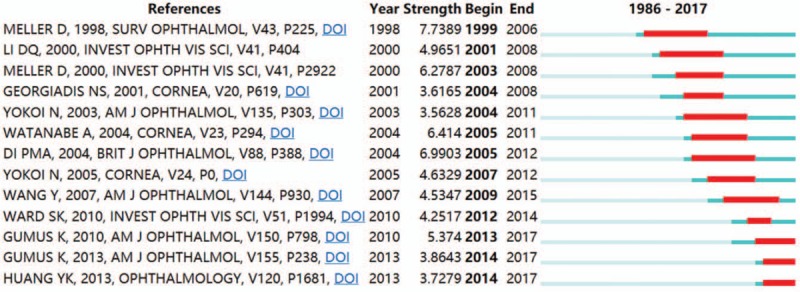
Top 13 references with the strongest citation bursts. The red bars represent some references cited frequently; the green bars represent references cited infrequently.
For the basic keywords analysis in terms of citation counts (Table 7), we can get hot spots in the field. Conjunctivochalasis was the hot topic. Keywords related to observation indicators, risk factors, treatment, graded diagnosis of conjunctivochalasis, etc. Evidently, contact lens wear was the common risk factor, it can be reflected in Mimura T literature; the observed indicators were cytokine (Representative documents were Ward SK published in 2010, Wang Y published in 2007) and fluorescein clearance (Representative documents was Maskin SL published in 2008); covered treatments included surgery and amniotic membrane. Among the amniotic membrane transplantation methods, representative literatures were Georgiadis[53,62], Meller[63,64], Kheirkhah[65], and Maskin[26]; in addition, fibroblast was research hotspot; properly, there were studies on lid-parallel conjunctival fold of conjunctivochalasis, and the typical literature was Fodor et al.[66] At length, the cluster map of keyword with the title term can reflected in the new research directions in conjunctivochalasis, they were divided into 7 categories, they were #0 lens-induced subconjunctival hemorrhage, and typical keyword was inflammation, etc.; #2 medical treatment; #5 chronic severe ocular surface disease, and important keywords were irritation, ocular surface, etc.; #4 high-frequency radio-wave electrosurgery, and representative keyword was cornea, it is also the anatomical location of this surgical treatment of disease; #6 tumor necrosis factor-alpha, and it is also the main indicator of clinical research; #7 cultured conjunctivochalasis fibroblast, and the main keywords were cell, dry eye, inflammation, expression, inhibitor, and cytokine, explained that the disease involved was dry eye, the main mechanism was the expression of cytokines, etc.; and dry eye symptom (#1, #3, and #8 were included), and pivotal keywords were optical coherence tomography, tear meniscus, dry eye, film dynamics, etc., indicating that the type of disease to be treated was dry eye, the examination means was optical coherence tomograph, the indicator of observation was tear meniscus. What's more, by time zone map of keyword on conjunctivochalasis, we can judge which keyword is more important in the same time zone, for example: ocular surface, expression, cell, amniotic membrane, amniotic membrane transplantation, matrix metalloproteinase, and this is their sorting in the time zone. However, there was a flaw in the study of keywords. Many clusters of keywords may not be linked to the results obtained. How to effectively filter out useful keywords is a knotty problem. There were also numerous synonyms in the “subject word” when searching data, which will bring huge challenges to our search. These are the key points we will overcome in future research.
5. Conclusion
In summary, through the bibliometric study of conjunctivochalasis, it can help us to extract useful information from complex data, such as: selection of submitted journals, major research institutions, key research countries, and overall development trends. Relevant clinicians and researchers benefited from these studies, these provided various possibilities for us to carry out new research later. At last, we can not only grasp research hotspots, frontiers, and research trends in this field, but also find new potential laws and present them in a visual form.
Author contributions
Data curation: Yanqing Zhao.
Formal analysis: Yanqing Zhao, Li Huang.
Funding acquisition: Zhengchi Lou and Qingsong Li.
Investigation: Yanqing Zhao, Minhong Xiang.
Methodology: Qingsong Li, Yanqing Zhao.
Software: Yanqing Zhao.
Supervision: Wanhong Miao.
Writing – original draft: Yanqing Zhao.
Writing – review & editing: Yanqing Zhao.
Footnotes
Abbreviations: CCh = conjunctivochalasis, IF = impact factor, NBUT= non-invasive break-up time, WoSCC = web of science core collection.
YZ and LH have contributed equally to this work and should be considered co-first authors.
This research was funded by the following funds: Medical Major Department of Ophthalmology of Shanghai (No. ZK2015A20), Independent Innovation Research Project of Putuo District Health System in Shanghai (No.2015PTKW001), Budget Project of Shanghai University of Chinese Medicine (No. 2015YSN50 and No. 18WK106), Hospital Personnel Training Plan-Yuying Plan of Putuo Hospital Affiliated to Shanghai University of Chinese Medicine (No.2017218A).
There is not any direct conflicts of interest between all authors.
References
- [1].Fu DH, Xie TH, Zhu J, et al. Epidemiologic study of Conjunctivochalasis in population 50 years old or more in Binhu district of Wuxi City, China. Chin J Exp Ophthalmol 2014;32:838–43. [Google Scholar]
- [2].Zhang X, Li Q, Zou H, et al. Assessing the severity of Conjunctivochalasis in a senile population: a community-based epidemiology study in Shanghai, China. BMC Public Health 2011;11:198. [DOI] [PMC free article] [PubMed] [Google Scholar]
- [3].Agarwal A, Durairajanayagam D, Tatagari S, et al. Bibliometrics: tracking research impact by selecting the appropriate metrics. Asian J Androl 2016;18:296–309. [DOI] [PMC free article] [PubMed] [Google Scholar]
- [4].Lu KN, Yu S, Yu M, et al. Bibliometric analysis of tumor immunotherapy studies. Med Sci Monit 2018;24:3405–14. [DOI] [PMC free article] [PubMed] [Google Scholar]
- [5].Borrett SR, Sheble L, Moody J, et al. Bibliometric review of ecological network analysis: 2010-2016. Ecol Model 2018;382:63–82. [Google Scholar]
- [6].Emmer A. GLOFs in the WOS: bibliometrics, geographies and global trends of research on glacial lake outburst floods (Web of Science, 1979-2016). Nat Hazards Earth Syst Sci 2018;18:813–27. [Google Scholar]
- [7].Boudry C, Baudouin C, Mouriaux F. International publication trends in dry eye disease research: a bibliometric analysis. Ocul Surf 2018;16:173–9. [DOI] [PubMed] [Google Scholar]
- [8].Schargus M, Kromer R, Druchkiv V, et al. The top 100 papers in dry eye - a bibliometric analysis. Ocul Surf 2017;16:180–90. [DOI] [PubMed] [Google Scholar]
- [9].Ramin S, Pakravan M, Habibi G, et al. Scientometric analysis and mapping of 20 years of glaucoma research. Int J Ophthalmol 2016;9:1329–35. [DOI] [PMC free article] [PubMed] [Google Scholar]
- [10].Caglar C, Demir E, Kucukler FK, et al. A bibliometric analysis of academic publication on diabetic retinopathy disease trends during 1980-2014: a global and medical view. Int J Ophthalmol 2016;9:1663–8. [DOI] [PMC free article] [PubMed] [Google Scholar]
- [11].Lin ZN, Chen J, Zhang Q, et al. The 100 most influential papers about cataract surgery: a bibliometric analysis. Int J Ophthalmol 2017;10:1586–91. [DOI] [PMC free article] [PubMed] [Google Scholar]
- [12].Wen PF, Dong ZY, Li BZ, et al. Bibliometric analysis of literature on cataract research in PubMed (2001-2013). J Cataract Refract Surg 2015;41:1781–3. [DOI] [PubMed] [Google Scholar]
- [13].Xu CT, Li SQ, Lu YG, et al. Development of biomedical publications on ametropia research in PubMed from 1845 to 2010: a bibliometric analysis. Int J Ophthalmol 2011;4:1–7. [DOI] [PMC free article] [PubMed] [Google Scholar]
- [14].Chen CM, Dubin R, Kim MC. Emerging trends and new developments in regenerative medicine: a scientometric update (2000--2014). Expert Opin Biol Ther 2014;14:1295–317. [DOI] [PubMed] [Google Scholar]
- [15].Di Pascuale MA, Espana EM, Kawakita T, et al. Clinical characteristics of Conjunctivochalasis with or without aqueous tear deficiency. Br J Ophthalmol 2004;88:388–92. [DOI] [PMC free article] [PubMed] [Google Scholar]
- [16].Wang Y, Dogru M, Matsumoto Y, et al. The impact of nasal conjunctivochalasis on tear functions and ocular surface findings. Am J Ophthalmol 2007;144:930–7. [DOI] [PubMed] [Google Scholar]
- [17].Yokoi N, Komuro A, Nishii M, et al. Clinical impact of conjunctivochalasis on the ocular surface. Cornea 2005;24:S24–31. [DOI] [PubMed] [Google Scholar]
- [18].Watanabe A, Yokoi N, Kinoshita S, et al. Clinicopathologic study of conjunctivochalasis. Cornea 2004;23:294–8. [DOI] [PubMed] [Google Scholar]
- [19].Mimura T, Yamagami S, Usui T, et al. Changes of conjunctivochalasis with age in a hospital-based study. Am J Ophthalmol 2009;147:171–7. [DOI] [PubMed] [Google Scholar]
- [20].Erdogan-Poyraz C, Mocan MC, Irkec M, et al. Delayed tear clearance in patients with Conjunctivochalasis is associated with punctal occlusion. Cornea 2007;26:290–3. [DOI] [PubMed] [Google Scholar]
- [21].Kheirkhah A, Casas V, Esquenazi S, et al. New surgical approach for superior Conjunctivochalasis. Cornea 2007;26:685–91. [DOI] [PubMed] [Google Scholar]
- [22].Meller D, Tseng SCG. Conjunctivochalasis: literature review and possible pathophysiology. Surv Ophthalmol 1998;43:225–32. [DOI] [PubMed] [Google Scholar]
- [23].Kheirkhah A, Casas V, Blanco G, et al. Amniotic membrane transplantation with fibrin glue for Conjunctivochalasis. Am J Ophthalmol 2007;144:311–3. [DOI] [PubMed] [Google Scholar]
- [24].Meller D, Li DQ, Tseng SCG. Regulation of collagenase, stromelysin, and gelatinase B in human conjunctival and conjunctivochalasis fibroblasts by Interleukin-1β and tumor necrosis factor-α. Invest Ophthalmol Vis Sci 2000;41:2922–9. [PubMed] [Google Scholar]
- [25].Ward SK, Wakamatsu TH, Dogru M, et al. The role of oxidative stress and inflammation in conjunctivochalasis. Invest Ophthalmol Vis Sci 2010;51:1994–2002. [DOI] [PubMed] [Google Scholar]
- [26].Maskin SL. Effect of ocular surface reconstruction by using amniotic membrane transplant for symptomatic Conjunctivochalasis on fluorescein clearance test results. Cornea 2008;27:644–9. [DOI] [PubMed] [Google Scholar]
- [27].Yokoi N, Komuro A, Maruyama K, et al. New surgical treatment for superior limbic keratoconjunctivitis and its association with Conjunctivochalasis. Am J Ophthalmol 2003;135:303–8. [DOI] [PubMed] [Google Scholar]
- [28].Zhang XR, Li QS, Zou HD, et al. Assessing the severity of Conjunctivochalasis in a senile population: a community-based epidemiology study in Shanghai, China. BMC Public Health 2011;11:198. [DOI] [PMC free article] [PubMed] [Google Scholar]
- [29].Liang YD, Li Y, Zhao J, et al. Study of acupuncture for low back pain in recent 20 years: a bibliometric analysis via CiteSpace. J Pain Res 2017;10:951–64. [DOI] [PMC free article] [PubMed] [Google Scholar]
- [30].Miao Y, Liu R, Pu YP, et al. Trends in esophageal and esophagogastric junction cancer research from 2007 to 2016: a bibliometric analysis. Medicine (Baltimore) 2017;96:e6924. [DOI] [PMC free article] [PubMed] [Google Scholar]
- [31].Zhang XM, Zhang X, Luo X, et al. Knowledge mapping visualization analysis of the military health and medicine papers published in the web of science over the past 10 years. Mil Med Res 2017;4:23. [DOI] [PMC free article] [PubMed] [Google Scholar]
- [32].Biglu M, Abotalebi P, Ghavami M. Breast cancer publication network: profile of co-authorship and co-organization. Bioimpacts 2016;6:211–7. [DOI] [PMC free article] [PubMed] [Google Scholar]
- [33].Zhang XR, Li QS, Xiang MH, et al. Analysis of tear mucin and goblet cells in patients with conjunctivochalasis. Spektrum Augenheilkd 2010;24:206–13. [Google Scholar]
- [34].Doss LR, Doss EL, Doss RP. Paste-Pinch-Cut conjunctivoplasty: subconjunctival fibrin sealant injection in the repair of conjunctivochalasis. Cornea 2012;31:959–62. [DOI] [PubMed] [Google Scholar]
- [35].Craig JP, Purslow C, Murphy PJ, et al. Effect of a liposomal spray on the pre-ocular tear film. Cont Lens Anterior Eye 2010;33:83–7. [DOI] [PubMed] [Google Scholar]
- [36].Akpek EK, Gottsch JD. Immune defense at the ocular surface. Eye (Lond) 2003;17:949–56. [DOI] [PubMed] [Google Scholar]
- [37].Lemp MA. Evaluation and differential diagnosis of keratoconjunctivitis sicca. J Rheumatol Suppl 2000;61:11–4. [PubMed] [Google Scholar]
- [38].Yen MT, Pflugfelder SC, Feuer WJ. The effect of punctal occlusion on tear production, tear clearance, and ocular surface sensation in normal subjects. Am J Ophthalmol 2001;131:314–23. [DOI] [PubMed] [Google Scholar]
- [39].Xie L, Shi W, Guo P. Roles of tumor necrosis factor-related apoptosis-inducing ligand in corneal transplantation. Transplantation 2003;76:1556–9. [DOI] [PubMed] [Google Scholar]
- [40].Rieger G. Contrast sensitivity in patients with keratoconjunctivitis sicca before and after artificial tear application. Arch Clin Exp Ophthalmol 1993;231:577–9. [DOI] [PubMed] [Google Scholar]
- [41].Bosniak SL, Cantisano-Zilkha M. Reconstructive blepharoplasty. Otolaryngol Clin North Am 2005;38:985–1007. [DOI] [PubMed] [Google Scholar]
- [42].Sun YS, Cheon JE, Hwang JM, et al. Ocular manifestations in Korean patients with multiple sclerosis. Neuro-Ophthalmology 2008;32:55–61. [Google Scholar]
- [43].Azuara-Blanco A, Reddy A, Wilkinson G, et al. Safe eye surgery: non-technical aspects. Eye 2011;25:1109–11. [DOI] [PMC free article] [PubMed] [Google Scholar]
- [44].Cho BJ, Djalilian AR, Holland EJ. Tandem scanning confocal microscopic analysis of differences between epithelial healing in limbal stem cell deficiency and normal corneal reepithelialization in rabbits. Cornea 1998;17:68–73. [DOI] [PubMed] [Google Scholar]
- [45].Prabhasawat P, Tesavibul N, Leelapatranura K, et al. Efficacy of subconjunctival 5-fluorouracil and triamcinolone injection in impending recurrent pterygium. Ophthalmology 2006;113:1102–9. [DOI] [PubMed] [Google Scholar]
- [46].Uchino M, Ikeuchi H, Bando T, et al. Ostomy creation with fewer sutures using tissue adhesives (cyanoacrylates) in inflammatory bowel disease: a pilot study. Ann R Coll Surg Engl 2017;100:1–4. [DOI] [PMC free article] [PubMed] [Google Scholar]
- [47].Pult H, Britta Helen RP. Impact of conjunctival folds on central tear meniscus height. Invest Ophthalmol Vis Sci 2015;56:1459–66. [DOI] [PubMed] [Google Scholar]
- [48].Huang YK, Sheha H, Tseng SCG. Conjunctivochalasis interferes with tear flow from Fornix to tear meniscus. Ophthalmology 2013;120:1681–7. [DOI] [PubMed] [Google Scholar]
- [49].Nakasato S, Uemoto R, Mizuki N. Thermocautery for inferior conjunctivochalasis. Cornea 2012;31:514–9. [DOI] [PubMed] [Google Scholar]
- [50].Pult H, Purslow C, Berry M, et al. Clinical tests for successful contact lens wear: relationship and predictive. Optom Vis Sci 2008;85:E924–9. [DOI] [PubMed] [Google Scholar]
- [51].Pult H, Purslow C, Murphy PJ. The relationship between clinical signs and dry eye symptoms. Eye (Lond) 2011;25:502–10. [DOI] [PMC free article] [PubMed] [Google Scholar]
- [52].Chen Q, Wang JH, Shen MX, et al. Lower volumes of tear menisci in contact lens wearers with dry eye symptoms. Invest Opthalmol Vis Sci 2009;50:3159–63. [DOI] [PubMed] [Google Scholar]
- [53].Georgiadis NS, Terzidou CD. Epiphora caused by conjunctivochalasis: treatment with transplantation of preserved human amniotic membrane. Cornea 2001;20:619–21. [DOI] [PubMed] [Google Scholar]
- [54].Sobrin L, Liu Z, Monroy DC, et al. Regulation of MMP-9 activity in human tear fluid and corneal epithelial cult. Invest Ophthalmol Vis Sci 2000;41:1703–9. [PubMed] [Google Scholar]
- [55].Clark AF. New discoveries on the roles of matrix metalloproteinases in ocular cell biology and pathology. Invest Ophthalmol Vis Sci 1998;39:2514–6. [PubMed] [Google Scholar]
- [56].Fini ME, Girard MT, Matsubara M, et al. Unique regulation of the matrix metalloproteinase, gelatinase B. Invest Ophthalmol Vis Sci 1995;36:622–33. [PubMed] [Google Scholar]
- [57].Uy HS, Reyes JM, Flores JD, et al. Comparison of fibrin glue and sutures for attaching conjunctival autografts after pterygium excision. Ophthalmology 2005;112:667–71. [DOI] [PubMed] [Google Scholar]
- [58].Duchesne B, Tahi H, Galand A. Use of human fibrin glue and amniotic membrane transplant in corneal perforation. Cornea 2001;20:230–2. [DOI] [PubMed] [Google Scholar]
- [59].Pfister RR, Sommers CI. Fibrin sealant in corneal stem cell transplantation. Am J Ophthalmol 2005;140:966–7. [DOI] [PubMed] [Google Scholar]
- [60].Korb DR, Herman JP, Greiner JV, et al. Lid wiper epitheliopathy and dry eye symptoms. Eye Contact Lens 2005;31:2–8. [DOI] [PubMed] [Google Scholar]
- [61].Nichols JJ, Mitchell GL, Nichols KK, et al. The performance of the contact lens dry eye questionnaire as a screening survey for contact lens-related dry eye. Cornea 2002;21:469–75. [DOI] [PubMed] [Google Scholar]
- [62].Georgiadis NS, Terzidou CD. Epiphora due to conjunctivochalasis treatment with transplantation of preserved human amniotic membrane. Investigative Ophthalmology & VisualL Science 2000;41:S452. [DOI] [PubMed] [Google Scholar]
- [63].Meller D, Tseng SCD. Amniotic membrane transplantation with or without limbal allografts for corneal surface reconstruction in limbal deficiency. Ophthalmologe 2000;97:100–7. [DOI] [PubMed] [Google Scholar]
- [64].Kruse FE, Meller D. Amniotic membrane transplantation for ocular surface reconstruction. Ophthalmologe 2001;98:801–10. [DOI] [PubMed] [Google Scholar]
- [65].Kheirkhah A, Casas V, Blanco G, et al. Amniotic membrane transplantation with fibrin glue for conjunctivochalasis. Am J Ophthalmol 2007;144:311–3. [DOI] [PubMed] [Google Scholar]
- [66].Fodor E, Barabino S, Montaldo E, et al. Quantitative Evaluation of ocular surface inflammation in patients with different grade of conjunctivochalasis. Curr Eye Res 2010;35:665–9. [DOI] [PubMed] [Google Scholar]


