



Abstract
IgA nephropathy (IgAN) is one of the most common recurrent glomerulonephritis after renal transplantation. Rarely, it is accompanied with the presence of crescents that leads to rapid deterioration of renal function and graft loss. We present a 54-year-old patient with IgAN that received a cadaveric kidney allograft, but developed biopsy proven recurrent IgAN 7 months after renal transplantation. He was treated with intravenous steroids and angiotensin-converting enzyme inhibitor and remission was achieved. 4 years later, he presented again with heavy proteinuria, hematuria and deterioration of renal function. Allograft biopsy revealed recurrent IgAN with crescents, which was successfully treated with pulse intravenous steroids and six monthly doses of intravenous cyclophosphamide. This regime resulted in long-term sustained remission with a stable functioning graft 3 years later. Although it is not an established treatment as in native kidneys, intravenous cyclophosphamide should probably be considered in kidney transplants with potentially reversible recurrent crescentic IgAN.
Keywords: IgA nephropathy, Renal transplantation, Recurrent glomerulonephritis, Crescents
Introduction
Immunoglobulin A nephropathy (IgAN) is the most common form of glomerulonephritis worldwide, characterized by mesangial proliferation and diffuse deposition of IgA [1]. Up to 25% of the patients will progress to end stage renal disease (ESRD) within 10 years after diagnosis [2], but usually such patients have little comorbidities and represent ideal candidates for transplantation [3]. IgAN is also one of the most common recurrent glomerulonephritis after renal transplantation. It has been reported that 9–61% of the patients with primary IgAN that receive kidney allograft, will develop recurrent disease [4]. The wide range of the recurrence rate is partly explained by the different policies regarding protocol biopsies as well as the indications for allograft biopsy in each center [3, 4]. More often the recurrences occur after 3 years post transplantation [3, 5] and they affect graft survival only in the long term, associated with a twofold increase in the risk for graft loss [6]. Patients usually develop persistent hematuria and low-grade proteinuria, following a benign course, but rarely rapid deterioration of renal function may occur that is related with the presence of crescents [7]. We present a patient that developed recurrent IgAN during the first year after renal transplantation that progressed 4 years later to crescentic glomerulonephritis.
Case report
Our patient was diagnosed with IgAN in 2001, at the age of 40. He underwent renal biopsy due to declining renal function (serum Creatinine—sCr: 3 mg/dl) over a period of 3 months, which revealed glomeruli with mesangial proliferation and depositions of IgA. There was extensive interstitial fibrosis and 50% of the glomeruli were sclerosed, but he still received steroids for a period of 6 months with no improvement. He reached ESRD after 2 years and commenced hemodialysis in Dec 2003.
7 years later, in Sept 2010 he received a cadaveric kidney allograft. Basiliximab was administered as induction treatment, while maintenance immunosuppression treatment consisted of tacrolimus (Tac) 2 mg twice daily, mycophenolic acid (MPA) 500 mg twice daily and prednisolone (Pred). No allograft biopsy was obtained at the time of implantation. He was discharged with sCr: 1.2 mg/dl and no antihypertensive medication other than amlodipine 10 mg. During the next 7 months renal function was stable, blood pressure was under control, Tac levels (C0) were around 6 ug/l and he experienced no rejection episodes or CMV infections.
In Apr 2011, he was found to have hypertension (160/85 mmHg) with microscopic hematuria and proteinuria of 5 g/24 h, while his renal function remained stable. Allograft biopsy showed recurrence of IgAN with mesangial proliferation in > 50% of the glomeruli, but no crescents. Interstitial fibrosis and tubular vacuolization in < 10% of the specimen indicated a small degree of CNI toxicity. He received three daily doses of intravenous methylprednisolone (MP) 500 mg, continued with daily 40 mg po Pred for 2 months and tampered over another 4 months down to 5 mg per day. He also received maximum dose of quinapril 40 mg. He remained on Tac 2 mg twice daily (C0 target: 6 ug/l) and MPA 500 mg twice daily. For the next 4 years, renal function remained stable (sCr: 1.5 mg/dl) with a small degree of proteinuria (0.3–0.4 g/24 h) and microscopic hematuria.
In Jan 2015, proteinuria 3 g/24 h was noted in a regular follow-up appointment. During the next month renal function deteriorated (sCr: 2.1 mg/dl), while proteinuria increased to 9.8 g/24 h, combined with microscopic hematuria. Allograft biopsy showed again recurrence of IgAN with diffuse mesangial proliferation combined with cellular crescent formation and necrosis in 60% of the glomeruli (Figs. 1, 2, 3). Glomerulosclerosis and interstitial fibrosis accounted for < 20% of the biopsy specimen. ANCA were negative. He received MP pulse therapy (500 mg × 3) and six doses of intravenous cyclophosphamide 650 mg (500 mg/m2) for the next 6 months, combined with small increase in Pred (30 mg) tampered over 3 months. During this 6 month period, MPA was halved to 250 mg twice daily, but not stopped, he remained on his usual dose of Tac (C0 target: 6 ug/l) and maximum dose of quinapril. No major side effect, such as pancytopenia or infection, was observed. Three years later, the patient is considered to be in remission with stable renal function (sCr: 1.6 mg/dl) and low-grade proteinuria (0.5–0.8 g/24 h), while receiving the initial regime Tac-MPA-Pred and maximum dose of quinapril 40 mg. The patient’s clinical course is summarized in Fig. 4.
Fig. 1.
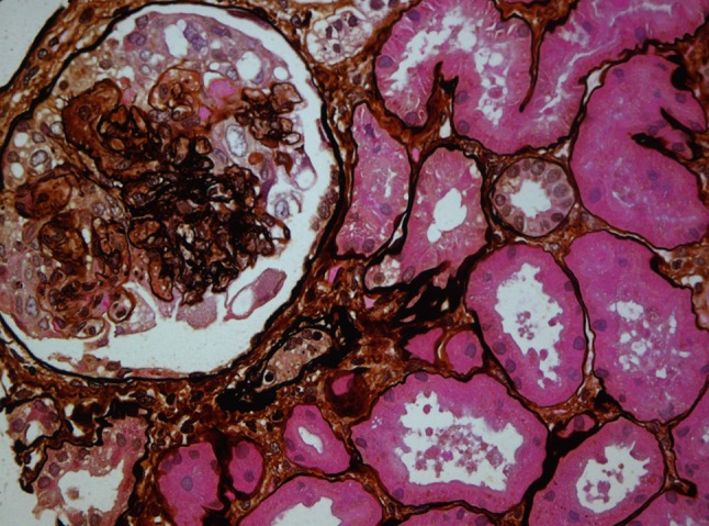
Glomerulus with mesangial proliferation and crescent formation (Methenamine silver-periodic acid–Schiff stain ×400)
Fig. 2.
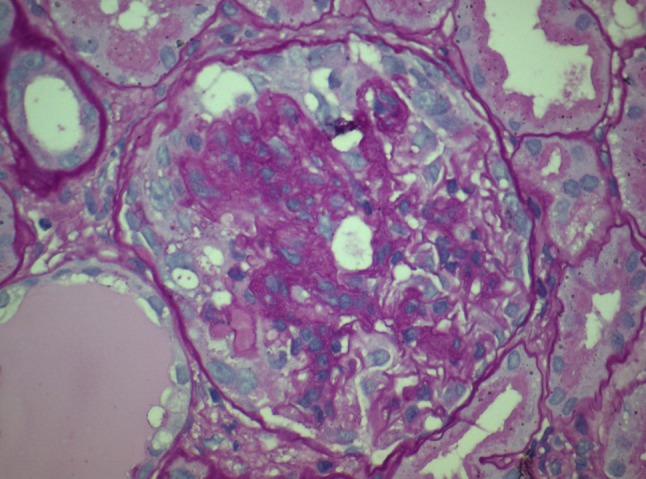
Mesangial hypercellularity and full cellular crescent (Periodic acid–Schiff stain ×600)
Fig. 3.
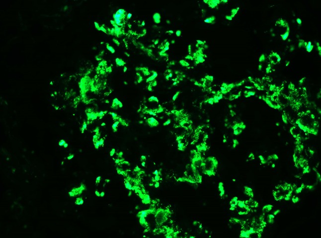
Immunoflorescence staining for IgA showing dominant mesangial distribution (×600)
Fig. 4.

Patient’s clinical course. Pred prednisolone, MMF mycophenolate mofetil, FK tacrolimus, MP methylprednisolone, CYC cyclophosphamide, sCr serum creatinine
Discussion
Crescentic IgAN is an uncommon finding in native kidneys, estimated to account for 3–5%, (when > 50% of glomeruli are involved by crescents) and 32% of IgAN cases (if any number of glomeruli are involved) [8]. The presence of crescents is even rarer in allograft kidneys, although it has been reported in 4–14% of recurrent or de novo allograft IgAN in Asian patients [9, 10]. Several case reports [11–20] and two case series have been published [7, 10] (Table 1). It appears to be a male predominance and the only factor associated with the development of crescentic IgAN is the presence of IgAN in native kidneys that led to ESRD [7, 10]. Mousson et al. speculated that the presence of crescentic IgAN in native kidneys may be associated with the recurrence of crescents in the allograft, but this has not been reported elsewhere [17]. Tang et al. suggested that while in the majority of cases the immunosuppressive regimen consisted of Calcineurin inhibitor (CNI) + Azathioprine (Azp) + Pred, the establishment of mycophenolate mofetil (MMF) therapy may prevent the formation of crescents [10]. Disappointingly, patients on MMF still experience crescentic IgAN recurrence [17–19].
Table 1.
Clinical features of transplant recipients with crescentic IgAN, findings in kidney biopsy, treatment and outcome
| References | Age (year) | Sex | Disease in native kidney | Time from Tx | Treatment protocol | Hematuria | Proteinuria (g/day) | Blood pressure (mmHg) | sCr (mg/dl) | Kidney Biopsy | Treatment | Outcome | |
|---|---|---|---|---|---|---|---|---|---|---|---|---|---|
| GS (%) | Crescents (%) | ||||||||||||
| [11] | 54 | M | IgAN | 10 mo | CsA + MP | Gross | 1.3 | HT | 9 | NA | 60 | PEX, po CYC, po Pred | HD after 2 months |
| [12] | 29 | M | MPGN | 5 mo | CsA + Pred | Microscopic | 0.68 | NA | 3.8 | NA | 80 | PEX, po CYC, MP pulse | HD after 4 months |
| [13] | 52 | M | IgAN | 4 y | Azp + Pred | NA | NA | NA | 7.1 | NA | 40 | PEX, MP pulse | sCr: 1.8 mg/dl after 30 Days |
| [14] | 51 | M | NA | 4 y | CsA + Pred | Microscopic | 10.6 | NA | 11.6 | NA | 50 | MP pulse | sCr: 2.5 after 12 months |
| [15] | 46 | M | NA | 27 mo | CsA + MMF + Pred | Microscopic | 10.13 | NA | 1.8 | NA | 66.7 | CsA to Tac, warfarin | HD after 4 months |
| [16] | 52 | M | IgAN | 10 mo | Tac + Azp + Pred | Microscopic | 4.48 | 180/100 | 2.7 | NA | 33 | Azp to MMF po Pred, ACEi |
sCr: 3.7 md/dl after 6 months but deteriorating |
| [7] | 43 | M | IgAN | 20 y | CYC + steroid | NA | 3+ dipstick | 140/90 | 4.3 | 10 | 7 | NA | HD after 12 months |
| [7] | 48 | M | IgAN | 17 y | Azp + steroid | Microscopic | 10.77 | 130/95 | 1.9 | 30–40 | 12.5 | Azp, po steroid, ASA, propranolol | HD after 6 months |
| [7] | 53 | F | IgAN | 6 y | Azp + CNI | NA | 3.5 | NA | 2.3 | 14 | 20 | NA | Unknown |
| [7] | 41 | M | IgAN | 22 mo | CNI + steroid | NA | NA | 140/84 | 5 | 28 | 30 | MP pulse, PEX | HD after 1 month |
| [7] | 59 | M | PIGN | 3 y | CNI + steroid | NA | NA | 175/92 | 2.7 | 25 | 17 | MP pulse | HD after 2 months |
| [7] | 38 | M | IgAN | 9 y | CNI + Azp + steroid | NA | NA | 140/88 | 2 | 15 | 7 | ACEi | sCr: 1.6 mg/dl after 4 years |
| [7] | 22 | F | IgAN | 3 y | CNI + Azp + steroid | Microscopic | 3+ dipstick | 136/90 | 6.5 | 13 | 13 | MP pulse, CNI, ACEi | sCr: 1.3 mg/dl after 5 years |
| [7] | 37 | M | reflux | 5 y | CNI + MMF | Microscopic | 1+ dipstick | NA | 2.7 | 4.8 | 14.3 | Po steroid | sCr: 1.7 mg/dl after 6 years |
| [17] | NA | NA | IgAN | 15 mo | NA | Microscopic | 0.8 | NA | 2.8 | NA | 30 | MP pulse | HD after 2 months |
| [17] | NA | NA | IgAN | 47 mo | NA | Microscopic | 2.4 | NA | 3.5 | NA | 20 | MP pulse | HD after 27 months |
| [10] | 26 | M | IgAN | 4 y | CsA + Azp + Pred | Gross | 3.2 | HT | 3.56 | 30 | 60 | MP pulse Azp to MMF |
Re-transplant after 6 months |
| [10] | 25 | M | IgAN | 5 y | CsA + Azp + Pred | Microscopic | 1.8 | HT | 1.79 | 15 | 20 | MP pulse Azp to MMF |
HD after 6 months |
| [10] | 28 | M | NA | 4 y | CsA + Azp + Pred | Microscopic | 3+ dipstick | HT | 4.68 | 25 | 50 | MP pulse CsA to Tac |
HD after 1 year |
| [10] | 28 | M | IgAN | 6.5 y | CsA + Azp + Pred | Microscopic | 3.14 | HT | 3.73 | 20 | 66.7 | MP pulse Azp to MMF |
HD after 2 years |
| [10] | 31 | M | NA | 6.5 y | CsA + Azp + Pred | Microscopic | 1.5 | HT | 6.25 | 50 | 20 | MP pulse CRRT |
HD after 2 months |
| [10] | 32 | M | MPGN | 6 y | CsA + Azp + Pred | Microscopic | 1.88 | Normal | 2.83 | 15 | 20 | MP pulse Azp to MMF |
HD after 6 months |
| [10] | 28 | M | IgAN | 3 y | CsA + MMF + Pred | Microscopic | 2.05 | HT | 2.94 | 25 | 35.5 | MP pulse CsA to Tac |
HD after 3 years |
| [10] | 48 | M | IgAN | 4 y | CsA + Azp + Pred | Microscopic | 2+ dipstick | HT | 2.65 | 10 | 40 | MP pulse Azp to MMF |
Re-transplant after 5 years |
| [10] | 69 | F | Vasculitis | 7 y | CsA + Azp + Pred | Microscopic | 1.54 | Normal | 1.45 | 20 | 10 | MP pulse CsA to Tac |
sCr: 2.5 mg/dl stable after 3 years |
| [10] | 45 | F | APKD | 4.5 y | CsA + MMF + Pred | Microscopic | 1.36 | HT | 3.8 | 40 | 50 | MP pulse CsA to Tac |
HD after 8 months |
| [18]a | 20 | M | Unknown | 1 y | CsA + MMF + Pred | Gross | NA | NA | NA | NA | 50 | - | HD after 6 months |
| [18]a | 25 | M | Unknown | 3 y | Tac + MMF + Pred | Gross | uPCR: 5.1 | NA | 5.2 | NA | 56 | MP pulse IV CYC |
sCr: 1.8 mg/dl after 6 months |
| [19] | 29 | M | IgAN | 2 mo | Tac + MMF + Pred | NA | 13 | NA | 3.8 | NA | 60 | IV CYC, po Pred Eculizumab |
HD after 5 months |
| [20] | 48 | M | Unknown | 10 y | Tac + MMF + MP | Microscopic | 1.0 | NA | 1.8 | 30 | 10 | Tonsillectomy, MP pulse | sCr: 1.7 mg/dl after 3 years |
IgAN IgA nephropathy, Tx transplantation, sCr serum creatinine, GS glomerulosclerosis, CsA cyclosporine A, MP methylprednisolone, HT hypertension, PEX plasma exchange, CYC cyclophosphamide, Pred prednisolone, HD hemodialysis, MPGN membranoproliferative glomerulonephritis, Azp azathioprine, MMF mycophenolate mofetil, Tac tacrolimus, ACEi angiotensin converting enzyme inhibitor, CNI calcineurin inhibitor, APKD autosomal polycystic kidney disease, Ref reference, M male, F female, NA not available
aSame patient who received second kidney allograft
The presence of crescents in the allograft with IgAN is predictor of poor outcome [9]. Kowalewska et al. reported eight patients with allograft IgAN recurrence and crescent formation in only 7–30% of the glomeruli. Nevertheless, half of them returned to HD [7]. In the case series reported by Tang et al., the percentage of glomeruli with crescents was greater, from 10 to 66.7%, and nine out of ten patients experienced graft loss [10]. Various treatment strategies have been tried with poor outcomes, including pulse steroids, cyclophosphamide per os, plasma exchange and intensification of immunosuppressive regimen [7, 10]. Recently, Herzog et al. reported the use of eculizumab with no effect [19], while Katsumata et al. presented a patient with crescents in only 10% of the glomeruli, that achieved remission with tonsillectomy and steroid pulse therapy [20]. Our patient received pulse steroids and six monthly doses of intravenous cyclophosphamide. Remission was achieved and sustained, with a functioning graft 3 years later. We decided to administer such intensive immunosuppressive treatment because we considered the recurrence potentially reversible, as crescents were found in more than 50% of nonsclerosed glomeruli and the degree of glomerulosclerosis was relatively small < 20%. To minimize the overall immunosuppressive effect, we decided to administer only 0.5 mg/kg Pred (30 mg), quickly tampered over 3 months and lower the dose of MPA. Intravenous cyclophosphamide is recommended for crescentic IgAN in native kidneys by KDIGO guidelines with low quality of evidence [21], but it is not an established treatment in allograft recurrence. The same treatment has been reported to be successful only once, by Gopalakrishnan et al., in a patient with great extent of crescents (56%) as well, although the duration of sustained remission is not stated [18]. Herzog et al., before the unsuccessful use of eculizumab, tried intravenous cyclophosphamide with no effect, however, apart from IgAN recurrence, their patient suffered a long course of repeated graft-related infectious complications [19].
Conclusion
Treatment with intravenous cyclophosphamide is not recommended routinely in recurrent crescentic IgAN after renal transplantation as in native kidneys. There is always concern about the pros and cons of intensive immunosuppressants in transplant patients. Nevertheless, it should probably be considered, especially in patients with minimal glomerulosclerosis and potentially viable allograft.
Conflict of interest
The authors have declared that no Conflict of interest exists.
Research involving human participants and/or animals
This article does not contain any studies with human participants or animals performed by any of the authors.
Informed Consent
Informed consent was obtained from all individual participants included in the study. Additional informed consent was obtained from all individual participants for whom identifying information is included in this article.
Contributor Information
Georgios Zagkotsis, Email: gzagkotsis@gmail.com.
Christina Vourlakou, Email: ch.vourlakou@yahoo.com.
Aristeidis Paraskevopoulos, Email: aparaskevopoulos@mesogeios.gr.
Theofanis Apostolou, Email: tapostolou@ath.forthnet.gr.
References
- 1.Wyatt RJ, Julian BA. IgA nephropathy. N Engl J Med. 2013;368(25):2402–2414. doi: 10.1056/NEJMra1206793. [DOI] [PubMed] [Google Scholar]
- 2.D’Amico G. Natural history of idiopathic IgA nephropathy and factors predictive of disease outcome. Semin Nephrol. 2004;24(3):179–96. doi: 10.1016/j.semnephrol.2004.01.001. [DOI] [PubMed] [Google Scholar]
- 3.Floege J. Recurrent IgA nephropathy after renal transplantation. Semin Nephrol. 2004;24(3):287–91. doi: 10.1016/j.semnephrol.2004.01.008. [DOI] [PubMed] [Google Scholar]
- 4.Ponticelli C, Glassock RJ. Posttransplant recurrence of primary glomerulonephritis. Clin J Ame Soc Nephrol. 2010;5(12):2363–2372. doi: 10.2215/cjn.06720810. [DOI] [PubMed] [Google Scholar]
- 5.Cosio FG, Cattran DC. Recent advances in our understanding of recurrent primary glomerulonephritis after kidney transplantation. Kidney Int. 2017;91(2):304–14. doi: 10.1016/j.kint.2016.08.030. [DOI] [PubMed] [Google Scholar]
- 6.Allen PJ, Chadban SJ, Craig JC, Lim WH, Allen RDM, Clayton PA, et al. Recurrent glomerulonephritis after kidney transplantation: risk factors and allograft outcomes. Kidney Int. 2017;92(2):461–469. doi: 10.1016/j.kint.2017.03.015. [DOI] [PubMed] [Google Scholar]
- 7.Kowalewska J, Yuan S, Sustento-Reodica N, Nicosia RF, Smith KD, Davis CL, et al. IgA nephropathy with crescents in kidney transplant recipients. Am J Kidney Dis. 2005;45(1):167–75. doi: 10.1053/j.ajkd.2004.09.030. [DOI] [PubMed] [Google Scholar]
- 8.Jennette JC. Rapidly progressive crescentic glomerulonephritis. Kidney Int. 2003;63(3):1164–1177. doi: 10.1046/j.1523-1755.2003.00843.x. [DOI] [PubMed] [Google Scholar]
- 9.Jeong HJ, Kim YS, Kwon KH, Kim SI, Kim MS, Choi KH, et al. Glomerular crescents are responsible for chronic graft dysfunction in post-transplant IgA nephropathy. Pathol Int. 2004;54(11):837–842. doi: 10.1111/j.1440-1827.2004.01751.x. [DOI] [PubMed] [Google Scholar]
- 10.Tang Z, Ji SM, Chen DR, Wen JQ, Chen JS, Liu ZH, et al. Recurrent or de novo IgA nephropathy with crescent formation after renal transplantation. Renal Fail. 2008;30(6):611–616. doi: 10.1080/08860220802134516. [DOI] [PubMed] [Google Scholar]
- 11.Diaz-Tejeiro R, Maduell F, Diez J, Esparza N, Errasti P, Purroy A, et al. Loss of renal graft due to recurrent IgA nephropathy with rapidly progressive course: an unusual clinical evolution. Nephron. 1990;54(4):341–343. doi: 10.1159/000185892. [DOI] [PubMed] [Google Scholar]
- 12.Robles NR, Gomez Campdera FJ, Anaya F, Niembro De Rasche E, Galan A, Rengel MA, et al. IgA nephropathy with rapidly progressive course after kidney transplantation. Nephron. 1991;58(4):487–488. doi: 10.1159/000186488. [DOI] [PubMed] [Google Scholar]
- 13.Streather CP, Scoble JE. Recurrent IgA nephropathy in a renal allograft presenting as crescentic glomerulonephritis. Nephron. 1994;66(1):113–114. doi: 10.1159/000187780. [DOI] [PubMed] [Google Scholar]
- 14.Park SB, Joo I, Park KA, Cho WH, Park CH, Kim SP, et al. Rapidly progressive glomerulonephritis in a renal transplant with recurrent IgA nephropathy. Transpl Proc. 1996;28(3):1529–1530. [PubMed] [Google Scholar]
- 15.Shimizu T, Tanabe K, Tokumoto T, Shimmura H, Koga S, Ishikawa N, et al. A case of rapid progressive glomerulonephritis with IgA deposits after renal transplantation. Clin Transpl. 2001;15(5):11–15. doi: 10.1034/j.1399-0012.2001.0150s5011.x. [DOI] [PubMed] [Google Scholar]
- 16.Benabdallah L, Rerolle JP, Peraldi MN, Noel LH, Bruneel MF, Carron PL, et al. An unusual recurrence of crescentic nephritis after renal transplantation for IgA nephropathy. Am J Kidney Dis. 2002;40(6):E20. doi: 10.1053/ajkd.2002.36931. [DOI] [PubMed] [Google Scholar]
- 17.Mousson C, Charon-Barra C, Funes de la Vega M, Tanter Y, Justrabo E, Martin L, et al. Recurrence of IgA nephropathy with crescents in kidney transplants. Transpl Proc. 2007;39(8):2595–2596. doi: 10.1016/j.transproceed.2007.08.025. [DOI] [PubMed] [Google Scholar]
- 18.Gopalakrishnan N, Murugananth S, Dineshkumar T, Dhanapriya J, Sakthirajan R, Balasubramaniyan T. Two consecutive recurrences of crescentic immunoglobulin a nephropathy in a renal transplant recipient. Indian J Nephrol. 2016;26(5):376–378. doi: 10.4103/0971-4065.169565. [DOI] [PMC free article] [PubMed] [Google Scholar]
- 19.Herzog A.L., Wanner C., Amann K., Lopau K. First Treatment of Relapsing Rapidly Progressive IgA Nephropathy With Eculizumab After Living Kidney Donation: A Case Report. Transplantation Proceedings. 2017;49(7):1574–1577. doi: 10.1016/j.transproceed.2017.02.044. [DOI] [PubMed] [Google Scholar]
- 20.Katsumata H, Yamamoto I, Komatsuzaki Y, Kawabe M, Okabayashi Y, Yamakawa T, et al. Successful treatment of recurrent immunoglobulin a nephropathy using steroid pulse therapy plus tonsillectomy 10 years after kidney transplantation: a case presentation. BMC Nephrol. 2018;19(1):64. doi: 10.1186/s12882-018-0858-9. [DOI] [PMC free article] [PubMed] [Google Scholar]
- 21.Radhakrishnan J, Cattran DC. The KDIGO practice guideline on glomerulonephritis: reading between the (guide) lines–application to the individual patient. Kidney Int. 2012;82(8):840–856. doi: 10.1038/ki.2012.280. [DOI] [PubMed] [Google Scholar]