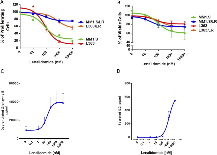
Figure 5. Lenalidomide enhances PBMC-mediated killing of both lenalidomide-sensitive and lenalidomide-resistant MM cells.
(A) 3H-thymidine incorporation in lenalidomide-sensitive MM1.S and L363 and their lenalidomide-resistant progeny cells (MM1.S/LR, L363/LR) following treatment with either vehicle control or lenalidomide (10–10000 nM). The results are presented as percent of the vehicle control. (B) The portion of viable MM cells as percent of the vehicle control in a co-culture assay with lenalidomide-pretreated PBMCs obtained from a healthy donor. CFSE-labeled lenalidomide-sensitive MM1.S and L363 and their lenalidomide-resistant progeny (MM1.S/LR, L363/LR) cells were cultured for 4 hours at 3:1 ratio with PBMCs, which were pretreated for 72 hours with muromonab-CD3 and lenalidomide (10–10000 nM). The viable MM cells were identified by Annexin-V/To-Pro-3 negative staining by flow cytometry. (C) ELISA measurement of granzyme B levels in the supernatant of the PBMC/MM cell co-culture as depicted in panel (B). (D) Secreted IL-2 from PBMCs that were treated with lenalidomide (0–10000 nM) for 72 hours. IL-2 levels were measured by ELISA from the supernatant of PBMCs that were subsequently used for the co-culture experiment as depicted in panel (B). Negative values were set at 0 (for 4 data points, their value in the ELISA readout was lower than the reference value 0). All figures shown are representatives of n = 3 experiments.

