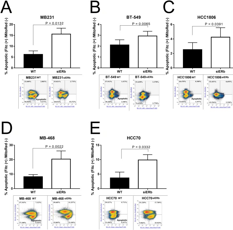
Figure 7. Knockdown of ERβ results in increased apoptosis.
TNBC cells were knockdown for ERβ and then subjected to flow cytometer as described in methods Representative images of flow cytometry data were presented. Percentage of apoptotic cells in (A) MB231, (B) BT-549, (C) MB-468, (D) HCC-70, and (E) HCC-1806 were showed in bar graphs as mean ±SD. The statistical differences between the indicated groups were determined by paired t-test.

