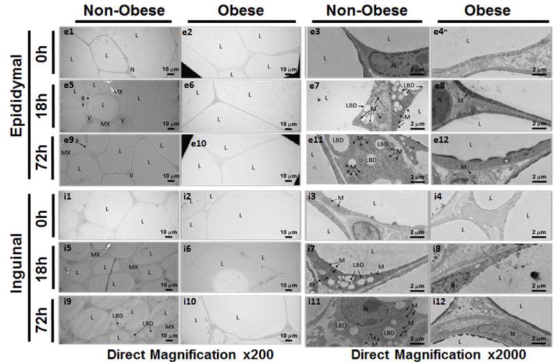
Figure 5. Morphological changes of WAT by Transmission Electron Microscopy in non-obese and obese mice at timepoints after CLP.

Epididymal and inguinal WAT in non-obese and obese mice at 0h, 18h, and 72h after sepsis at 200× and 2000× magnification. All mice were 12 weeks of age at the time of harvest. L=lipid droplet, N=nucleus, MX=extracellular matrix, LBD=lipid breakdown, B=budding, M=mitochondria, V=blood vessel.
