Sir,
Anagen effluvium is known to occur postchemotherapy with cytotoxic as well as noncytotoxic drugs and postradiation therapy. Anagen hair loss occurs when the metabolic and mitotic activity of hair is suppressed by the cytotoxic drug.[1] Trichoscopic features of anagen effluvium include black dots, yellow dots, Pohl Pinkus constriction, and tapering hair.[2] These trichoscopic features are not specific to anagen effluvium and are also seen in other trichological disorders. Herein, we describe a new trichoscopic feature, “tulipoid hair” specific to anagen effluvium and also explain its genesis.
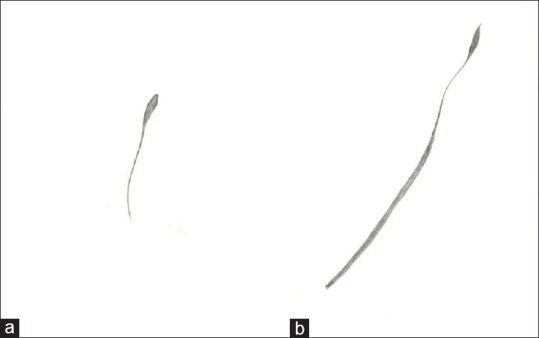
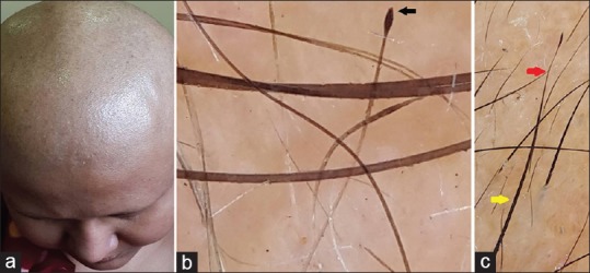
The first case presented to us with diffuse hair loss over the entire scalp along with partial loss of eyebrows and eyelashes. She was initiated on chemotherapy 1 month ago. On the basis of history and clinical examination, a diagnosis of anagen effluvium was made. On trichoscopic examination, one field showed a new trichoscopic feature, “tulipoid hair” which resembles a tulip hair but is characteristically different. Tulip hair has a light-colored hair shaft and a distal dark tip owing to the oblique fracture of the distal end of the hair shaft, thus resembling a “tulip flower.” It is a nonspecific feature seen in alopecia areata, trichotillomania, etc [Figures 1a,b and 2a,b].[3]
Figure 1.

(a) Tulip hair. (b) Tulipoid hair
Figure 2.

(a) Anagen effluvium in a patient following chemotherapy. (b) Tulip hair as a result of oblique fracture to the distal tip of the hair shaft. (c) Tulipoid hair in anagen effluvium. Distal portion of the hair shaft (red arrow) marks the onset of chemotherapy causing constriction and tapering, resulting in the distal tip falling off to form the tulip-like tip. Proximal portion of the hair shaft (yellow arrow) indicates the cessation of chemotherapy and resumption of follicular activity as the hair shaft is of normal thickness
On the other hand, tulipoid hair is specific to anagen effluvium. It is normal in color and thickness in its proximal portion. The distal portion of the tulipoid hair has an acute constriction, beyond which the shaft progressively narrows ultimately to end as a dark tip resembling a “tulip flower.” The tulipoid hair is so named because it resembles tulip hair but is characteristically different. The genesis of tuilpoid hair is interesting as its formation is attributed to commencement and cessation of chemotherapy in anagen effluvium. The constriction of the tulipoid hair in anagen effluvium corresponds to induction of chemotherapy. There is progressive narrowing of the hair shaft beyond the constriction causing the distal most part of the hair to fall off resulting in a tulip like tip. The proximal portion of the tulipoid hair signifies the cessation of chemotherapy and resumption of follicular activity resulting in normal thickness of the shaft [Figure 2c].[2]
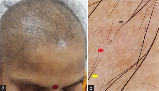
Our second case presented to us with variable loss of hair on the scalp within 3 weeks of induction of chemotherapy [Figure 3a].
Figure 3.

(a) Second case of anagen effluvium following chemotherapy. (b) Tulipoid hair in case 2 of anagen effluvium. Distal portion (red arrow) indicating the onset of chemotherapy resulting in a tulip-like tip while proximal portion (yellow arrow) indicating stoppage of chemotherapy. Also note Pohl pinkus constriction in anagen effluvium
On the basis of history and clinical examination, a diagnosis of anagen effluvium was made. Trichoscopy showed common features of anagen effluvium such as yellow dots, tapering hair, Pohl Pinkus constriction, etc. However, one trichoscopic field again demonstrated the presence of tulipoid hair [Figure 3b].
The distal part of the hair with the constriction beyond which the hair tapers ultimately to end as a dark “tulip like” tip marks the beginning of chemotherapy. The proximal part of the tulipoid hair is normal in thickness and color, thus signifying the cessation of chemotherapy and resumption of follicular activity.
We propose that tulipoid hair is a specific trichoscopic marker of anagen effluvium. Its recognition is of great importance as it can be easily confused with tulip hair which is a nonspecific feature of alopecia areata or trichotillomania. The genesis of tulipoid hair in anagen effluvium is of vital significance as it serves as a prognostic marker indicating the commencement and cessation of chemotherapy and resumption of follicular activity.
Declaration of patient consent
The authors certify that they have obtained all appropriate patient consent forms. In the form, the patients have given their consent for their images and other clinical information to be reported in the journal. The patients understand that their names and initials will not be published and due efforts will be made to conceal their identity, but anonymity cannot be guaranteed.
Financial support and sponsorship
Nil.
Conflicts of interest
There are no conflicts of interest.
REFERENCES
- 1.Trüeb RM. Chemotherapy-induced alopecia. Curr Opin Support Palliat Care. 2010;4:281–4. doi: 10.1097/SPC.0b013e3283409280. [DOI] [PubMed] [Google Scholar]
- 2.Malakar S, Chowdhary B. Anagen effluvium. In: Malakar S, Chandrashekhar BS, Mukherjee S, Mehta P, Pradhan P, editors. Trichoscopy – A Text and Atlas. New Delhi: Jaypee; 2017. pp. 193–206. [Google Scholar]
- 3.Rakowska A, Olszewska M, Rudnicka L, editors. Atlas of Trichoscopy. London: Springer; 2012. Anagen effluvium; p. 247. [Google Scholar]


