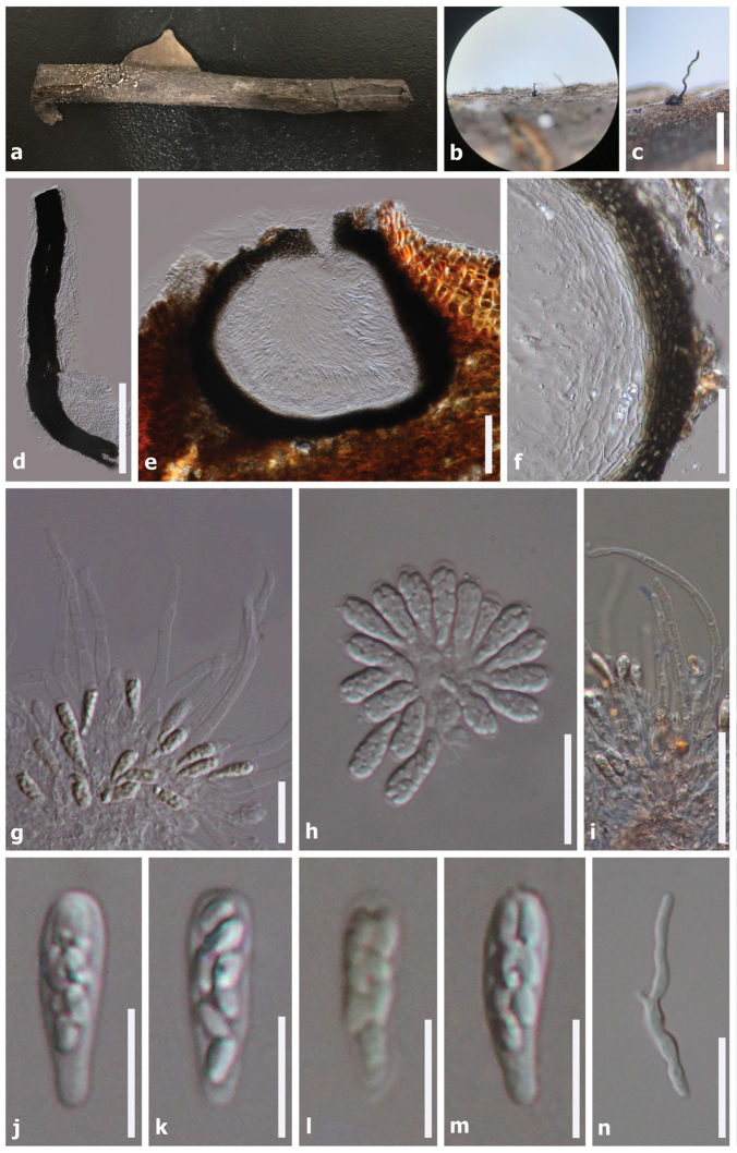
Figure 2.
Phaeoacremoniumovale (HKAS99550, holotype). a Substrate b, c Ascoma on host d Squashed neck e Ascoma in vertical section fPeridiumg Asci surrounded by paraphyses h Asci i Septate paraphyses j–m Asci with ascospores n Germinating ascospores. Note: Fig. i stained in Congo red reagent, fig l stained in Melzer’s reagent. Scale bars: 500 µm (c); 200 µm (d); 100 µm (e); 50 µm (f, i); 30 µm (n); 20 µm (g–h); 10 µm (j–m)

