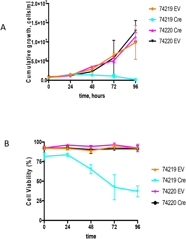
Figure 4. Growth analyses of PCT cells in vitro upon Cre-mediated deletion of the Igh3’RR.
(A) Shown are the cumulative growth curves after normalization for dilution at each subculture. PCT 74219 has a translocation between transgenic Igh and Myc, and PCT 74220 has translocation between endogenous Igh and Myc. Transgenic Igh has loxP sites flanking Igh3’RR enhancers. PCT cell lines were infected with an empty vector (EV) or Cre-expressing (Cre) retroviruses. 4-OHT was added in the culture medium and growth was monitored by cell counting for 96 h. (B) Cell viability curve. Time course effect of Cre-mediated Igh3’RR deletion on cell viability was determined by counting cells using trypan blue staining. All values are presented as mean± SD.

