

Abstract
Background:
Chemical pleurodesis is generally accepted palliative dyspnea therapy and preventive of re-accumulation of pleural fluid in patients with malignant pleural effusions.
Aim:
Comparative analyses of efficiency of chemical pleurodesis between Video Assisted Thoracoscopic Surgery (VATS) and standard thoracostomy.
Methods:
From 01.01.2016-01.01.2017 at the Clinic for Thoracic Surgery of University Clinical Center (UCC) Sarajevo retrospective analysis was performed. Studied patients underwent VATS pleurodesis (G1) and standard thoracostomy pleurodesis (G2), with 60 in each group, respecting defined inclusion and exclusion criteria. Pleurodesis success was examined radiologically over the next three months.
Results:
Average age of all patients was 63.97±8.75 years. Gender related, 45% were men and 55% were women (F/M=1.47:1). Average hospitalization was 7.22±1.37 (G1: 6.68±1.16; G2: 7.44±1.40; Mann-Whitney U-test: p=0.0016) days. Average thoracic drainage duration was 5.45±1.69, (G1: 4.28±1.15,G2: 6.05±1.58; Mann-Whitney U-test p<0.0001) days. Pleurodesis success after first month was 98.30% in G1, 91.60% in G2 (G1 vs. G2; p=0.2089); after second month was 98.30% in G1, 78.30% in G2 (G1 vs. G2; p=0.0011) and after three months was 91.60% in G1, 63.30% in G2(G1 vs. G2; p=0.0006). Average dyspnea degree (0-5) after the pleurodesis was 0.050±0.22 in G1 and 0.62±0.76 in G2 (Mann-Whitney U-test; p=0.0001). Complication were noticed in 9.2% patients, in G1 3.3%, 15.0% in G2.
Conclusion:
Difference in pleurodesis efficiency between the G1 and G2 was established after second month and was even more evident after third month in favor of G1. Results show the significant statistical improvement of the degree of dyspnea in G1 as opposite to the G2.
Keywords: VATS pleurodesis, standard thoracostomy pleurodesis
1. INTRODUCTION
Patients with malignant pleural effusions (MPE) are treated mainly symptomatically. Efficient palliation of dyspnea is the main objective of the therapy. Chemical pleurodesis (CP) is generally accepted palliative therapy for patients with recurrent MPE. The main objective of CP is to create adhesions between the visceral and parietal pleura and preventing re-accumulation of fluid.
2. OBJECTIVES
The comparative analyses of CP with talc instilled through the standard thoracic drainage or Video Assisted Thoracoscopic Surgery (VATS) pleurodesis. Analyses of the duration of hospitalization, thoracic drainage, success of pleurodesis, appearance of complications and assessment of the dyspnea prior and after pleurodesis.
3. PATIENTS AND METHODS
At the Clinic for Thoracic Surgery of UCC Sarajevo, in the period from January 1, 2016 to January 1, 2017 retrospective analysis was performed and it included 60 patients with the CP conducted with talc through the standard thoracic drainage and 60 patients with VATS pleurodesis. Patients were divided into two groups: group G1 (VATS pleurodesis) and group G2 (standard thoracostomy and pleurodesis). The inclusion criteria were: both genders, MPE, Karnofsky index ≥50 %, life expectancy over 30 days, better subjective and general condition after the thoracentesis. The criteria for exclusion were: Karnofsky index <50%, poor general condition with life expectancy of <30 days, atelectasis due to endobronchial obstruction, visceral pleural thickening, pleural empyema, inability to achieve the full lung re-expansion.
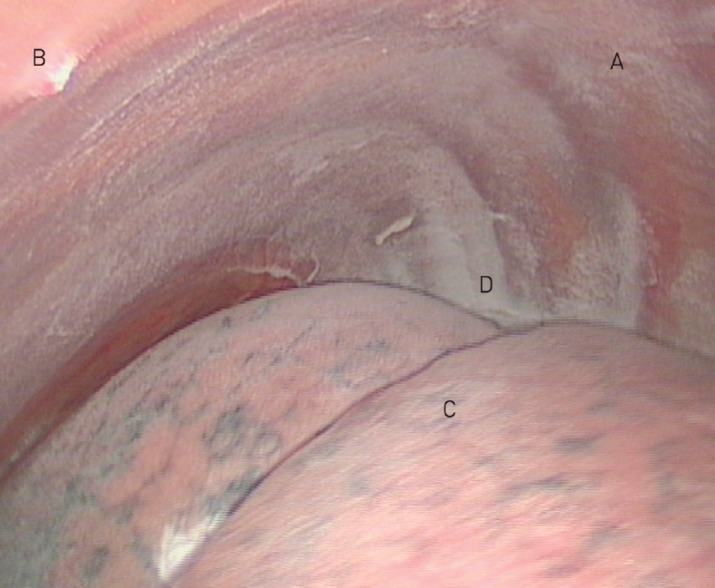
The technique of VATS pleurodesis: After standard preoperative preparation, VATS was conducted in an operation room with appropriate video equipment and thoracoscopic instruments. After evacuation of the pleural fluid and pleural space examination, followed the unification of the eventually divided areas and taking of bioptic samples. Intrapleural, 1-2 thoracic drains (20-32F) were placed. The sclerosans was instilled through the drain and dispersed on the visceral and parietal pleura (Figure 1).
Image 1. VATS procedure. (22).

The technique of inserting the standard thoracic drain: In the local anesthesia the thoracic drain was inserted (20-32F) and the pleural fluid was evacuated. After the evacuation the re-expansion of lungs was checked radiographically. When the conditions were met (secretion was reduced below 100 mL/24h or 3-5 mL/kg of body weight) the pleurodesis by talc was performed. After the instillation of agent, the drain was closed. For the next 6 hours, patients changed the position in bed every 15 minutes in order to have the agent equally distributed all over the pleural surfaces, and then the drainage system was connected to active suction for next 72h. After the reduction/elimination of secretion the drain was removed.
Criteria defined by the American Thoracic Society as well as the European Respiratory Society, categorize success of CP as: complete (no re-accumulation within 3 months post intervention), partial (less re-accumulation in comparison to the pre-drainage level) and unsuccessful (same/larger re-accumulation), all on the basis of radiological findings during the next 3 months.
The success of pleurodesis was controlled once a month the next three months. Assessment of the dyspnea prior and after pleurodesis pursuant to the Scale for determining the degree of dyspnea (0-5).
Statistical analysis of data has been performed by SPSS (Statistical package for Social Sciences-v.19) with application of appropriate statistical methods. Determined level of significance was p<0.05. The obtained results were compared to the data of relevant research in literature with the presentation of discussion and comments of individual results.
4. RESULTS
In relation to gender, 45% (54/120) were men and 55% (66/120) were women (F:M=1.47:1). In the groups: G1[F=58.3% (35/60); M=41.6% (25/60); F:M=1.4:1];G2 (F=52.4% (31/60); M=47.5% (29/60); F:M=1.06:1). The average age of all patients was 63.97±8.75 (31-80) years, and for groups was: G1: 63.92±8.53 (34-80), G2: 64.56±6.45 (47-80) years. Student t-test assuming equal variances for independent samples showed that there is no statistically significant difference between patient in relation to control group (t=0.466, p=0.6424). The average hospitalization duration of all patients was 7.22±1.37 (5-13) days, and for groups: G1: 6.68±1.16 (5-12) days, G2: 7.44±1.40 (6-13) days. In application of Mann-Whitney U-test independent samples showed that there is statistically significant difference in duration of hospitalization between the G1 and G2 group (z=3.151, p=0.0016).
The average thoracic drainage duration was 5.45±1.69 (3-12) days, and for groups: G1: 4.28±1.15, (3-10) days; G2: 6.05±1.58 (5-12) days. In application of Mann-Whitney U-test independent samples showed that there is statistically significant difference in duration of thoracic drainage between the G1 and G2 group (z=7.574, p<0.0001). Success of pleurodesis after first month pursuant to the chest x-ray has shown no significant statistical difference in the outcome of the pleurodesis between the groups after first month (χ2=1.579, p=0.2089). Success of pleurodesis after second month has shown the statistically significant difference in efficiency of pleurodesis after second month between the groups G1 and G2 (χ2=10.734, p=0.0011). Success of pleurodesis after three months has shown the statistically significant difference in the efficiency of pleurodesis after the third month between the groups G1 and G2 (χ2=14.994, p=0.0006).
Image 2. VATS talc pleurodesis. (23).
Descriptive statistics in relation to dyspnea prior to and after the pleurodesis by the groups have shown that the average value of the degree of dyspnea in group G1 prior to pleurodesis was 3.28±0.64, and after the pleurodesis it was 0.050±0.22 and with the application of Wilcoxon test (paired sample) of the ranks for dependent samples, the significant reduction of the symptoms of difficult breathing after the performance of pleurodesis was determined (z=6.736, p<0.0001). In group G2, the average value of the dyspnea degree was 4.10±0.68 prior to pleurodesis, and 0.62±0.76 after, while with the application of Wilcoxon test (paired sample) of the ranks for dependent samples showed the significant reduction of the symptoms of dyspnea after the pleurodesis was also determined (z=6.791, p<0.0001). With the application of Mann-Whitney U test, the statistically significant difference was determined in the degree of dyspnea after the pleurodesis between the G1 and G2 group (z=5.122, p=0.0001). Morbidity and mortality after pleurodesis are shown on the Table 2. Complications were documented in9.2% (11/120) of all patients [G1: 3.3% (2/60); G2: 15.0% (9/60)].
Table 2. The complications after pleurodesis.
| Complications: | G1 | G2 | Total |
|---|---|---|---|
| Pain related with procedure | 3,3% (2/60) | 1,6% (2/120) | |
| Fever | 3,3% (2/60) | 5,0% (3/60) | 4,1% (5/120) |
| Dyspnea related with procedure | - | 1,6% (1/60) | 0,8% (1/120) |
| Repeated pleurodesis | - | 5,0% (3/60) | 2,5% (3/120) |
| Total: | 3,3% (2/60) | 15,0% (9/60) | 10% (12/120) |
5. DISCUSSION
Despite the achievements in the treatment of carcinoma, treatment of MPE remains palliative with average patient survival being3-12 months from the moment of diagnosis (1-3).Treatment of patients with initial or recurrent MPE may be very simple and efficient but also very difficult and complex. Since the limited survival is provided to majority of such patients, useful and efficient relief of symptoms, minimization of time of hospitalization and life quality improvement must be ensured. The best therapeutic results are achieved in case of patients with relatively good general condition, when the basic disease is localized and when patient survival expectancy is >6 months. CP is conducted by different medical procedures such as thoracentesis, thoracostomy-standard or with pleural catheter, thoracoscopy (VATS), thoracotomy and pleuro-peritoneal shunt (1-9). Recently, VATS pleurodesis is used more and it showed as a safe procedure with low complication rate and gradually it replaces the standard modes of pleurodesis. In that manner, some authors try to inaugurate it as a procedure of choice in the treatment of MPE (9-13). Even through numerous papers of related literature were published recently, in relation to application of the VATS pleurodesis, there is still no universal standing on these issues (11, 14-20).
In our study, analysis between groups demonstrated no statistical significance in relation to the patients’ age (t=0.46, p=0.64), nor in relation to patients’ gender (χ2=3.234, p=0.199).
The average duration of hospitalization of all patients was 7.22±1.37 (G1: 6.68±1.16; G2: 7.44±1.40) days. Mann-Whitney U-test showed that there is statistically significant difference in hospitalization days number between the G1 and G2 group (z=3.151, p=0.0016). VATS pleurodesis showed quantitative advantage and it may be concluded that the average hospitalization when applying the VATS pleurodesis was shorter. Abouzgheib et al. 2009 mentioned in their retrospective study that the average duration of hospitalization was 9 days after the VATS pleurodesis. Likewise, Fortin et al. 2015, mentioned that the hospitalization period was 4-7 days for VATS pleurodesis. Troteret al. 2005, and Mitrofan et al. 2005 mentioned that the hospitalization period for VATS pleurodesis with talc was 7-10 days. The comparison of results of the hospitalization duration obtained in the conducted research with the results obtained in the aforementioned literature, leads to a conclusion that in majority of cases those results do in fact correspond (1, 7, 8).
Our average duration of thoracic drainage was 5.45±1.69 (G1: 4.28±.15; G2:6.05±1.58) days. Mann-Whitney U-test showed that there is statistically significant difference in duration of thoracic drainage between the G1 and G2 group (z=7574, p<0.0001). The average duration of thoracic drainage for patients of G1 group was shorter in relation to the group G2. Khalil et al. 2016 mention that the average duration of thoracic drainage was 4.73±0.3 days when applying the VATS pleurodesis. Chen et al. 2015 published that the average postoperative duration of drainage was 4.74±1.56 days after the thorascopic pleurodesis with talc, conducted in 1061 patients. Vermaet al. 2013 mention that the average duration of thoracic drainage for patients with thorascopic pleurodesis was 4.5 (1–6) days. Basso et al. 2012 mention duration of thoracic drainage from 9.4±4.1 days in VATS pleurodesis with talc. Luh et al. 2006 compared the average duration of pleural drainage between the pleurodesis conducted with standard thoracic drainage (lasted 9.1±3.3 days) and average duration of pleural drainage with VATS pleurodesis (lasted 6.2 ±2.3 days) and established that there is statistically significant difference (p< 0.01). The comparison of results of duration of thoracic drainage from the conducted research with the results mentioned in the available literature, it may be concluded that such data are in similar ranges of duration of drainage, whereby it is evident that duration of pleural drainage was shorter in case of VATS pleurodesis.
Our success of pleurodesis after first month pursuant to the chest x-ray was98.3% in group G1 (59/60) and 91.6% in group G2 (55/60). χ2 test has shown no significant statistical difference in the outcome of pleurodesis between the groups after the first month(G1 vs. G2; χ2=1.579; p=0.2089). Our success of pleurodesis after second month was98.3% in group G1 (59/60) and 78.3% in group G2 (47/60).χ2 test has shown the statistically significant difference in efficiency of pleurodesis after second month between the groups (G1 vs. G2 χ2=10.734, p=0.0011). Success of pleurodesis after three months is presented in the Table 1, namely 91.60% in group G1 (55/60) and 63.30% in group G2 (38/60). χ2 test has shown the statistically significant difference in the efficiency of pleurodesis after the third month between the groups (G1 vs. G2; χ2=14.994, p = 0.0006).
Table 1. Success of pleurodesis after third month.
| Success of pleurodesis | G1 | G2 |
|---|---|---|
| Complete | 91%(55/60) | 64%(38/60 |
| Partial | 7%(4/60) | 32%(20/60) |
| Unsuccessful | 2%(1/60) | 4(2/60) |
| Total | 100%(60/60) | 100%(60/60) |
Analysis of obtained results shows that after first month after the pleurodesis, there was no difference in the efficiency between the groups. After second month, the difference in the efficiency between the G1 and G2 group was established. After third month, the difference between the groups was even more evident in favor of group G1. According to the available literature data the efficiency of pleurodesis, according to Zarogoulidis et al. 2013 was between 60-100%. Chen et al. 2015 report that they achieved complete pleurodesis success with 65.41% (694/1061), partial success with 22.62% (240/1061), and no success with 11.97% (127/1061) patients, noting best results with talc thorascopic pleurodesis (90-96% success). Reddy et al. 2011 mention the 92% pleurodesis success in 30 patients after the thorascopic procedure with application of talc. Ferreiro et al. 2017 cite Kennedy et al. 1994 who, in their report, mention the 91% success although many following reports never achieved such result. Shulze et al. 2001 mention success of 93% in the series of 105 VATS talc pleurodesis. Verma et al. 2013 report success of thorascopic pleurodesis as follows: 30 days after conducted pleurodesis 77.8%, after 60 days it was 80.0% and after 90 days it was also 80.0%. Koledin et al. 2001 report that the success of talcthorascopic pleurodesis was 97%. Dresler et al. 2005 present that the success of thorascopic pleurodesis as 78% and 71% by standard drainage, on the series of 501 patients, month after procedure. Lee et al. 2012 mention 94% success pleuroscopic talc pleurodesis 3 months after procedure. Our results show that the percentage of pleurodesis success in relation to defined time interval of 3 months is more stable in cases of G1 patients (98.30%, after first and second month and 91.60% after third month). The results obtained were in accordance with the majority of reviewed available literature data in which there are, however, extensive variations of success results in relation to individual techniques of pleurodesis (1, 7, 14-19).
Dyspnea was analyzed as it is main indication as well as an important indicator of procedure success. The average value of the degree of dyspnea in G1 prior to pleurodesis was 3.28±0.64, and 0.050±0.22 after (Wilcoxon test; z=6.736, p<0.0001, according to reduction of dyspnea). In group G2, the average value of the degree of dyspnea prior to pleurodesis was 4.10±0.68, and 0.62±0.76 after (Wilcoxon test; z=6.791, p<0.0001,according to reduction of dyspnea). The degree of dyspnea after the pleurodesis differed between the G1 and G2 group, showing the significant statistical improvement of the degree of dyspnea in group G1 (Mann-Whitney U test; z=5.122, p=0.0001). Boujaoude et al. 2015 mention that there is statistically significant reduction of degree of dyspnea from 8.34 to 3.2 (p <0.001) measured by Borg scale, and after the pleuroscopic talc pleurodesis. Basso et al. 2012 mention that statistically significant difference was determined (p<0.001) in scores for dyspnea before (4.2±0.8) and after VATS talc pleurodesis with (2.7±1.0). In their paper Koledin et al. 2001 emphasize that dyspnea determined that 98% of patients had dyspnea before VATS talc pleurodesis and only 2% after. Not a small number of studies seem to report only about the simple“reduction of dyspnea” such as Van den Toorn 2005or about the “symptomatic improvement” as published by Efthymiou 2009 without presenting the measure of statistical significance. The meta-analysis of twelve studies conducted by the Van Meter 2011 reports about the symptomatic improvement in 95.6% patients after procedure and the fact that in those researches standardized forms for quality of life were not used (for example dyspnea score). Reddy et al. 2011 mention that in application of VATS talc pleurodesis, dyspnea before the pleurodesis was 4.9, and 1.6 seven days after pleurodesis, where by for the assessment of dyspnea degree the Borg’s scale was used. The results achieved in this research showed significant reduction of dyspnea degree for all examinees regardless of the applied pleurodesis technique, which is in accordance with the results from the available reports in literature (1, 14-20).
The complications of thoracoscopy were usually moderate and include the pain, dyspnea, respiratory insufficiency associated with the procedure, haemorrhage, hypotension, heart arrhythmia, prolonged air leak, subcutaneous emphysema, postoperative fever, pleural empyema, operative wound infection and malignancy dissemination on the thoracic wall (1-7). Morbidity and mortality after pleurodesis are shown on the Table 2. Complications were documented in 9.2% (11/120) of all patients (G1: 3.3% (2/60); G2 15.0% (9/60). The treatment was completed by the primary procedure for all G1 patients while inG2, pleurodesis was repeated for 5.0% (3/60). Procedure related pain was reported in 1.6% (2/120) of examinees, who were all in G2. Fever was reported in total of 4.1% (5/120) cases, out of which 3.3% (2/60) were in G1 and 5.0% (3/60) were in G2. Procedure related dyspnea was reported in total of 0.8% (1/120) cases, all of which were in G2 (1.6%). Among the cases there were no severe morbidities nor lethal outcomes in the three months of following. The frequency of complications with patients included in this research corresponds to quotations of other authors form the available literature sources (1. 13-18). Verma et al. 2013 published that the complications in thorascopic pleurodesis occurred in 14.6% patients and included also subcutaneous emphysema (n=2), pneumothorax (n=3) and hypoxemia (n=1). British thoracic society in 2010 reported 16/4736 lethal cases from 47 studies (mortality rate was 0.3%). Hooper et al. 2010 mention that the mortality in case of conventional thoracoscopy performed by the rigid instrument was 0.09%-0.24%, and in case of VATS procedure was 0.34% with very low rate of complications (<1%). Lee et al. 2012 reported that the mortality during 30 days after the pleuroscopic talc pleurodesis was 0%. Chen et al. 2015 mention that the most frequent complications after the thorascopic talc pleurodesis performed on 1061 patients were chest pain (68%), fever (47%), lung edema (occurring only in 0.28% patients). Trotter et al. 2005 and Mitrofan et al. 2005 reported that the complications in thorascopic talc pleurodesis amounted to 3-25% (chest pain 25%, fever 15%, air fistula 4% and empyema from 1.5-4.5%). Khalil et al. 2017 reported febrility registered at 13%, chest pain at 26%, air fistula at 13% and pleural empyema in 6.6% patients after thorascopic pleurodesis. VATS pleurodesis in comparison with other techniques is generally considered safe because complications are same or less compared to other techniques.
6. CONCLUSIONS
The results show quantitative advantage of VATS pleurodesis because the average duration of hospitalization period after the VATS pleurodesis was shorter.
Duration of pleural drainage was shorter in case of VATS pleurodesis (G1) than in G2.
After second month, the difference in the efficiency between the G1 and G2 was established, even more evident after third month in favor of G1.
The results show the significant statistical improvement of dyspnea degree G1.
The rate of complications after VATS pleurodesis was same or less in comparison to other techniques.
Author’s contribution:
AAP conceptualized this study, designed the methodology for data collection, collected and analyzed the data, and wrote the manuscript. VM extensively supported the development of the study concept, data analysis, and the writing, editing and finalizing of the manuscript. AH intensively supported the process of designing the field study, planning data collection, and critically reviewed the manuscript. AP provided valuable support in data collection. KG assisted in data entry and analysis. All authors read and approved the final manuscript.
Conflict of interest:
none declared.
REFERENCES
- 1.Penz E, Watt KN, Hergott CA, Rahman NM, Psallidas I. Management of malignant pleural effusion: challenges and solutions. Cancer Manag Res. 2017;9:229–241. doi: 10.2147/CMAR.S95663. [DOI] [PMC free article] [PubMed] [Google Scholar]
- 2.Chen H, Brahmer J. Management of malignant pleural effusion. Curr Oncol Rep. 2008;10:287–293. doi: 10.1007/s11912-008-0045-4. [DOI] [PubMed] [Google Scholar]
- 3.Roberts ME, Neville E, Berrisford RG, Antunes G, Ali NG. Management of malignant pleural effusion. British Thoracic Society Pleural Disease Guideline Thorax. 2010;65:32–40. doi: 10.1136/thx.2010.136994. [DOI] [PubMed] [Google Scholar]
- 4.Kolschmann S, Ballin A, Gillissen A. Clinical efficacy and safety of thorascopic talc pleurodesis in malignant pleural effusions. Chest. 2008;128:1431–1435. doi: 10.1378/chest.128.3.1431. [DOI] [PubMed] [Google Scholar]
- 5.Kastelik JA. Management of malignant pleural effusion. Lung. 2013 Apr;191(2):165–175. doi: 10.1007/s00408-012-9445-1. [DOI] [PubMed] [Google Scholar]
- 6.Davies HE, Lee YC. Management of malignant pleural effusions: questions that need answers. Curr opin pulm Med. 2013 Jul;19(4):374–379. doi: 10.1097/MCP.0b013e3283615b67. [DOI] [PubMed] [Google Scholar]
- 7.Clive AO, Jones HE, Bhatnagar R, et al. Interventions for the management of malignant pleural effusions: a network meta-analysis. Cochrane Database Syst Rev. 2016:CD010529. doi: 10.1002/14651858.CD010529.pub2. [DOI] [PMC free article] [PubMed] [Google Scholar]
- 8.Lee P. Point. Should thorascopic talc pleurodesis be the first choice management for malignant effusion? Yes. Chest. 2012 Jul;142(1):15–17. doi: 10.1378/chest.12-1085. [DOI] [PubMed] [Google Scholar]
- 9.Trotter D, Aly A, Siu L, Knight S. Video-assisted thorascopic (VATS) pleurodesis for malignant effusion: an Australian teaching hospital’s experience. Heart Lung Circ. 2005;14:93–97. doi: 10.1016/j.hlc.2005.02.004. [DOI] [PubMed] [Google Scholar]
- 10.Mitrofan C, Aldea A, Grigorescu C, et al. Pleurodeza Toracoscopica in pleureziile maligne. [Thoracoscopic pleurodesis in malignant pleural effusions] Rev Med Chir Soc Med Nat Iasi. 2005;109:799–803. [PubMed] [Google Scholar]
- 11.Luh SP, Chen CY, Tzao CY. Malignant pleural effusion treatment outcomes: pleurodesis via video-assisted thoracic surgery (VATS) versus tube thoracostomy. Thorac Cardiovasc Surg. 2006;54:332–336. doi: 10.1055/s-2006-923931. [DOI] [PubMed] [Google Scholar]
- 12.Cardillo G, Facciolo F, Carbone L, et al. Long-term follow-up of video-assisted talc pleurodesis in malignant recurrent pleural effusions. Eur J Cardiothorac Surg. 2002;21:302–306. doi: 10.1016/s1010-7940(01)01130-7. [DOI] [PubMed] [Google Scholar]
- 13.Basso SM, Mazza F, Marzano B, Santeufemia DA, Chiara GB, Lumachi F. Inproved quality of life in patients with malignant pleural effusion following videoassisted thorascopic talc pleurodesis. Preliminary results. Anticancer Res. 2012 Nov;32(2):5131–5134. [PubMed] [Google Scholar]
- 14.Ferreiro L, Suarez-Antelo J, Valdes L. Pleural procedures in the management of malignant effusions. An Thorac med. 2017;12:3–10. doi: 10.4103/1817-1737.197762. [DOI] [PMC free article] [PubMed] [Google Scholar]
- 15.Anevlavis S, Kouliatsis G, Sotiriou I, et al. Prognostic factors in patients presenting with pleural effusion revealing malignancy. Respiration. 2014;87:311–316. doi: 10.1159/000356764. [DOI] [PubMed] [Google Scholar]
- 16.Zamboni MM, da Silva CT, Jr, Baretta R, Cunha ET, Cardoso GP. Important prognostic factors for survival in patients with malignant pleural effusion. BMC Pulm Med. 2015;15:29. doi: 10.1186/s12890-015-0025-z. [DOI] [PMC free article] [PubMed] [Google Scholar]
- 17.Yoon DW, Cho JH, Choi YS, et al. Predictors of survival in patients who underwent video-assisted thoracic surgery talc pleurodesis for malignant pleural effusion. Thorac Cancer. 2016;7:393–398. doi: 10.1111/1759-7714.12354. [DOI] [PMC free article] [PubMed] [Google Scholar]
- 18.Koledin M, Đurić D, Bijelović M, Baroš B. Pleurodeza talkom kod pleuralnih izliva maligne etiologije. Pneumon. 2000;38:37–43. [Google Scholar]
- 19.Psallidas I, Kalomenidis I, Porcel JM, Robinson BW, Stathopoulos GT. Malignant pleural effusion: from bench to bedside. Eur Respir Rev. 2016;25:189–198. doi: 10.1183/16000617.0019-2016. [DOI] [PMC free article] [PubMed] [Google Scholar]
- 20.Dixit R, Agarwal KC, Gokhroo A, Patil CB, Meena M, Shah NS, et al. Diagnosis and management options in malignant pleural effusions. Lung India. 2017;34:160–166. doi: 10.4103/0970-2113.201305. [DOI] [PMC free article] [PubMed] [Google Scholar]
- 21.Terra RM, Teixeira LR, Bibas BJ, Pego-Fernandes PM, Vargas FS, Jatene FB. Effectiveness and safety of outpatient pleurodesis in patients with recurrent malignant pleural effusion and low perfomance status. Clinics (Sao Paulo) 2011;66(2):211–216. doi: 10.1590/S1807-59322011000200005. [DOI] [PMC free article] [PubMed] [Google Scholar]
- 22.Bhatnagar R, Corcoran JP, Maldonado F, Feller-Kopman D, Janssen J, Astoul P, Rahman NM. Advanced medical interventions in pleural disease. European Respiratory Review. 2016;25:199–213. doi: 10.1183/16000617.0020-2016. [DOI] [PMC free article] [PubMed] [Google Scholar]
- 23.Bibby AC, Tsim S, Kanellakis N, Ball H, Talbot DC, Blyth KG, et al. Malignant pleural mesothelioma: an update on investigation, diagnosis and treatment. Eur Respir Rev. 2016;25:472–486. doi: 10.1183/16000617.0063-2016. [DOI] [PMC free article] [PubMed] [Google Scholar]