Abstract
This study investigated biceps brachii distal myotendinous junction (MTJ) displacement during maximal eccentric elbow flexor contractions to test the hypothesis that muscle length change would be smaller (less MTJ displacement) during the second than the first exercise bout. Ten untrained men performed two eccentric exercise bouts (ECC1 and ECC2) with the same arm consisting of 10 sets of six maximal isokinetic (60°/s) eccentric elbow flexor contractions separated by 4 wk. Biceps brachii distal MTJ displacement was assessed using B-mode ultrasonography, and changes in the displacement (muscle length change) from the start to the end of each contraction during each set and over 10 sets were compared between bouts by two-way repeated-measures ANOVA. Several indirect muscle damage markers were also measured and compared between bouts by two-way repeated-measures ANOVA. The magnitude of MTJ displacement (average of six contractions) increased from set 1 (8.2 ± 4.7 mm) to set 10 (16.4 ± 4.7 mm) during ECC1 (P < 0.05), but no significant changes over sets were evident during ECC2 (set 1: 8.5 ± 4.0 mm; set 10: 9.3 ± 3.1 mm). Changes in maximal voluntary isometric contraction strength, range of motion, muscle thickness, ultrasound echo intensity, serum creatine kinase activity, and muscle soreness (visual analog scale) were smaller (P < 0.05) following ECC2 than ECC1, showing less damage in the repeated bout. These results indicate that the magnitude of muscle lengthening was less during the second than the first eccentric exercise bout, which appears to be a mechanism underpinning the repeated-bout effect.
Keywords: repeated-bout effect, B-mode ultrasound, biceps brachii, myotendinous junction, delayed-onset muscle soreness, muscle strength
muscle damage is often induced after performing unaccustomed eccentric exercise (10, 24). The alterations in muscle structure observed after eccentric exercise are also considered to represent a process of muscle remodeling (33). Typical symptoms of muscle damage include delayed onset muscle soreness (DOMS) and prolonged losses in muscle strength and range of motion (ROM), which are most prominent 1–3 days after exercise and can negatively impact daily activities and athletic performances (4, 24). To develop strategies to minimize these symptoms, it is important to understand how it is induced, yet the factors influencing the magnitude of eccentric contraction-induced muscle damage have not been fully elucidated (10, 24).
Previous evidence indicates that one of the key events in the muscle damage process is an increased intracellular Ca2+ concentration, mediated through stretch-activated channels stimulated by coincident muscle activation and lengthening (1). Another possibility is that damage results directly from the imposition of mechanical strain, triggering inflammation-dependent catabolic processes that weaken the muscle and trigger pain responses (2, 1, 6, 20). Morgan and Proske (22) stated that damage was strongly dependent on the sarcomere lengths during eccentric contractions, and damage would occur when sarcomeres were beyond optimum lengths. If these mechanisms are indeed pivotal, then a greater muscle fiber lengthening during eccentric contractions should result in greater muscle damage and soreness. Indeed, previous animal (11, 18) and human studies (13, 25) have shown that muscle fiber length change is a key factor influencing the magnitude of muscle damage induced by eccentric contractions.
Interestingly, a repeated bout of the same exercise performed within several weeks or months results in less muscle damage than the first bout, which is typically indicated by a faster recovery of muscle function and smaller increases in DOMS and creatine kinase (CK) activity in the blood (20, 24). This protective adaptation is referred to as the repeated-bout effect and has been investigated using models of eccentric exercise in the knee extensors (14, 21) and elbow flexors (8, 23), with a clearer and stronger repeated-bout effect being reported using elbow flexor exercise (9, 12).
The repeated-bout effect has been speculated to be associated with neural, mechanical, and cellular adaptations, although its underpinning mechanisms have yet to be fully described (20). McHugh and Pasiakos (19) reported that an initial bout of eccentric contractions at a short muscle length did not confer protection for a repeated bout at a longer muscle length. Together with the evidence presented above, it seems logical to predict that muscle fiber length change would be less during the second eccentric exercise bout than the initial bout, because the magnitude of muscle damage should be largely determined by the magnitude of muscle fiber lengthening during the exercise bout. Nonetheless, this fundamental hypothesis has never been explicitly tested.
The present study used the B-mode ultrasound technique to estimate biceps brachii muscle length changes during maximal eccentric elbow flexor contractions and compared these length changes between first and second bouts. It was assumed that the movement distance (displacement) of the distal biceps brachii myotendinous junction (MTJ) from the onset to the end of each eccentric contraction would represent biceps brachii muscle fascicle, and presumably muscle fiber, length changes, thus a greater distal MTJ displacement would indicate a greater muscle fiber length change.
A remarkable characteristic of the repeated-bout effect is that its effects last for more than a month after the first bout without any exercise in between. Therefore, the present study imposed the second bout 4 wk after the initial bout to specifically test the hypothesis that the magnitude of the distal MTJ displacement (muscle length) changes over contractions would be less during the second bout when compared with the first eccentric exercise bout. Changes in muscle length were examined between contractions performed within a set, as well as across 10 complete sets of the exercise to examine whether the magnitude of muscle fiber lengthening would be associated with the magnitude of changes in indirect markers of muscle damage.
METHODS
Experimental Design
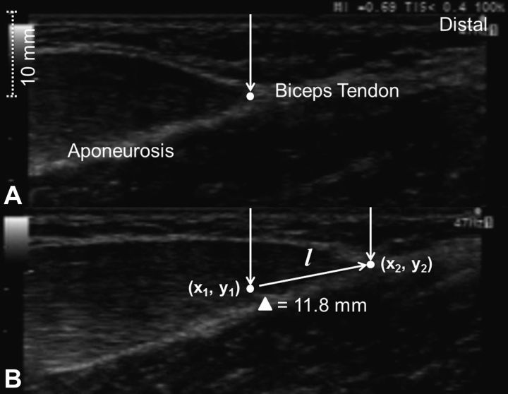
Ten healthy men performed two exercise bouts consisting of 10 sets of 6 maximal isokinetic (60°/s) eccentric elbow flexor contractions using a randomly chosen arm separated by 4 wk. Indirect markers of muscle damage included maximal voluntary isometric contraction (MVC) torque, range of motion (ROM), muscle thickness, ultrasound echo intensity, muscle soreness, and serum CK activity. All of these markers except for CK activity were measured before, immediately after, and 1, 2, 3, 4, 5, and 7 days after exercise, and serum CK activity was measured before, and 4, 5, and 7 days after exercise. Changes in these variables over time were compared between bouts. Biceps brachii distal MTJ displacement during eccentric contractions (Fig. 1) was recorded using B-mode ultrasonography, and the MTJ displacement from the beginning to the end of each eccentric contraction was assessed as explained below, and its changes within and over sets were compared between bouts.
Fig. 1.
A typical B-mode ultrasound image of biceps brachii immediately before an eccentric contraction (maximal isometric contractions at 60° elbow flexion; A) and at the end of the eccentric contraction (at 0° elbow flexion; B). The displacement of the myotendinous junction (as shown by the white dot) was calculated from the two pictures based on the formula; , showing the myotendinous junction (as shown by an arrow) moved. The displacement (l) is 11.8 mm in this example.
Subjects
This study was approved by the Institutional Human Research Ethics Committee and complied with the Declaration of Helsinki. Ten young men (21–39 yr) who had no current or previous upper arm injuries and who had not performed upper limb resistance training for at least 6 mo prior to the present study were invited to participate. The number of subjects was determined by a sample size estimation using the data of a previous study (3), which reported on the repeated-bout effect after maximal elbow flexors eccentric exercise. On the basis of an α-level of 0.05 and a power (1-β) of 0.80, and an expected 20% difference in the recovery of MVC torque at 90° elbow flexion at 3 days postexercise between the first and second bouts, the analysis indicated that at least 10 subjects were required. Their mean (± SD) body mass, height, and MVC torque were 69.2 ± 8.3 kg, 170 ± 6 cm, and 60.0 ± 12.0 Nm, respectively. All subjects provided informed written consent, and a medical questionnaire was completed before participating in the study. Subjects were requested not to change their lifestyle and diet, not to take any anti-inflammatory drugs or nutritional supplements, and not to perform unaccustomed exercise during the experimental period.
Eccentric Exercise
The exercise consisted of 10 sets of 6 maximal voluntary eccentric elbow flexor contractions on an isokinetic dynamometer (Cybex 6000, Ronkonkoma, NY) using one arm that was randomly chosen without considering arm dominance. Each subject was positioned on a seated preacher arm curl bench that secured the shoulder joint at 45° flexion in front of the body, with the elbow being aligned with the axis of rotation of the dynamometer and the lever arm of the dynamometer being attached to the subject's wrist in a supinated position. For each eccentric contraction, the elbow joint was forcibly extended from a flexed (60°) to a fully extended position (0°) in 1 s at an angular velocity of 60°/s, while the subjects were verbally encouraged to generate maximal force at the flexed position and to maximally resist against the elbow-extending action throughout the full range of motion. The smaller range of motion (60°) than that generally used in previous studies [e.g., starting angle of eccentric contractions is 90° (16) or greater (12)] was set in the present study to obtain better ultrasound images during eccentric contractions, as explained below. After each eccentric contraction, the isokinetic dynamometer was programmed to return the arm to the flexed position at a velocity of 6°/s, giving 10 s of rest between contractions. The rest period between sets was 3 min. Torque signals were recorded via a data acquisition system (PowerLab with a Chart 7 software; ADInstruments, Bella Vista, Australia) at a sampling rate of 200 Hz, and real-time visual torque feedback was displayed on a computer monitor. The body position was the same between bouts, and the torque during the eccentric exercise was carefully checked to make sure that the two eccentric exercise bouts were performed similarly for all subjects.
Indirect Muscle Damage Markers
Maximal voluntary isometric contraction torque.
MVC torque of the elbow flexors was measured using the isokinetic dynamometer with the same positioning of the subjects as that for the eccentric exercise described above. Each subject performed two 3-s maximal voluntary isometric contractions at an elbow joint angle of 90° with a 30-s rest between contractions. The higher peak torque of the two contractions was used as the MVC torque. The test-retest reliability of this measure was established from the two baseline measures separated by 1 day, and intraclass correlation (r), and coefficient of variation (CV) values were 0.97 and 3.8%, respectively.
Range of motion.
A plastic goniometer was used to measure extended (EANG) and flexed elbow joint angles (FANG). The EANG was determined when subjects attempted to fully extend the elbow joint while standing and hanging the arm by their side, and the FANG was determined when subjects attempted to fully flex the elbow joint to touch the shoulder of the same side with the palm. A semipermanent ink pen was used to mark the lateral epicondyle of the humerus, the acromion process and the midpoint of the styloid process of the ulna and radius. Measurements were taken twice for each joint angle and the mean value of the two measurements was used to calculate the ROM by subtracting FANG from EANG (16, 23). The r and CV values for the two baseline measures were 0.99 and 0.4%, respectively.
Muscle thickness and echo intensity.
B-mode ultrasound images were obtained using an Aloka SSD-α10 ultrasound system (Aloka, Tokyo, Japan) using a frame rate of 47 Hz with a 10-MHz electronic flat T-head probe (6 cm, UST-5713) from the biceps brachii midbelly at 9 cm above the elbow crease. The examiner placed the probe on this site to obtain longitudinal images. Images were recorded by the ultrasound system and transferred to a portable computer (Dell Laptop, MSK 1750), and a software program (ImageJ, version 1.47, National Institutes of Health, Bethesda, MD) was used to determine muscle thickness and echo intensity. Elbow flexor muscle thickness was measured as the distance between the subcutaneous fat layer and the edge of the humerus (27, 26). B-mode echo intensity of each image was determined by selecting a region of interest (1 cm × 1 cm) within biceps brachii in each image, and the histogram of gray scale (0: black, 255: white) for the region was quantified using a software program (27). Previous animal (7) and human (27) studies have reported that an increased echo intensity indicated inflammatory responses, such as edema. The r and CV values for the two baseline measures were 0.98 and 2.6%, respectively for muscle thickness, and 0.96 and 4.4%, respectively for echo intensity.
Serum CK activity.
Approximately 8 ml of blood was drawn from the antecubital vein by a standard venipuncture technique. Since previous studies have shown that CK activity peaks 4–5 days after eccentric elbow flexor exercise (16, 28), blood samples were taken immediately before, and 4, 5, and 7 days after exercise. The samples were allowed to clot at room temperature and then centrifuged for 10 min at 4°C to obtain serum. Serum CK activity was determined by a Hitachi Modular PT automated clinical chemistry analyzer (Roche, Mannheim, Germany) with a commercially available Roche Diagnostics Reagent. The normal reference range using this method is <200 IU/l.
Visual analog scale.
The level of muscle soreness was assessed using a 100-mm visual analog scale (VAS) in which 0 indicated no pain and 100 represented extreme pain. The subjects were asked to mark the level of perceived soreness on the VAS when the elbow flexors were palpated by the investigator before, immediately after, and 1–5 and 7 days postexercise. In the palpation, the investigator placed his index and middle fingers over the midbelly of biceps brachii at 3, 9 and 15 cm above the elbow crease and applied pressure and palpated with the tips of the finger toward the deeper tissues for ∼3 s, while the subject was lying on the massage table with his forearm in an armrest position. The measurement at the 3-cm site was performed first followed by the measurements at the 9- and 15-cm sites. One measurement was taken from each site with a 10-s interval between measurements. The pressure given to the sites was kept as constant as possible between days and among subjects, and the measurements were taken by the same investigator throughout the study. The mean of the three sites was used for further analysis (16). The r and CV values for the two baseline measures were 0.98 and 2.2%, respectively.
Biceps Brachii Distal Myotendinous Junction Displacement
The movements of the distal biceps brachii MTJ were captured by a real time B-mode ultrasound apparatus with the specifications described above and recorded on a data acquisition system (PowerLab with a Chart 7 software, ADInstruments). It was assumed that the distal MTJ displacement represented muscle fiber-length changes of biceps brachii, and thus muscle fiber strain, although this technique does not include the observation of the proximal MTJ, and the relationship between the MTJ displacement and muscle fiber-length change have not been clarified. The ultrasound probe was firmly attached to the distal portion of the muscle over the MTJ above the elbow crease by tape and bandage. The investigator identified the probe position in a familiarization session, and a semipermanent ink pen was used to mark this position on biceps brachii to achieve consistent measurements over two bouts of eccentric exercise. The video images were displayed in real time on the ultrasound and computer monitors during the exercise. The ultrasound images captured by a frame rate of 47 Hz and torque data on the LabChart 7.0 were synchronized and recorded by a computer (Dell Laptop, MSK 1750) for further analysis. Changes in MTJ displacement from the beginning to the end of each contraction were analyzed by computer software (DartFish Prosuite 5.0; DartFish, Alpharetta, GA), and the corresponding elbow joint angles were checked using the LabChart 7.0 computer software program to ensure that a full extension was achieved. During eccentric contractions, MTJ displacement (l) was determined by the following formula: , where l is the MTJ displacement, x1 and y1 are the MTJ coordinates at the beginning of the contraction (60°), and x2 and y2 are the coordinates at the end of the contraction (0°) as depicted in Fig. 1. The test-retest reliability of this measure was examined by changes in the MTJ displacement during a passive movement of the elbow joint from 60° to 0° performed twice before the eccentric exercise, and r and CV values were 0.96 and 4.6%, respectively.
Statistical Analysis
A one-way repeated-measures ANOVA was used to compare the changes in the biceps brachii MTJ displacement between contractions within each set. A two-way repeated-measures ANOVA was then used to compare the changes in displacement over the 10 sets between the first (ECC1) and second (ECC2) bouts and for the changes in muscle damage markers (MVC, ROM, muscle thickness, echo intensity, serum CK activity, and muscle soreness by VAS) over time (before, immediately after, and 1–5, and 7 days postexercise). When the ANOVA revealed significant time or interaction effects, a Tukey's post hoc test was used for multiple comparisons. Relationships between the within-bout changes in MTJ displacement (i.e., between the 1st and 10th sets) and changes in muscle damage markers (MVC torque and ROM at 1 day postexercise, muscle thickness, and peak ultrasound echo intensity, peak serum CK activity, peak muscle soreness) were examined by computing Pearson's product moment correlation coefficients. The specific time points were chosen as they were considered to represent the magnitude of muscle damage most clearly. For some cases, curvilinear (logarithmic) relationship was also calculated. Statistical significance was set at P < 0.05, and all data are presented as means ± SD.
RESULTS
Peak Torque and Total Work During Eccentric Exercise
From the observation, the two bouts of eccentric exercise were performed similarly for all subjects. No significant difference in the changes in peak torque (P = 0.12) or total work (P = 0.35) over six contractions in a set, and more than 10 sets of six eccentric contractions was evident between two bouts. The total peak torque over the 10 sets (average of 10 subjects) was 367.8 ± 35.6 Nm for the ECC1 and 357.1 ± 23.7 Nm for the ECC2; and the total work over the 10 sets (average of 10 subjects) was 1,816.3 ± 170.5 J for the ECC1 and 1,797.8 ± 135.4 J for the ECC2, without significant differences between bouts.
Muscle Damage Markers
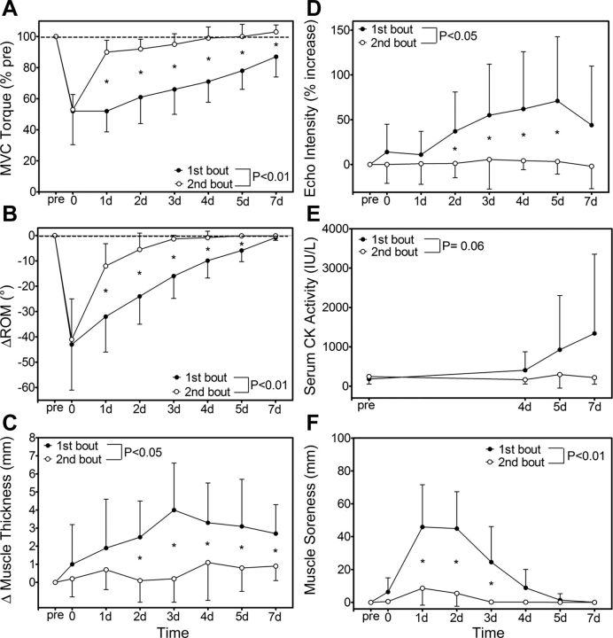
There were no significant differences in preexercise values between bouts; baseline MVC torque was 60.2 ± 12.2 Nm for ECC1 and 56.3 ± 10.8 Nm for ECC2, ROM was 139.5 ± 6.6° for ECC1 and 139.3 ± 6.7° for ECC2, and muscle thickness was 27.3 ± 5.6 mm for ECC1 and 27.8 ± 5.2 mm for ECC2. MVC torque decreased significantly immediately after exercise by ∼50% in both bouts, but recovered significantly faster following ECC2 when compared with ECC1 (Fig. 2A). ROM also decreased similarly between bouts immediately after exercise, but the recovery was significantly faster after ECC2 than ECC1 (Fig. 2B). A significant increase in muscle thickness was observed after exercise; however, the magnitude of increase was significantly less in ECC2 than ECC1 at 2–7 days postexercise (Fig. 2C). Figure 2D shows the relative changes in ultrasound echo intensity from baseline (100%), which was not significantly different between bouts. In the figure, it is clear that echo intensity increased significantly from preexercise values after ECC1, but did not change after ECC2 (Fig. 2D). The baseline serum CK activity was not significantly different between bouts, but there was a tendency (P = 0.06) for the increases in serum CK activity to be smaller after ECC2 compared with ECC1 (Fig. 2E). Muscle soreness increased significantly after both bouts, but the magnitude of muscle soreness was significantly less after ECC2 than ECC1 (Fig. 2F).
Fig. 2.
Changes (means ± SD) in maximal voluntary isometric contraction torque (MVC; A), range of motion (ROM; B), elbow flexor muscle thickness (C), B-mode ultrasound echo intensity from baseline (D), serum CK activity (E), and muscle soreness by visual analog scale (F) before (pre), immediately after (0), and 1–7 days following the first and second eccentric exercise bouts. A significant (P < 0.05 or P < 0.01) interaction effect is shown for all variables. *Significant difference (P < 0.05) difference between bouts.
Distal MTJ Displacement
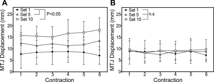
Figure 3 compares the changes in the biceps brachii distal MTJ displacement from the beginning to the end of each contraction over six contractions in sets 1, 5, and 10 for ECC1 and ECC2. No significant changes over six contractions were evident within each set, and this was also the case for other sets that are not included in the figure (i.e., sets 2–4 and sets 6–9). During ECC1, the displacement was significantly greater during sets 5 and 10 when compared with set 1, and for set 10 compared with set 5 (Fig. 3A); however, no significant difference between the sets was evident during ECC2 (Fig. 3B). No significant difference between ECC1 and ECC2 was evident in set 1, but there was a significant difference between bouts in sets 3–10.
Fig. 3.
Changes (means ± SD) in the magnitude of biceps brachii myotendinous junction (MTJ) displacement during eccentric contractions 1–6 in the 1st (set 1), 5th (set 5), and 10th sets (set 10) for the first (A) and second (B) bouts. A significant (P < 0.05) effect of set was found for the first bout, but not for the second bout.
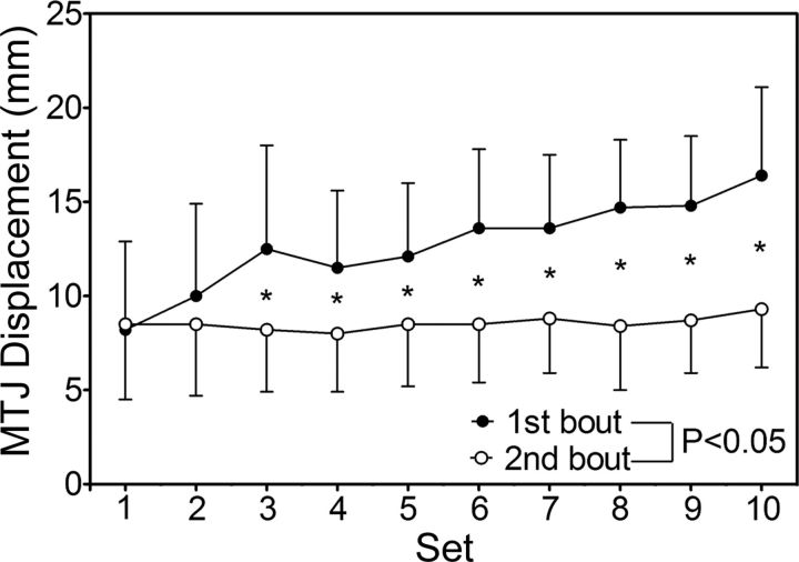
Figure 4 shows the changes in the magnitude of MTJ displacement over 10 sets (average of each set) during ECC1 and ECC2. No significant difference between bouts was evident for the change in the set 1 (ECC1: 8.2 ± 4.7 mm; ECC2: 8.5 ± 4.0 mm). Displacement in ECC1 significantly increased over sets, but this was not the case for ECC2, and a significant difference between bouts was evident from set 3 onward. In ECC1, the MTJ displacement doubled from set 1 to set 10 (16.4 ± 4.7 mm); however, there were no significant changes in ECC2 from set 1 to set 10 (9.3 ± 3.1 mm).
Fig. 4.
Changes (means ± SD) in the magnitude of biceps brachii MTJ displacement in a set (average of six contractions) over 10 sets for the first and second bouts. A significant (P < 0.05) interaction effect is shown. *Significant different between bouts.
Correlation Between the Change in MTJ Displacement and Muscle Damage Markers
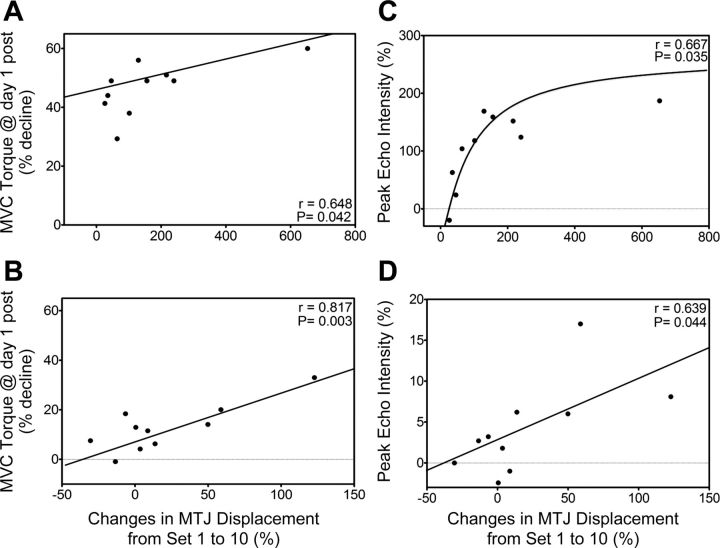
A significant correlation was found between the percent change in distal MTJ displacement from set 1 to set 10 during the first exercise bout and the magnitude of decrease in MVC torque from baseline to 1 day postexercise (Fig. 5A) and the change in MTJ displacement and the magnitude of change in peak ultrasound echo intensity (Fig. 5C); however, no significant correlation was found between the MTJ displacement and changes in other variables. Similarly, a significant correlation between the change in the distal MTJ displacement and the magnitude of change in MVC torque at 1 day postexercise (Fig. 5B), as well as the MTJ displacement and the change in peak ultrasound echo intensity (Fig. 5D), was evident for the second bout. However, a significant relationship was not observed for the other markers (ROM: r = 0.149; muscle thickness: r = 0.110; CK: r = 0.260, muscle soreness: r = 0.497).
Fig. 5.
Correlation between the normalized changes in the distal MTJ displacement from the 1st to 10th sets and the magnitude of change in MVC torque at one-day postexercise from the preexercise value (A, B), and the magnitude of change in peak echo intensity from baseline (C, D) for the first (upper figures) and the second exercise (lower figures) bouts. For the regression line, the model (either liner or curvilinear) that showed a greater r value is shown.
DISCUSSION
The present study tested the hypothesis that the magnitude of change in MTJ displacement (muscle lengthening) over 10 sets of six maximal eccentric contractions would be less during the second than the first eccentric exercise bout separated by 4 wk and that this reduction in lengthening would be associated with a decrease in indirect muscle damage marker changes. Interestingly, no difference in the distal MTJ displacement was observed between bouts when comparing only the first set of exercise. However, an important and novel finding of the present study was that, while there was a 50% increase in the distal MTJ displacement from the 1st to the 10th set during the first eccentric exercise bout, the displacement was constant over sets 1 to 10 during the second eccentric exercise bout (Fig. 4). Thus, it seems likely that the increase in muscle lengthening seen in the first bout was absent in the second bout. Importantly, there was less evidence of muscle damage (i.e., there was a faster recovery of MVC torque and ROM, lack of increase in muscle thickness, ultrasound echo intensity and serum CK activity, and minimal DOMS) after the second bout of exercise, despite significant evidence of damage being observed after the first bout (Fig. 2), indicating a typical repeated-bout effect. From the present study, it is not known whether the reduced muscle damage after the second bout was due to the smaller changes in the distal MTJ displacement over sets. However, it appears that the finding of a lesser muscle lengthening as exercise progressed in the repeated bout supports the hypothesis that the repeated-bout effect results from a lesser muscle lengthening being imposed even though muscular torque and joint range of motion (i.e., total work) were not significantly different between bouts. As evidence of a potential causal link between the minimization of muscle length change and the reduction in muscle damage in the second bout, individuals who showed greater increases in the distal MTJ displacement from the 1st to the 10th set also showed greater decreases in MVC torque after exercise, and greater increases in ultrasound echo intensity following both exercise bouts (Fig. 5). These results suggest that the repeated bout is associated with a reduction in muscle lengthening during eccentric contractions and that less damage is induced probably when muscle fiber lengthening does not increase as exercise continues during repetitive eccentric contractions.
It is important to note that no significant differences in any of the baseline-dependent variables were evident between bouts, and the exercise appeared to be performed similarly for the initial and secondary bouts, as indicated by the similar peak torque production and work performed during the two exercise bouts. Thus, it seems reasonable to assume that the significant differences between bouts for the changes in all dependent variables except serum CK activity after eccentric exercise were due to the different changes in the MTJ displacement over sets between bouts. For serum CK activity, the lack of difference between bouts was likely to have resulted from the large variability in the changes after ECC1 among the subjects. It is possible that the large variability among subjects for their responses to eccentric exercise (e.g., changes in ROM, muscle thickness, and CK activity) was associated with the difference in muscle lengthening during eccentric contractions among subjects (Fig. 5). It is also interesting to note that the extent of changes in some variables (e.g., MVC torque, ROM) was similar between bouts immediately postexercise, but recovery was facilitated after the second exercise bout. It may be that the changes immediately postexercise were due to the combination of muscle damage and fatigue, and it is possible that fatigue was similarly induced between bouts. Muscle damage is indicated better at 1 day postexercise and beyond (32). Thus, it seems that the magnitude of muscle lengthening is a factor determining the magnitude of muscle damage.
As shown in Fig. 3, there was no change in the magnitude of the distal MTJ displacement between contractions within each set, although it was significantly different between sets in the first bout of eccentric exercise (Fig. 4). It is interesting that the displacement did not increase between contractions within a set, but was greater in the subsequent set after 3 min of passive rest (Fig. 3). It may be practically important to determine the cause of this in future research, because the mechanisms influencing the muscle behavior appear to act distinctly between sets rather than between contractions; the resting phase is, therefore, an important point of future examination.
Since the torque, which was produced over 60 eccentric contractions, was similar between bouts, it does not appear that the constant muscle length changes over sets during the second bout could be explained by torque. It appears that the muscle was not stretched by the same lengthening torque during the second bout, especially after the third set, as much as that during the first eccentric exercise bout. It is not known how this happened; however, it could be speculated that the muscle became stiffer, or the tendon became more compliant after performing ECC1. Although the current study was not designed to examine the mechanisms responsible for the effect, it is possible that changes in intramuscular pressure (i.e., changes in blood flow), afferent feedback (particularly via type III and type IV afferent pathways), and rapid effects of calpain-mediated protein degradation are important. Regardless, it is important to note that a greater decrease in MVC torque and an increase in ultrasound echo intensity after the first and second exercise bouts were evident in subjects who showed the greatest increase in the distal MTJ displacement as the sets progressed (Fig. 5). This is the best evidence, thus far, that the magnitude of muscle damage is associated with the muscle behavior during eccentric exercise, and the magnitude of the increase in muscle length during eccentric exercise determines the magnitude of muscle damage.
To the best of our knowledge, the present study was the first study to investigate the distal MTJ displacement changes during elbow flexor eccentric contractions, and no previous studies have investigated either proximal region or both proximal and distal regions simultaneously during eccentric exercise. It is difficult to investigate the proximal region of biceps brachii muscle, because it has two heads; thus, this study focused on the distal region. It should be noted that the present study was not able to measure the changes in fascicle length and pennation angle during eccentric contractions, because biceps brachii pennation angle is small, and its fascicle length is long. It is also not known whether the changes in pennation angle during eccentric contractions may result in unequal length changes of muscle and fascicle with the MTJ displacement. Thus, the actual muscle fiber length changes during eccentric contractions are not known, but it seems likely that the distal MTJ displacement reflects muscle fiber length changes. It is necessary to investigate the relationship between the distal MTJ displacement and muscle fiber length, as well as sarcomere length in the future studies.
The present study focused only on biceps brachii, and other elbow flexor muscles, brachialis, and brachioradialis behaviors were not investigated. Thus, it is hasty to conclude that the reduced muscle damage after the second eccentric exercise bout was solely due to the smaller biceps brachii muscle length changes over repeated eccentric contractions in the second than the first bout. If the present study results are interpreted that muscle fiber lengthening was reduced in the second exercise bout, another question to be answered in future research is how muscle fiber lengthening is reduced in the second bout, even when 4 wk are allowed between bouts. In line with the finding of the present study, Peñailillo et al. (29) have recently reported that vastus lateralis fascicle elongation during 10-min eccentric cycling was 16% less during the second than the first exercise bout separated by 2 wk, and estimated maximal tendinous tissue (TT) length occurred at a smaller knee joint angle during the second than the first bout. They concluded that a lesser fascicle elongation and earlier TT elongation were associated with reduced muscle soreness after the second than the first eccentric cycling exercise, and changes in muscle-tendon behavior might be an important mechanism underpinning the repeated-bout effect.
Proske and Morgan (30) suggested that increases in sarcomere number in series were associated with the repeated-bout effect, and this cellular adaptation theory is indirectly supported by a shift of optimum angle toward a longer muscle length. In the present study, the elbow joint angle to produce the largest isometric torque (optimum angle) was not assessed; however, Chen et al. (3) used a similar eccentric exercise model of the elbow flexors to that of the present study, and reported 4° shift toward a longer muscle length remaining at 2–3 wk after the first maximal eccentric exercise bout. They also found in their study that the repeated-bout effect was conferred by submaximal (40–80%) eccentric exercise without any shift of the optimum angle after submaximal eccentric exercise and stated that the shift of the optimum angle did not appear to be directly related to the mechanisms underpinning the repeated-bout effect. It seems that the longitudinal addition of sarcomeres fits well to explain the observation of less muscle fiber lengthening in the second eccentric exercise bout, but it is not known whether sarcomere number in a series increased in biceps brachii muscle fibers between the first and second eccentric exercise bouts in the present study. Thus, further study is warranted to examine this speculation.
McHugh et al. (20) speculated that increases in extensibility of relaxed muscle (passive stiffness) and active muscle (dynamic stiffness), remodeling of the intermediate filament system, and increased intramuscular connective tissue following eccentric training are mechanical adaptations that could protect against damage from a repeated bout. However, they also pointed out the evidence against the mechanical adaptation theory, such as the fact that muscle with greater passive stiffness was more susceptible to damage. In the present study, the magnitude of muscle length changes in the first set was the same between bouts, but muscle lengthening appeared to be much less in the third set and beyond (Fig. 4). This may suggest that an increase in dynamic stiffness is more likely responsible for the repeated-bout effect. Lapier et al. (15) examined the intramuscular connective tissue of rat extensor digitorum longus muscles after 3 wk of immobilization in either a shortened or lengthened position, and they found that the intramuscular connective tissue concentration was increased under both conditions and that muscle damage was attenuated in these muscles after electrically stimulated eccentric plantar flexor contractions. This finding indicates that, regardless of how it is induced, changes in connective tissue are associated with a decrease in muscle damage. Crameri et al. (5) found increases in the staining of human vastus lateralis intramuscular connective tissue (tenascin C) after voluntary, as well as electrically stimulated eccentric contractions of the knee extensors. Also, Raastad et al. (31) reported that tenascin-C and NH2-terminal propeptide of procollagen type III increased in the endomysium after 300 eccentric knee extensor contractions, and Mackey et al. (17) showed that laminin-β1 and types I and III collagen were elevated after the initial eccentric exercise. They concluded that remodeling and strengthening of extracellular matrix (ECM) played a role in the protective effect. These findings are suggestive of the possibility that a single bout of eccentric exercise remodels the ECM and/or connective tissues to make the muscle more resilient to eccentric contraction-induced muscle damage. It remains to be seen whether such changes remain prominent at least 1 mo after an initial bout of eccentric exercise and whether the magnitude of these changes is related to the magnitude of the repeated-bout effect.
In conclusion, the present study showed that the magnitude of biceps brachii distal MTJ displacement during maximal eccentric contractions increased over 10 sets during the first eccentric elbow flexors bout but did not increase during a second bout performed 4 wk later. Muscle damage markers showed a typical repeated-bout effect, including a faster recovery of muscle function, minimal change in ultrasound echo intensity, attenuated DOMS and a lack of increase in serum CK activity. Notably, individuals who displayed the greatest increase in MTJ displacement over the 10 sets in both first and second bouts also showed the greatest loss in isometric torque and greater increase in ultrasound echo intensity (i.e., muscle damage). It seems possible that the smaller changes in MTJ displacement during the second eccentric exercise imposed less mechanical strain on biceps brachii muscle and presumably to its muscle fibers, inducing less damage. This may, thus, be one factor influencing the repeated-bout effect; however, the mechanisms that might underpin the resistance to lengthening after the first bout are not known and need to be elucidated in future research.
DISCLOSURES
No conflicts of interest, financial or otherwise, are declared by the authors.
AUTHOR CONTRIBUTIONS
Author contributions: W.Y.L., A.J.B., M.J.N., and K.N. conception and design of research; W.Y.L., S.S.X.W., and K.N. performed experiments; W.Y.L., S.S.X.W., and K.N. analyzed data; W.Y.L., A.J.B., and K.N. interpreted results of experiments; W.Y.L. prepared figures; W.Y.L. and K.N. drafted manuscript; W.Y.L., A.J.B., M.J.N., and K.N. edited and revised manuscript; W.Y.L., A.J.B., M.J.N., S.S.X.W., and K.N. approved final version of manuscript.
REFERENCES
- 1.Allen DG, Whitehead NP, Yeung EW. Mechanisms of stretch-induced muscle damage in normal and dystrophic muscle: role of ionic changes. J Physiol : 723–735, 2005. [DOI] [PMC free article] [PubMed] [Google Scholar]
- 2.Allen DG. Eccentric muscle damage: mechanisms of early reduction of force. Acta Physiol Scand : 311–319, 2001. [DOI] [PubMed] [Google Scholar]
- 3.Chen TC, Nosaka K, Sacco P. Intensity of eccentric exercise, shift of optimum angle, and the magnitude of repeated-bout effect. J Appl Physiol : 992–999, 2007. [DOI] [PubMed] [Google Scholar]
- 4.Clarkson PM, Hubal MJ. Exercise-induced muscle damage in humans. Am J Phys Med Rehabil : S52–S69, 2002. [DOI] [PubMed] [Google Scholar]
- 5.Crameri RM, Aagaard P, Ovortrup K, Langberg H, Olesen JK. Myofibre damage in human skeletal muscle: effects of electrical stimulation versus voluntary contraction. J Physiol : 365–380, 2007. [DOI] [PMC free article] [PubMed] [Google Scholar]
- 6.Féasson L, Stockholm D, Freyssenet D, Richard I, Duguez S, Beckmann JS, Denis C. Molecular adaptations of neuromuscular disease-associated proteins in response to eccentric exercise in human skeletal muscle. J Physiol : 297–306, 2002. [DOI] [PMC free article] [PubMed] [Google Scholar]
- 7.Fujikake T, Hart R, Nosaka K. Changes in B-mode ultrasound echo intensity following injection of bupivacaine hydrochloride to rat hind limb muscles in relation to histologic changes. Ultrasound Med Biol : 687–96, 2009. [DOI] [PubMed] [Google Scholar]
- 8.Hosseninzadeh M, Andersen OK, Adrendt-Nielsen L, Madeleine P. Pain sensitivity is normalized after a repeated bout of eccentric exercise. Eur J Appl Physiol : 2595–2602, 2013. [DOI] [PubMed] [Google Scholar]
- 9.Howatson G, van Someren K, Hortobáguyi T. Repeated bout effect after maximal eccentric exercise. Int J Sports Med : 557–563, 2007. [DOI] [PubMed] [Google Scholar]
- 10.Howatson G, van Someren KA. The prevention and treatment of exercise-induced muscle damage. Sports Med : 483–503, 2008. [DOI] [PubMed] [Google Scholar]
- 11.Hunter KD, Faulkner DA. Plyometric contraction-induced injury of mouse skeletal muscle: effect of initial length. J Appl Physiol : 278–283, 1997. [DOI] [PubMed] [Google Scholar]
- 12.Jamurtas AZ, Theocharis V, Tofas T, Yfanti C, Paschalis V, Koutedakis Y, Nosaka K. Comparision between leg and arm eccentric exercises of the same relative intensity on indices of muscle damage. Eur J Appl Physiol : 179–185, 2005. [DOI] [PubMed] [Google Scholar]
- 13.Jones DA, Newham DJ, Torgan C. Mechanical influences on long-lasting human muscle fatigue and delayed-onset pain. J Physiol : 415–427, 1989. [DOI] [PMC free article] [PubMed] [Google Scholar]
- 14.Kamandulis S, Skurvydas A, Brazaitis M, Škikas L, Duchateau J. The repeated bout effect of eccentric exercise is not associated with changes in voluntary activation. Eur J Appl Physiol 1065–1074, 2010. [DOI] [PubMed] [Google Scholar]
- 15.Lapier TK, Burton HW, Cerny F. Alterations in intramuscular connective tissue after limb casting affect contraction-induced muscle injury. J Appl Physiol : 1065–1069, 1995. [DOI] [PubMed] [Google Scholar]
- 16.Lau WY, Nosaka K. Effect of vibration treatment on symptoms associated with eccentric exercise-induced muscle damage. Am J Phys Med Rehabil : 648–657, 2011. [DOI] [PubMed] [Google Scholar]
- 17.Mackey AL, Brandstetter S, Schjerling P, Bojsen-Moller J, Qvortrup K, Pedersen MM, Doessing S, Kjaer M, Peter Magnusson S, Langberg H. Sequenced response of extracellular matric deadhesion and fibrotic regulators after muscle damage is involved in protection against future injury in human skeletal muscle. FASEB J : 1943–1059, 2011. [DOI] [PMC free article] [PubMed] [Google Scholar]
- 18.Macpherson PC, Dennis RG, Faulkner JA. Sarcomere dynamics nd contraction-induced injury to maximally activated single muscle fibers from soleus muscles of rats. J Physiol : 523–533, 1997. [DOI] [PMC free article] [PubMed] [Google Scholar]
- 19.McHugh MP, Pasiakos S. The role of exercising muscle length in protective adaptation to a single bout of eccentric exercise. Eur J Appl Physiol : 286–293, 2005. [DOI] [PubMed] [Google Scholar]
- 20.McHugh MP. Recent advances in the understanding of the repeated bout effect: the protective effect against muscle damage from a single bout of eccentric exercise. Scand J Med Sci Sports : 88–97, 2003. [DOI] [PubMed] [Google Scholar]
- 21.Miyama M, Nosaka K. Protection against muscle damage following 50 drop jumps conferred by 10 drop jumps. J Strength Cond Res : 1087–1092, 2007. [DOI] [PubMed] [Google Scholar]
- 22.Morgan DL, Proske U. Popping sarcomere hypothesis explains stretch-induced muscle damage. Clin Exp Pharmacol Physiol : 541–545, 2004. [DOI] [PubMed] [Google Scholar]
- 23.Muthalib M, Lee H, Millet GY, Ferrari M, Nosaka K. The repeated-bout effect: influence on biceps brachii oxygenation and myoelectrical activity. J Appl Physiol : 1390–1399, 2011. [DOI] [PubMed] [Google Scholar]
- 24.Nosaka K. Exercise-induced muscle damage and delayed onset muscle soreness. In: Strength and Conditioning: Biological Principles and Practical Applications, edited by Cardinale M, Newton R, and Nosaka K. Chichester, UK: Wiley-Blackwell, 2011, p. 179–192. [Google Scholar]
- 25.Nosaka K, Newton M, Sacco P, Chapman D, Lavender A. Partial protection against muscle damage by eccentric actions at short muscle lengths. Med Sci Sports Exerc : 746–753, 2005. [DOI] [PubMed] [Google Scholar]
- 26.Nosaka K, Newton M. Difference in the magnitude of muscle damage between maximal and submaximal eccentric loading. J Strength Cond Res : 202–208, 2002. [PubMed] [Google Scholar]
- 27.Nosaka K, Clarkson PM. Changes in indicators of inflammation after eccentric exercise of the elbow flexors. Med Sci Sports Exerc : 953–961, 1996. [DOI] [PubMed] [Google Scholar]
- 28.Nosaka K, Sakamoto K. Effect of elbow joint angle on the magnitude of muscle damage to the elbow flexors. Med Sci Sports Exerc : 22–29, 2001. [DOI] [PubMed] [Google Scholar]
- 29.Peñailillo L, Blazevich AJ, Nosaka K. Muscle fascicle behavior during eccentric cycling and its relation to muscle soreness. Med Sci Sports Exerc : 708–717, 2015. [DOI] [PubMed] [Google Scholar]
- 30.Proske U, Morgan DL. Muscle damage from eccentric exercise: mechanism, mechanical signs, adaptation and clinical applications. J Physiol : 333–345, 2001. [DOI] [PMC free article] [PubMed] [Google Scholar]
- 31.Raastad T, Owe SG, Paulsen G, Enns D, Overgaard K, Crameri R, Kill S, Belcastro A, Bergersen L, Hallen J. Changes in calpain activity, muscle structure, and function after eccentric exercise. Med Sci Sports Exerc : 86–95, 2010. [DOI] [PubMed] [Google Scholar]
- 32.Warren GL, Lowe DA, Armstrong RB. Measurement tools used in the study of eccentric contraction-induced injury. Sports Med : 43–59, 1992. [DOI] [PubMed] [Google Scholar]
- 33.Yu JG, Furst DO, Thornell LE. The mode of myofibril remodelling in human skeletal muscle affected by DOMS induced by eccentric contractions. Histochem Cell Biol : 383–393, 2003. [DOI] [PubMed] [Google Scholar]


