Cerebral autosomal dominant arteriopathy with subcortical infarcts and leukoencephalopathy (CADASIL) is a rare, hereditary form of small-vessel disease caused by mutations in the NOTCH3 gene at q13.1 on chromosome 19, which result in strokes in young adults. Multiple subcortical lacunar infarcts and diffuse leukoencephalopathy are typical findings in patients with CADASIL. Anterior temporal pole and external capsule lesions are known to have high sensitivity for CADASIL, with anterior temporal pole lesions showing higher specificity.1
SyMRI is a MRI technique that enables rapid simultaneous quantification of T1 and T2 relaxation times, and proton density.2 Based on these absolute values, any contrast-weighted image with optional inversion recovery time (TI) can be created by synthetic MRI. The volume of myelin can also be estimated from the acquired quantitative values. SyMRI has been evaluated for use in patients with diseases such as multiple sclerosis and Sturge-Weber Syndrome, with promising results.3,4
Here, we present a case of a 40-year-old man with proven NOTCH3 mutation who was referred to our hospital with an episode of dysarthria. Radiological investigations were performed, including diffusion-weighted MRI (image not shown), which revealed an acute infarct in the left centrum semiovale, explaining the patient’s symptom. As part of our hospital’s routine protocol, SyMRI was also performed using a 3T MRI system (Discovery MR750w 3.0T; GE Healthcare, Milwaukee, WI, USA). Acquisition time was 7 min 12 s. Synthetic images were created using SyMRI software (ver 8.0, SyntheticMR AB, Linköping, Sweden).
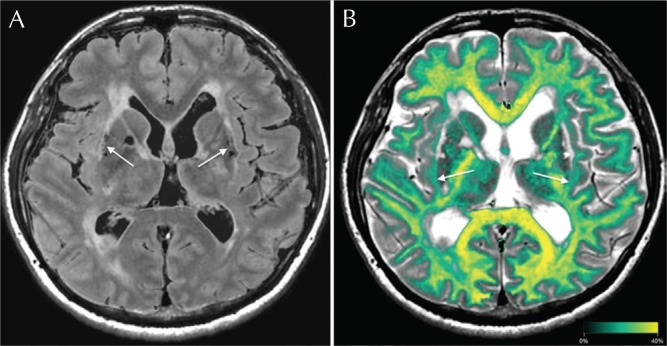
Synthetic fluid attenuated inversion recovery (FLAIR) (Fig. 1A; TR 15,000 ms; TE 100 ms; TI 3000 ms) and double inversion recovery (DIR) (Fig. 1B; TR 15,000 ms; TE 100 ms; initial TI 470 ms; second TI 3750 ms) images showed hyperintense lesions in both anterior temporal poles, with these lesions more clearly visualized, particularly in the left anterior temple pole, in the DIR images. These lesions were also visualized on synthetic T1-weighted (Fig. 1C; TR 500 ms; TE 10 ms) and phase-sensitive inversion recovery (PSIR) (Fig. 1D; TR 6000 ms; TE 10 ms; TI 500 ms) images, with the PSIR image clearer than the T1-weighted image, especially in the left anterior pole. A myelin map overlaid on the synthetic T2-weighted image (Fig. 1E; TR 4500 ms; TE 100 ms) clearly showed the decrease in volume of myelin in both anterior temporal poles. The synthetic FLAIR image showed bilateral external capsule lesions (Fig. 2A). The decrease in volume of myelin was clearly visualized bilaterally in the external capsules using the myelin map overlaid on the T2-weighted image (Fig. 2B).
Fig. 1.
We used synthetic magnetic resonance imaging to visualize brain changes in a patient with cerebral autosomal dominant arteriopathy with subcortical infarcts and leukoencephalopathy (CADASIL). A synthetic fluid attenuated inversion recovery (FLAIR) image (A) shows hyperintense lesions in both anterior temporal poles. Synthetic double inversion recovery (DIR) imaging (B) is superior to FLAIR imaging for detection of these hyperintense lesions, particularly in the left anterior temporal pole (arrow). Synthetic T1-weighted (C) and phase-sensitive inversion recovery (PSIR) (D) images show hypointense lesions in both anterior temporal poles, visualized more clearly on the PSIR image, particularly in the left anterior temporal pole (arrow). Decrease in volume of myelin in these lesions is evident on the myelin map overlaid on the T2-weighted image (E).
Fig. 2.
A synthetic fluid attenuated inversion recovery (FLAIR) image (A) shows bilateral external capsule lesions and bilateral putaminal infarcts (arrows). The decrease in volume of myelin is clear in both external capsules on the myelin map overlaid on the T2-weighted image (B: arrows).
This case demonstrates the usefulness of SyMRI in generating contrast-weighted images and enabling measurement of myelin volume after MRI acquisition. Synthetic DIR and PSIR may increase sensitivity for detecting anterior temporal pole lesions, and decreases in volume of myelin can be visualized on a myelin map. Detection of these lesions may be useful for differential early diagnosis of CADASIL.
Acknowledgment
This work was supported by grants from MEXT-Supported Program for the Private University Research Branding Project.
Footnotes
Conflicts of Interest
The authors declare that they have no conflicts of interest.
References
- 1.Di Donato I, Bianchi S, De Stefano N, et al. Cerebral Autosomal Dominant Arteriopathy with Subcortical Infarcts and Leukoencephalopathy (CADASIL) as a model of small vessel disease: update on clinical, diagnostic, and management aspects. BMC Med 2017; 15:41. [DOI] [PMC free article] [PubMed] [Google Scholar]
- 2.Hagiwara A, Warntjes M, Hori M, et al. SyMRI of the brain: rapid quantification of relaxation rates and proton density, with synthetic MRI, automatic brain segmentation, and myelin measurement. Invest Radiol 2017; 52: 647–657. [DOI] [PMC free article] [PubMed] [Google Scholar]
- 3.Andica C, Hagiwara A, Nakazawa M, et al. The advantage of synthetic MRI for the visualization of early white matter change in an infant with sturge-weber syndrome. Magn Reson Med Sci 2016; 15:347–348. [DOI] [PMC free article] [PubMed] [Google Scholar]
- 4.Hagiwara A, Nakazawa M, Andica C, et al. Dural enhancement in a patient with sturge-weber syndrome revealed by double inversion recovery contrast using synthetic MRI. Magn Reson Med Sci 2016; 15: 151–152. [DOI] [PMC free article] [PubMed] [Google Scholar]


