



Summary
Clinical treatment of Candida albicans infections has become more difficult due to the limited development of antifungal agents and the rapid emergence of drug resistance. In this study, we demonstrate the synthesis of a series of piperazine derivatives and the evaluation of their inhibitory activity against C. albicans virulence. Thirty‐four (1‐aryloxy‐2‐hydroxypropyl)‐phenylpiperazine derivatives, including 25 new compounds, were synthesized and assessed for their efficacy against the physiology and pathogenesis of C. albicans. Several compounds strongly inhibited the morphological transition and virulence of C. albicans cells, although they did not influence the growth rate of the fungal pathogen. A leading novel compound, (1‐(4‐ethoxyphenyl)‐4‐(1‐biphenylol‐2‐hydroxypropyl)‐piperazine), significantly attenuated C. albicans virulence by interfering with the process of hyphal development, but it showed no cytotoxicity against human cells at a micromolar level. These findings suggest that (1‐aryloxy‐2‐hydroxypropyl)‐phenylpiperazine derivatives could potentially be developed as novel therapeutic agents for the clinical treatment of C. albicans infections by interfering with morphological transition and virulence.
Introduction
Candida albicans has become the most frequently encountered fungal pathogen, causing mild mucosal infections and superficial but often persistent oral or vaginal candidiasis in humans (Poulain, 2015). It can also induce life‐threatening systemic infections known as candidemia in immunocompromised patients (Miller et al., 2001; Pappas et al., 2003). C. albicans has a distinguishing feature, the yeast‐to‐hyphae dimorphism, which is the most important virulence factor that enables C. albicans to infect humans (Madhani and Fink, 1998; Brown and Gow, 1999; Staib et al., 2000). During the initial stage of infection, C. albicans cells exhibit a planktonic yeast morphology that is avirulent, and a subsequent transition from yeast to hyphae leads to tissue invasion in patients (Sudbery et al., 2004; Saville et al., 2003; Lo et al., 1997; Finkel and Mitchell, 2011). This ability to switch between yeast and hyphae is indispensable for the pathogenesis of C. albicans (Noble et al., 2017).
The clinical treatment of candidiasis caused by C. albicans relies on limited drugs, which are usually composed of three major classes: polyenes, azoles and echinocandins (Odds et al., 2003; Pierce et al., 2013). However, problems such as selectivity, toxicity and the development of resistance have led to an emergent need for new strategies and novel drugs to efficiently treat C. albicans infections (Chandra et al., 2001; Ramage et al., 2002; Tobudic et al., 2010). Several categories of natural products and synthetic chemicals have been screened and evaluated for their efficacy against C. albicans adhesion and morphological transition in recent years. Carvone and perillaldehyde were found to inhibit the formation of C. albicans filamentous structures (McGeady et al., 2002). Quorum sensing molecules such as farnesoic acid and cis‐2‐dodecenoic acid (BDSF) inhibit C. albicans hyphae formation and appear to play a key role in regulating the C. albicans morphological transition (Oh et al., 2001; Kim et al., 2002; Boon et al., 2008; Deng et al., 2010). The recently identified small molecule filastatin exhibits excellent inhibitory activity against the cell adhesion, morphogenesis and pathogenesis of C. albicans (Fazly et al., 2013).
Piperazine derivatives were reported to exhibit extensive pharmacological activities, such as antifungal, antioxidative and antitumor activities and the inhibition of cardiovascular disease (Huang et al., 2015, 2016; Gettys et al., 2017). These previous findings motivated us for the further exploration of the pharmacological activity of piperazine derived compounds. In this study, we reported the chemical synthesis of 34 piperazine derivatives, including some novel compounds, for the first time and investigated their ability to inhibit C. albicans virulence. Some derivatives showed excellent efficacy in preventing yeast‐to‐hyphae transition, biofilm formation and virulence but did not interfere with the growth rate of C. albicans cells. Intriguingly, these compounds were nontoxic to human cells, even at a high concentration. Together, our methods focus on evaluating multiple pathogenesis‐related functions that do not involve the death of fungal pathogen cells to promote the development of novel antifungal therapeutics against C. albicans.
Results
Chemical synthesis
Piperazine derivatives 1c‐34e were synthesized following the methods outlined in Fig. 1 (Pollard and Christie, 1958; Huang et al., 2014). Phenol, 2,4‐dichlorophenol and 4‐hydroxybiphenyl were the initial precursors, which were then converted into epoxides through nucleophilic substitution with epichlorohydrin, followed by substitution with different piperazines in 2‐propanol to yield the target compounds 1c‐34e (Fig. 1, Table S1). The derivatives were characterized based on ESI‐MS, HRESI‐MS and 1H NMR spectral data, the results of which were completely consistent with their depicted structures (Fig. 1, Table S1).
Figure 1.
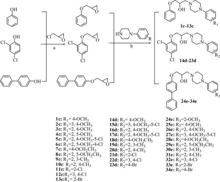
Chemical synthesis of piperazine compounds 1c‐34e.
A. NaOH, 0–60°C.
B. 2‐propanol, reflux.
Piperazine derivatives inhibit hyphae formation in C. albicans
Yeast‐to‐hyphae transition is essential and required for C. albicans to infect human mucosal tissue (Lo et al., 1997; Saville et al., 2003; Sudbery et al., 2004; Finkel and Mitchell, 2011). The effects of piperazine derivatives on C. albicans morphological transition were then evaluated in vitro under hyphae induction conditions at 37°C. After induction for 4 h, the majority of C. albicans cells formed germ tubes in the control group, while hyphae formation was obviously inhibited with addition of many piperazine derivatives (Fig. 2). At least 16 compounds reduced hyphae formation in C. albicans cells by more than 50% when they were supplemented at a final concentration of 100 μM (Fig. 2).
Figure 2.
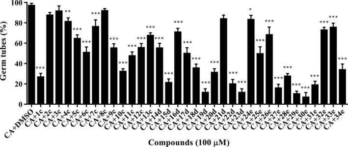
Effects of piperazine derivatives on Candida albicans hyphae formation. Each experiment was performed at least three times in triplicate, and each time at least 400 cells were counted for per treatment. CA: C. albicans. Compounds were dissolved in DMSO, and the same amount of DMSO as used as the solvent for compounds was used as control. Data are the mean ± standard deviation of three independent experiments. *P < 0.05; **P < 0.01; ***P < 0.001 (unpaired t test).
Piperazine derivatives inhibit C. albicans biofilm formation
Biofilm formation by fungal pathogens on implanted medical devices is a serious medical problem (Ganguly and Mitchell, 2011). Cell adhesion and biofilm formation by C. albicans cells on host surfaces are also the first step in establishing infection. As piperazine derivatives showed excellent efficacy in preventing C. albicans hyphae formation, we then evaluated their inhibitory activity on C. albicans cell adhesion and biofilm formation on polystyrene. Among the 34 derivatives, 15 compounds strongly inhibited C. albicans biofilm formation by more than 70% (Fig. 3). Surprisingly, compounds 17d and 19d exhibited notably high suppression of C. albicans biofilm formation by more than 85% when they were supplemented at a final concentration of 100 μM (Fig. 3), suggesting piperazine derivatives are excellent anti‐biofilm agents against C. albicans. In addition, it was further indicated that different substitutions have various anti‐biofilm activities. Phenol derivatives (1c‐13c) exerted weak inhibitory activity, 2,4‐dichlorophenol derivatives (14d‐23d) showed strong inhibition, and 4‐hydroxybiphenyl derivatives (24e‐34e) exhibited moderate activity (Fig. 3), suggesting that the introduction of a halogen group improves the anti‐biofilm activity of piperazine compounds. Additionally, substitution of the benzene ring clearly influenced the activity of these compounds. Derivatives with 2,3‐CH3, 2,4‐CH3, 3,4‐Cl, 4‐OCH2CH3, 2‐OCH3 and 4‐Br (19d, 23d, 24d, 28e, 30e, 31e, 32e) exhibited strong inhibitory activity towards biofilm formation by C. albicans (Fig. 3, Table S1). In addition, the inhibitory activity of piperazine derivatives during hyphae formation correlated with their inhibition of biofilm formation by C. albicans, possibly indicating the vital role of hyphae formation in the C. albicans biofilm formation process.
Figure 3.
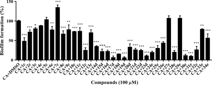
Effects of piperazine derivatives on Candida albicans biofilm formation. C. albicans cells were plated in the presence of compounds at a final concentration of 100 μM. CA: C. albicans. Compounds were dissolved in DMSO, and the same amount of DMSO as used as the solvent for compounds was used as control. C. albicans biofilm formed in the control group was arbitrarily defined as 100% and was used to normalize the biofilm of C. albicans formed with addition of compounds. Data are the mean ± standard deviation of three independent experiments. *P < 0.05; **P < 0.01; ***P < 0.001 (unpaired t test).
Piperazine derivatives attenuate C. albicans virulence
To determine the effects of piperazine derivatives on the pathogenicity of C. albicans, we then investigated whether these compounds influenced C. albicans virulence in a cell line. Cytotoxicity was measured by quantifying the release of lactate dehydrogenase (LDH) into the supernatants of cultured A549 cells. It was found that most derivatives showed no toxic effects on A549 cells at a final concentration of 100 μM (Fig. 4A). However, the exogenous addition of certain piperazine derivatives led to a reduction in C. albicans cytotoxicity to A549 cells (Fig. 4B). Although 2,4‐dichlorophenol derivatives exhibited strong inhibition of biofilm formation, their toxicity to the cell line was also very high (Fig. 4A). Compounds 1c, 2c, 8c and 28e were highly effective at attenuating C. albicans cytotoxicity by more than 80% when they were supplemented at a final concentration of 100 μM, while they exerted no toxic effects on the cell line at 8‐h postinoculation (Fig. 4A and B). Considering that compounds 2c and 8c only weakly inhibited hyphae formation in C. albicans, compounds 1c (1‐(4‐methoxyphenyl)‐4‐(3‐phenoxy‐2‐hydroxypropyl)‐piperazine) and 28e (1‐(4‐ethoxyphenyl)‐4‐(1‐biphenylol‐2‐hydroxypropyl)‐piperazine) were then selected for further investigation. Interestingly, compounds 1c and 28e also inhibited the virulence of other clinical isolated Candida species (Fig. S1). However, some commercialized piperazine compounds showed no inhibition on C. albicans virulence even when they were added at a final concentration of 400 μM (Fig. S2).
Figure 4.
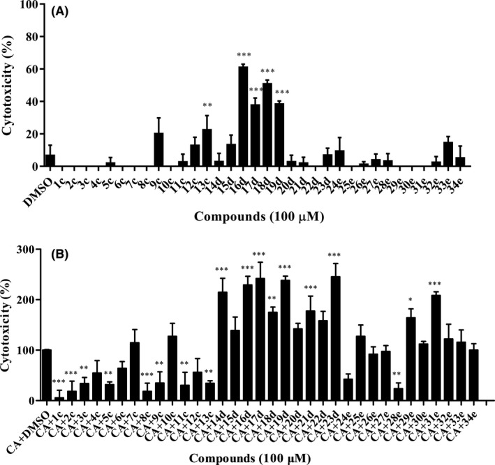
Effects of piperazine derivatives on Candida albicans virulence using a cell line.
A. Analysis of the toxicity of compounds to A549 cells. Compounds were dissolved in DMSO, and the same amount of DMSO as used as the solvent for compounds was used as control.
B. Analysis of the effect of compounds on the cytotoxicity of C. albicans to A549 cells. CA: C. albicans. Cell cytotoxicity was detected and measured as LDH release. The LDH released by A549 cells in (B) after inoculation with C. albicans in the absence of compounds was defined as 100% and used to normalize the LDH release ratios of other treatments. Data are the mean ± standard deviation of three independent experiments. *P < 0.05; **P < 0.01; ***P < 0.001 (unpaired t test).
The inhibitory activity of piperazine derivatives on C. albicans hyphae formation, biofilm formation and virulence is dose dependent
To determine whether the effects of piperazine derivatives on C. albicans are related to their dosage, different concentrations of compounds 1c and 28e were assessed for their inhibitory activity during C. albicans morphological transition, biofilm formation and virulence (Fig. 5A). We also examined the effects of different concentrations of fluconazole, which is always used for clinical treatment. Both compounds 1c and 28e exhibited dose‐dependent activity in which they reduced C. albicans cytotoxicity by more than 50% at a final concentration of 50 μM but did not affect the growth rate of pathogenic cells (Fig. 5B and C). As a control, fluconazole severely inhibited the growth rate of C. albicans cells, suggesting that compounds 1c and 28e might be good candidates for development as novel anti‐virulence agents against C. albicans infections. The addition of compounds 1c and 28e at final concentrations of 6.75–100 μM decreased C. albicans biofilm formation by 10% to 50% and 13% to 85%, respectively, but the addition of fluconazole had no detectable effect (Fig. 5D). Additionally, compounds 1c and 28e inhibited hyphae formation by 20% to 78% and 16% to 65%, respectively (Fig. 5E), while most C. albicans cells formed short stick mycelia that were considered hyphae after the addition of fluconazole.
Figure 5.
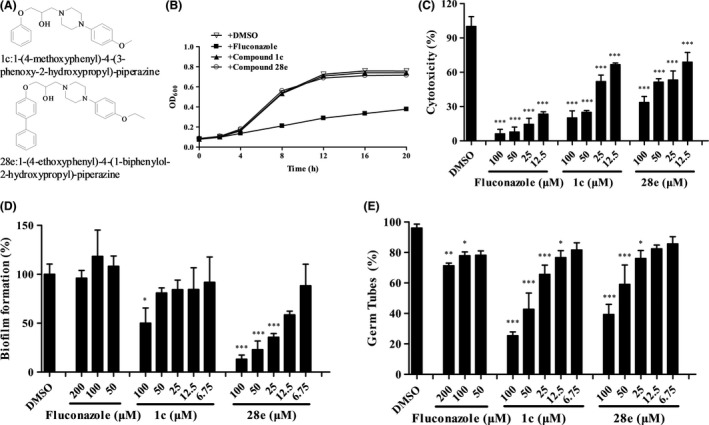
Effects of piperazine derivatives on C. albicans at different concentrations. (A) Structures of compounds 1c and 28e. (B) Effects of fluconazole and compounds 1c and 28e (100 μM) on the growth rate of C. albicans cells. The effects of different concentrations of fluconazole and compounds 1c and 28e on the virulence (C), biofilm formation (D) and hyphae formation (E) of C. albicans. Data are the mean ± standard deviation of three independent experiments. *P < 0.05; **P < 0.01; ***P < 0.001 (unpaired t test).
Piperazine derivatives interfere with the morphological transition of C. albicans through multiple pathways
Besides the statistics of hyphae formation per cent, the photograph of hyphae formation was also provided to reflect the effect of the candidate compounds. After adding compounds 1c and 28e, the count of hyphae declined obviously (Fig. 6A). In good agreement with the inhibition of hyphae formation, compound 28e also obviously affected the colony morphology of C. albicans (Fig. 6B). After addition of compound 28e, the colonies were changed from wrinkled to slippery (Fig. 6B). To explore the working model of piperazine derivatives on the morphological transition of C. albicans, we continued to test whether these compounds interfered with the signalling pathways of the hyphae development process (Sudbery et al., 2004; Lu et al., 2011; Shareck and Belhumeur, 2011; Nobile et al., 2012). Real‐time PCR analysis showed that exogenous addition of the leading compound 28e inhibited the expression levels of CDC35, EFG1, TEC1 and HWP1, which are regulators involved in the cAMP‐PKA pathways (Fig. 6C and D) (Sudbery, 2011). In addition, some regulators of the MAPK cascade (Zhao et al., 2013), such as HST7, CEK1 and CPH1, were also downregulated by the exogenous addition of compound 28e (Fig. 6C and D). Collectively, these results demonstrated that compound 28e influenced complex signal transduction pathways to interfere with the C. albicans filamentation process.
Figure 6.
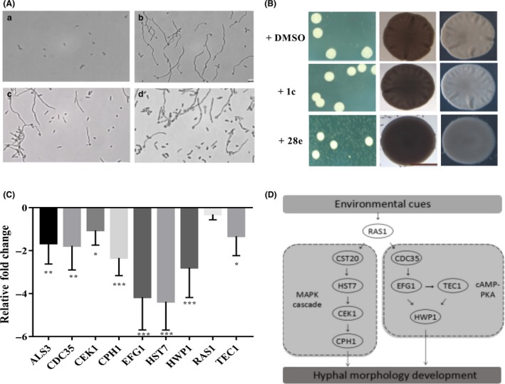
Influence of piperazine derivatives on Candida albicans morphology and hyphae formation.
A. Effects of compounds 1c and 28e on hyphae formation of C. albicans. C. albicans cells were grown under non‐induction conditions (30°C) (a), or under induction conditions (37°C) (b). In (c and d), the cells were grown under the same condition as in (b) but treated with 100 μM of compound 1c and compound 28e respectively. The photographs were taken 4 h after induction.
B. Effects of compounds 1c and 28e (100 μM) on colony morphology of C. albicans.
C. Comparison of relative fold changes of regulator encoding genes between C. albicans cells with and without addition of compound 28e. qRT‐PCR results were normalized using the C t values obtained for the GSP1 amplifications run in the same plate. The relative levels of gene transcripts were determined from standard curves. Data are the mean ± standard deviation of three independent experiments. *P < 0.05; **P < 0.01; ***P < 0.001 (unpaired t test).
D. Schematic diagram of the signalling pathways that govern hyphal morphogenesis in C. albicans affected by compound 28e.
Discussion
Our findings demonstrated for the first time that (1‐aryloxy‐2‐hydroxypropyl)‐phenylpiperazine compounds could be developed as potential antifungal agents to attenuate C. albicans virulence by targeting the filamentation process. The yeast‐to‐hyphae transition is always thought to be an indispensable step for invasion, and C. albicans mutants defective in morphological transitions are avirulent during infection (Braun et al., 2000; Saville et al., 2003; Zheng et al., 2003). The current clinical treatments against candidiasis caused by C. albicans almost always rely on limited antifungal agents, which are compromised by the limitation of drug development and the rapid emergence of drug‐resistant pathogens. Developing new strategies to treat fungal pathogens is a key research area for these reasons. As morphological transitions between yeast cells and filamentous forms play a vital role in pathogenesis, recent studies have focused on determining how to inhibit hyphae formation in fungal pathogens. Interestingly, some cell–cell communication signals, including farnesol, a quorum sensing signal produced by C. albicans and a BDSF quorum sensing signal from Burkholderia cenocepacia, show an excellent ability to inhibit C. albicans hyphae formation (Boon et al., 2008; Shirtliff et al., 2009). Our study here provided additional evidence that designing antifungal drugs using a function‐based approach that relies on the inhibition of hyphae formation but not the direct killing of pathogens is a feasible strategy.
Biofilms formed by C. albicans cells usually increase resistance to commonly used antifungal drugs, such as azoles and amphotericin B (Walker et al., 2010; Fiori et al., 2011). It is important to develop new antifungal drugs that efficiently control biofilm formation. Our results indicated that some piperazine derivatives showed a strong ability to inhibit C. albicans biofilm formation (Fig. 3). This ability was also consistent with their inhibition of hyphae formation (Fig. 2). As hyphae formation plays a vital role in C. albicans biofilm formation (Lu et al., 2014; Gulati and Nobile, 2016), the strategies to target morphological transition and biofilm development may be closely interlinked.
Piperazine is an important scaffold associated with several biological activities (Shaquiquzzaman et al., 2015; Singh et al., 2015). Many synthetic piperazine derivatives have been reported to exhibit promising antifungal activity by causing the death of fungal cells (Moraca et al., 2014; Thamban et al., 2017). However, our study verified for the first time that (1‐aryloxy‐2‐hydroxypropyl)‐phenylpiperazine scaffolds could be developed as antifungal agents against C. albicans using an anti‐virulence strategy. Another interesting characteristic of piperazine derivatives is that substitution on the benzene ring notably influences their activity (Figs 1, 2, 3, 4). We found that 2,4‐dichlorophenol derivatives showed strong anti‐biofilm activity but exhibited high toxicity against cell lines (Figs 3 and 4). Conversely, 4‐hydroxybiphenyl derivatives had high anti‐biofilm activity and low cytotoxicity, suggesting that they are appropriate candidates for further development. The novel compound 28e containing 4‐hydroxybiphenyl and the 4‐OCH2CH3 moiety might be a promising potential agent against C. albicans and other clinical isolated Candida species given its excellent efficacy (Figs 5 and 6, S1). In general, our findings demonstrated that the high efficacy of (1‐aryloxy‐2‐hydroxypropyl)‐phenylpiperazine compounds in terms of both hyphae formation and biofilm formation by C. albicans increases their advantages for their development as new antifungal drugs.
Experimental procedures
Chemistry
1H NMR (500 MHz) spectra were recorded in CDCl3 or DMSO on a Bruker Avance spectrometer using tetramethylsilane (TMS) as an internal standard. Electrospray ionization (ESI) mass spectra (electron ionization (EI), 70 eV) were recorded on an Agilent 6330 ion trap LC/MS system. HRESI‐MS spectra were recorded using a Shimadzu LCMS‐IT‐TOF mass spectrometer. Thin‐layer chromatography (TLC) was performed on an aluminium plate precoated with silica gel and a fluorescence indicator (Merck, Darmstadt, Germany). The compounds were detected on the TLC plates using UV light (254 nm). All other reagents and chemicals were obtained from commercial sources and used as received unless otherwise stated.
Strains, culture and agents
The C. albicans strain used in this study was the standard wild‐type strain SC5314 (ATCC® MYA‐2876TM), which was grown either in 6.7 g l−1 yeast nitrogen broth without amino acids (YNB) with 2% glucose or in YPD medium (1% yeast extract, 2% peptone and 2% dextrose) at 30°C (non‐hyphae induction conditions) or 37°C (hyphae induction conditions). Human lung epithelial A549 cells were incubated in Dulbecco's modified Eagle's medium (DMEM) containing 10% fetal bovine serum (FBS).
Biofilm formation assays
Candida albicans SC5314 strain was cultured overnight in YNB + 2% glucose media at 30°C and inoculated in the same medium to an OD600 of 0.5 in the presence of compounds at different concentrations, as indicated. One hundred microlitres of inoculated culture was grown in each well of a 96‐well polystyrene plate at 37°C for 4 h without shaking. The cultures were removed, and 0.02% crystal violet was added for 45 min. The plate was rinsed six to eight times with iced distilled water and decolorized with 200 μl of 75% ethanol. The quantity of crystal violet was determined by measuring the absorbance at 590 nm.
Hyphal formation assays
An overnight culture of C. albicans SC5314 grown at 30°C was diluted in fresh YNB + 2% glucose to an OD600 of 0.1. Compounds were then added separately as indicated, and the cells were induced for 6 h at 37°C. Cells were centrifuged for 10 min at 5000 rpm, and the pellet was resuspended in 100 μl of fresh medium. Images of cells were captured using a Leica inverted fluorescence microscope with 20× magnification.
Colony morphology
Spider medium agar plates (1% peptone, 1% mannitol, 0.2% K2HPO4 and 1.5% agar) were supplemented with different concentrations of compounds as indicated. C. albicans SC5314 cells were grown in the plates at 37°C for 24–30 h. Images of the colonies were obtained using a Leica DMi8 microscope and a Nikon Coolpix digital camera.
Cell growth analysis
For the cell growth assay, C. albicans cells were cultured in YNB + 0.2% glucose and inoculated in the same medium to an OD600 of 0.05 in the absence or presence of compounds at final concentrations as indicated. Three hundred microlitres of inoculated culture was grown in each well at 30°C in a low‐intensity shaking model using a Bioscreen‐C Automated Growth Curves Analysis System (OY Growth Curves AB, Finland).
Cytotoxicity assays
Cytotoxicity was assessed by measuring the release of LDH from A549 cells. The A549 cells were routinely grown in DMEM medium supplemented with 10% FBS in a 96‐well tissue culture plate with 1.5 × 10⁴ cells/well. Confluent A549 cells were washed and incubated with DMEM containing 1% FBS before infection. Overnight C. albicans cells were diluted to an OD600 of 0.1 with DMEM containing 1% FBS in the absence or presence of compounds at a final concentration as indicated. A549 cells were infected with fungal cells for 8 h. LDH in the supernatant was measured, and cytotoxicity was calculated relative to that of the uninfected control. Different strains and compounds were tested as the same way.
Quantitative real‐time PCR
Real‐time PCR assays were conducted as described previously (Li et al., 2013, 2014). Overnight cultures of C. albicans cells grown in YNB + 0.2% glucose at 30°C were inoculated in the same medium to an OD600 of 0.1 in the absence or presence of compounds at a final concentration of 100 μM. After incubation for 6 h at 37°C, the cell samples were collected and washed with PBS. Total RNA was extracted using TRIzol (InVitrogen, Carlsbad, California, America) and quantified. cDNA was obtained through a reverse transcription reaction using a reverse transcription kit (TaKaRa Biotechnology) with the primers shown in Table S2, and real‐time PCR was performed with a 7300 plus real‐time PCR system (Applied Biosystems, America). The expression level of each gene was normalized to that of GSP1, which is a housekeeping gene in C. albicans cells (Clément et al., 2000). The relative expression levels of the target genes were calculated using the quantitation‐comparative CT(ΔΔC t) method.
Conflict of interests
The authors declare no conflict of interests.
Supporting information
Table S1. Analysis of chemical structures of piperazine derivatives.
Table S2. PCR primers used in this study.
Fig. S1. Effects of piperazine derivatives on different clinic isolated C. albicans strains and other Candida species virulence using a cell line.
Fig. S2. Influences of different piperazine compounds on C. albicans SC5314 virulence using a cell line.
Acknowledgements
This work was supported financially by grants from the Guangdong Natural Science Funds for Distinguished Young Scholars (No. 2014A030306015), the National Key Project for Basic Research of China (973 Project, 2015CB150600), the National Natural Science Foundation of China (No. 31571969) and the Introduction of Innovative R&D Team Program of Guangdong Province (No. 2013S034), China.
Microbial Biotechnology (2018) 11(6), 1080–1089
Funding Information
This work was supported financially by grants from the Guangdong Natural Science Funds for Distinguished Young Scholars (No. 2014A030306015), the National Key Project for Basic Research of China (973 Project, 2015CB150600), the National Natural Science Foundation of China (No. 31571969) and the Introduction of Innovative R&D Team Program of Guangdong Province (No. 2013S034), China.
Contributor Information
Fei He, Email: hefei@scsio.ac.cn.
Yinyue Deng, Email: yinyue_deng@hotmail.com.
References
- Boon, C. , Deng, Y. , Wang, L.‐H. , He, Y. , Xu, J.‐L. , Fan, Y. , et al (2008) A novel DSF‐like signal from Burkholderia cenocepacia interferes with Candida albicans morphological transition. ISME J 2: 27–36. [DOI] [PubMed] [Google Scholar]
- Braun, B.R. , Head, W.S. , Wang, M.X. , and Johnson, A.D. (2000) Identification and characterization of TUP1‐regulated genes in Candida albicans . Genetics 156: 31–44. [DOI] [PMC free article] [PubMed] [Google Scholar]
- Brown, A.J. , and Gow, N.A. (1999) Regulatory networks controlling Candida albicans morphogenesis. Trends Microbiol 7: 333–338. 10.1016/S0966-842X(99)01556-5. [DOI] [PubMed] [Google Scholar]
- Chandra, J. , Mukherjee, P.K. , Leidich, S.D. , Faddoul, F.F. , Hoyer, L.L. , Douglas, L.J. , and Ghannoum, M.A. (2001) Antifungal resistance of candidal biofilms formed on denture acrylic in vitro. J Dent Res 80: 903–908. [DOI] [PubMed] [Google Scholar]
- Clément, M. , Fournier, H. , Repentigny, L. , and Belhumeur, P. (2000) Characterization of CaGSP1, the Candida albicans RAN/GSP1 homologue. Gene 250: 159–169. [DOI] [PubMed] [Google Scholar]
- Deng, Y. , Wu, J. , Eberl, L. , and Zhang, L.‐H. (2010) Structural and functional characterization of diffusible signal factor family quorum‐sensing signals produced by members of the Burkholderia cepacia complex. Appl Environ Microbiol 76: 4675–4683. [DOI] [PMC free article] [PubMed] [Google Scholar]
- Fazly, A. , Jain, C. , Dehner, A.C. , Issi, L. , Lilly, E.A. , Ali, A. , et al (2013) Chemical screening identifies filastatin, a small molecule inhibitor of Candida albicans adhesion, morphogenesis, and pathogenesis. Proc Natl Acad Sci USA 110: 13594–13599. [DOI] [PMC free article] [PubMed] [Google Scholar]
- Finkel, J.S. , and Mitchell, A.P. (2011) Genetic control of Candida albicans biofilm development. Nat Rev Microbiol 9: 109–118. [DOI] [PMC free article] [PubMed] [Google Scholar]
- Fiori, B. , Posteraro, B. , Torelli, R. , Tumbarello, M. , Perlin, D.S. , Fadda, G. , and Sanguinetti, M. (2011) In vitro activities of anidulafungin and other antifungal agents against biofilms formed by clinical isolates of different Candida and Aspergillus species. Antimicrob Agents Chemother 55: 3031–3035. [DOI] [PMC free article] [PubMed] [Google Scholar]
- Ganguly, S. , and Mitchell, A.P. (2011) Mucosal biofilms of Candida albicans . Curr Opin Microbiol 14: 380–385. 10.1016/j.mib.2011.06.001. [DOI] [PMC free article] [PubMed] [Google Scholar]
- Gettys, K. , Ye, Z. , and Dai, M. (2017) Recent advances in Piperazine synthesis. Synthesis 49: 2589–2604. [Google Scholar]
- Gulati, M. , and Nobile, C.J. (2016) Candida albicans biofilms: development, regulation, and molecular mechanisms. Microbes Infect 18: 310–321. 10.1016/j.micinf.2016.01.002. [DOI] [PMC free article] [PubMed] [Google Scholar]
- Huang, J.‐J. , Huang, Y.‐J. , Zhu, L. , Yuan, M. , and Huang, L. (2014) Design, synthesis and alpha1‐adrenoreceptor blocking activity of new arylpiperazines containing acetophenone substituents. Pharmazie 69: 578–584. [PubMed] [Google Scholar]
- Huang, J.‐J. , He, F. , Huang, M. , Liu, X. , Xiong, Y. , Huang, Y. , et al (2015) Novel naftopidil‐related derivatives and their biological effects as alpha1‐adrenoceptors antagonists and antiproliferative agents. Eur J Med Chem 96: 83–91. 10.1016/j.ejmech.2015.04.005. [DOI] [PubMed] [Google Scholar]
- Huang, J.‐J. , Cai, Y. , Huang, M.‐Y. , Zhu, L. , He, F. , Liu, X.‐W. , et al (2016) Pharmaceutical evaluation of naftopidil enantiomers: rat functional assays in vitro and estrogen/androgen induced rat benign prostatic hyperplasia model in vivo. Eur J Pharmacol 791: 473–481. 10.1016/j.ejphar.2016.09.009. [DOI] [PubMed] [Google Scholar]
- Kim, S. , Kim, E. , Shin, D.S. , Kang, H. , and Oh, K.B. (2002) Evaluation of morphogenic regulatory activity of farnesoic acid and its derivatives against Candida albicans dimorphism. Bioorg Med Chem Lett 12: 895–898. 10.1016/S0960-894X(02)00038-0. [DOI] [PubMed] [Google Scholar]
- Li, D.‐D. , Wang, Y. , Dai, B.‐D. , Li, X.‐X. , Zhao, L.‐X. , Cao, Y.B. , et al (2013) ECM17‐dependent methionine/cysteine biosynthesis contributes to biofilm formation in Candida albicans . Fungal Genet Biol 51: 50–59. [DOI] [PubMed] [Google Scholar]
- Li, D.‐D. , Zhao, L.‐X. , Mylonakis, E. , Hu, G.‐H. , Zou, Y. , Huang, T.‐K. , et al (2014) In vitro and in vivo activities of pterostilbene against Candida albicans biofilms. Antimicrob Agents Chemother 58: 2344–2355. [DOI] [PMC free article] [PubMed] [Google Scholar]
- Lo, H.J. , Köhler, J.R. , DiDomenico, B. , Loebenberg, D. , Cacciapuoti, A. , and Fink, G.R. (1997) Nonfilamentous Candida albicans mutants are avirulent. Cell 90: 939–949. 10.1016/S0092-8674(00)80358-X. [DOI] [PubMed] [Google Scholar]
- Lu, Y. , Su, C. , Wang, A. , and Liu, H. (2011) Hyphal development in Candida albicans requires two temporally linked changes in promoter chromatin for initiation and maintenance. PLoS Biol 9: e1001105 10.1371/journal.pbio.1001105. [DOI] [PMC free article] [PubMed] [Google Scholar]
- Lu, Y. , Su, C. , and Liu, H.‐P. (2014) Candida albicans hyphal initiation and elongation. Trends Microbiol 22: 707–714. 10.1016/j.tim.2014.09.001. [DOI] [PMC free article] [PubMed] [Google Scholar]
- Madhani, H.D. , and Fink, G.R. (1998) The control of filamentous differentiation and virulence in fungi. Trends Cell Biol 8: 348–353. 10.1016/S0962-8924(98)01298-7. [DOI] [PubMed] [Google Scholar]
- McGeady, P. , Wansley, D.L. , and Logan, D.A. (2002) Carvone and perillaldehyde interfere with the serum‐induced formation of filamentous structures in Candida albicans at substantially lower concentrations than those causing significant inhibition of growth. J Nat Prod 65: 953–955. [DOI] [PubMed] [Google Scholar]
- Miller, L.G. , Hajjeh, R.A. , and Edwards, J.E. Jr (2001) Estimating the cost of nosocomial candidemia in the United States. Clin Infect Dis 32: 1110 10.1086/319613. [DOI] [PubMed] [Google Scholar]
- Moraca, F. , Vita, D.D. , Pandolfi, F. , Santo, R.D. , Costi, R. , Cirilli, R. , et al (2014) Synthesis, biological evaluation and structure–activity correlation study of a series of imidazol‐based compounds as Candida albicans inhibitors. Eur J Med Chem 83: 665–673. 10.1016/j.ejmech.2014.07.001. [DOI] [PubMed] [Google Scholar]
- Nobile, C.J. , Fox, E.P. , Nett, J.E. , Sorrells, T.R. , Mitrovich, Q.M. , Hernday, A.D. , et al (2012) A recently evolved transcriptional network controls biofilm development in Candida albicans . Cell 148: 126–138. 10.1016/j.cell.2011.10.048. [DOI] [PMC free article] [PubMed] [Google Scholar]
- Noble, S.M. , Gianetti, B.A. , and Witchley, J.N. (2017) Candida albicans cell‐type switching and functional plasticity in the mammalian host. Nat Rev Microbiol 15: 96–108. [DOI] [PMC free article] [PubMed] [Google Scholar]
- Odds, F.C. , Brown, A.J. , and Gow, N.A. (2003) Antifungal agents: mechanisms of action. Trends Microbiol 11: 272–279. 10.1016/S0966-842X(03)00117-3. [DOI] [PubMed] [Google Scholar]
- Oh, K.B. , Miyazawa, H. , Naito, T. , and Matsuoka, H. (2001) Purification and characterization of an autoregulatory substance capable of regulating the morphological transition in Candida albicans . Proc Natl Acad Sci USA 98: 4664–4668. [DOI] [PMC free article] [PubMed] [Google Scholar]
- Pappas, P.G. , Rex, J.H. , Lee, J. , Hamill, R.J. , Larsen, R.A. , Powderly, W. , et al (2003) A prospective observational study of candidemia: epidemiology, therapy, and influences on mortality in hospitalized adult and pediatric patients. Clin Infect Dis 37: 634–643. 10.1086/376906. [DOI] [PubMed] [Google Scholar]
- Pierce, C.G. , Srinivasan, A. , Uppuluri, P. , Ramasubramanian, A.K. , and Lopez‐Ribot, J.L. (2013) Antifungal therapy with an emphasis on biofilms. Curr Opin Pharmacol 13: 726–730. 10.1016/j.coph.2013.08.008. [DOI] [PMC free article] [PubMed] [Google Scholar]
- Pollard, C. , and Christie, J.B. (1958) Derivatives of Piperazine. Reactions of 1‐Arylpiperazines with epoxides. J Org Chem 23: 1333–1335. [Google Scholar]
- Poulain, D. (2015) Candida albicans, plasticity and pathogenesis. Crit Rev Microbiol 41: 208–217. [DOI] [PubMed] [Google Scholar]
- Ramage, G. , Saville, S.P. , Wickes, B.L. , and Lopez‐Ribot, J.L. (2002) Inhibition of Candida albicans biofilm formation by farnesol, a quorum‐sensing molecule. Appl Environ Microbiol 68: 5459–5463. [DOI] [PMC free article] [PubMed] [Google Scholar]
- Saville, S.P. , Lazzell, A.L. , Monteagudo, C. , and Lopez‐Ribot, J.L. (2003) Engineered control of cell morphology in vivo reveals distinct roles for yeast and filamentous forms of Candida albicans during infection. Eukaryot Cell 2: 1053–1060. [DOI] [PMC free article] [PubMed] [Google Scholar]
- Shaquiquzzaman, M. , Verma, G. , Marella, A. , Akhter, M. , Akhtar, W. , Khan, M.F. , et al (2015) Piperazine scaffold: a remarkable tool in generation of diverse pharmacological agents. Eur J Med Chem 102: 487–529. 10.1016/j.ejmech.2015.07.026. [DOI] [PubMed] [Google Scholar]
- Shareck, J. , and Belhumeur, P. (2011) Modulation of morphogenesis in Candida albicans by various small molecules. Eukaryot Cell 10: 1004–1012. [DOI] [PMC free article] [PubMed] [Google Scholar]
- Shirtliff, M.E. , Krom, B.P. , Meijering, R.A. , Peters, B.M. , Zhu, J. , Scheper, M.A. , et al (2009) Farnesol‐induced apoptosis in Candida albicans . Antimicrob Agents Chemother 53: 2392–2401. [DOI] [PMC free article] [PubMed] [Google Scholar]
- Singh, K. , Siddiqui, H.H. , Shakya, P. , Bagga, P. , and Kumar, A. (2015) Piperazine – a biologically active scaffold. Int J Pharm Sci Res 6: 4145–4158. [Google Scholar]
- Staib, P. , Kretschmar, M. , Nichterlein, T. , Hof, H. , and Morschhäuser, J. (2000) Differential activation of a Candida albicans virulence gene family during infection. Proc Natl Acad Sci USA 97: 6102–6107. [DOI] [PMC free article] [PubMed] [Google Scholar]
- Sudbery, P.E. (2011) Growth of Candida albicans hyphae. Nat Rev Microbiol 9: 737–748. [DOI] [PubMed] [Google Scholar]
- Sudbery, P. , Gow, N. , and Berman, J. (2004) The distinct morphogenic states of Candida albicans . Trends Microbiol 12: 317–324. 10.1016/j.tim.2004.05.008. [DOI] [PubMed] [Google Scholar]
- Thamban, C.N. , Shrestha, S.K. , Ngo, H.X. , Tsodikov, O.V. , Howard, K.C. , and Garneau‐Tsodikova, S. (2017) Alkylated piperazines and piperazine‐azole hybrids as antifungal agents. J Med Chem. 10.1021/acs.jmedchem.7b01138. [DOI] [PMC free article] [PubMed] [Google Scholar]
- Tobudic, S. , Lassnigg, A. , Kratzer, C. , Graninger, W. , and Presterl, E. (2010) Antifungal activity of amphotericin B, caspofungin and posaconazole on Candida albicans biofilms in intermediate and mature development phases. Mycoses 53: 208–214. [DOI] [PubMed] [Google Scholar]
- Walker, L.A. , Gow, N.A.R. , and Munro, C.A. (2010) Fungal echinocandin resistance. Fungal Genet Biol 47: 117–126. 10.1016/j.fgb.2009.09.003. [DOI] [PMC free article] [PubMed] [Google Scholar]
- Zhao, L.‐X. , Li, D.‐D. , Hu, D.‐D. , Hu, G.‐H. , Yan, L. , Wang, Y. , and Jiang, Y.‐Y. (2013) Effect of tetrandrine against Candida albicans biofilms. PLoS ONE 8: e79671. [DOI] [PMC free article] [PubMed] [Google Scholar]
- Zheng, X.‐D. , Wang, Y.‐M. , and Wang, Y. (2003) CaSPA2 is important for polarity establishment and maintenance in Candida albicans . Mol Microbiol 49: 1391–1405. [DOI] [PubMed] [Google Scholar]
Associated Data
This section collects any data citations, data availability statements, or supplementary materials included in this article.
Supplementary Materials
Table S1. Analysis of chemical structures of piperazine derivatives.
Table S2. PCR primers used in this study.
Fig. S1. Effects of piperazine derivatives on different clinic isolated C. albicans strains and other Candida species virulence using a cell line.
Fig. S2. Influences of different piperazine compounds on C. albicans SC5314 virulence using a cell line.