Soon after Mary Hatten became the first female professor at New York City’s Rockefeller University in 1992, she gave a talk to the university’s board of directors about her life’s work on neuron migration during brain development. David Rockefeller, the university’s benefactor and honorary chairman, was there and, after listening attentively, asked, “But why would the young neuron want to leave the place where it was born?”
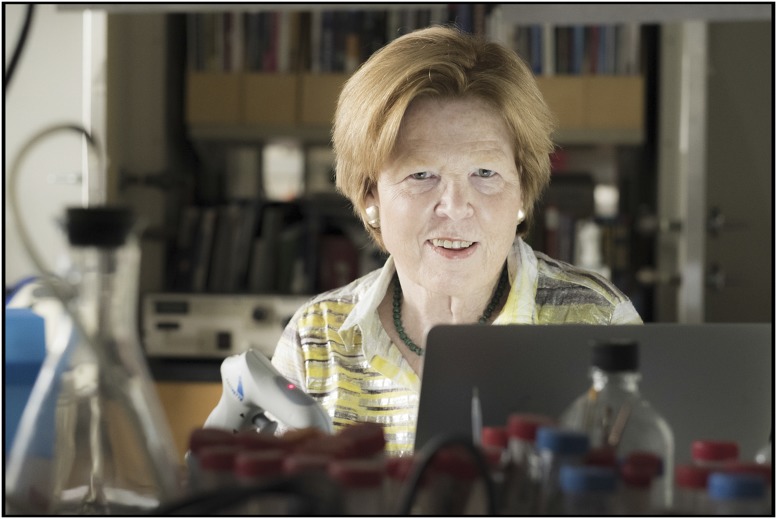
Mary E. Hatten. Image courtesy of Mario Morgado (photographer).
More than 25 years later, Hatten is working toward a complete answer. The Frederick P. Rose professor and head of the laboratory of developmental neurobiology at Rockefeller has spent her career on a methodical search for clues.
Hatten’s Inaugural Article (1) furnishes one piece of the puzzle by solving a mystery about how an adhesion protein binds to glial cells to allow neurons to pull themselves along during central nervous system development.
Throughout Hatten’s career, she has earned accolades and awards for her often-groundbreaking work. She received the McKnight Endowment Fund for Neuroscience Investigator Award, the Javits Neuroscience Investigator Award, the Cowan-Cajal Award for outstanding work in developmental neuroscience, and the Ralph W. Gerard Prize of the Society for Neuroscience for lifetime achievement in neuroscience. Hatten was elected to the US National Academy of Sciences in 2017.
Early Fascination with Science
The 2016 film Hidden Figures brings back memories for Hatten. She grew up in Newport News, Virginia, close to NASA’s Langley Research Center, where much of the movie’s plot unfolds. In high school and college Hatten conducted her own research at Langley during the summers. “I was the only girl,” she says. “And it was exactly like it was portrayed in the movie.”
“I loved what I was doing and so just did it, somewhat oblivious to the challenges,” she recalls. “Certainly it’s been frustrating at times. There was a lot of bias in science and still is. On the other hand, I was really lucky to have the opportunities I’ve had. It never occurred to me that I couldn’t be part of this world, so I always tried.”
It was Hatten’s self-driven fascination with science and her mother’s unflinching support that encouraged her as a child. Her father was an obstetrician, and her mother indulged her interest in science, taking her to science events and allowing her to conduct experiments at home, including some precocious studies on bacteria that involved using rabbits to generate antibodies.
For her summer job at NASA, Hatten designed a study to examine how gravity affects bacterial growth. Later, the experiment was sent into orbit with one of the Apollo moon missions. “We learned that gravity changed the shape of the bacterial colony, but did not change the rate of growth of the bacteria,” says Hatten. It was the beginning of a lifelong interest in how cellular interactions influence development.
Hatten attended a small women’s college in Roanoke, Virginia, called Hollins College. It was one of the few options for women in the 1960s and it was filled with inspiring women.
“I took the long view, knowing I would go on to graduate school,” recalls Hatten, who majored in chemistry. “I finished my chemistry requirements quickly and then took a lot of philosophy, literature, and art. It enriched me and made me a good writer, which has made me a better scientist.”
Hatten was graduated in 1971 and moved to Princeton University to study with biochemist Max Burger. When Burger moved to the University of Basel, Switzerland, Hatten moved with him to finish her research on the biophysics of cancer cell membranes. “I worked out methods to change the lipid composition of cells, which in turn changed the fluid properties of the membrane in the domains around receptors for plant lectins,” says Hatten. “Working on cancer cell membrane properties is what got me interested in metastasis and cell migration.”
The experience and her interest led her to Harvard Medical School for postdoctoral research in neuroscience with Richard Sidman, who was studying cell migration in the brain. Hatten’s work focused on developing a cell culture system for studying the cerebellum. She found the cerebellum and its abundant granule cells to be ideal for exploring nerve cell migration in cortical regions of the developing brain.
Working on cerebellar granule cells formed the foundation of Hatten’s career, which has helped piece together how brain development is orchestrated in time and space. “Every single neuron in the brain undergoes these long migrations equivalent to me walking from New York City to Chicago,” explains Hatten. “The histogenesis of the cortex is like making a six-story building: first you get all the floors in place, and then all the circuitry can form. I think the dynamics are amazing. And the fact that it works is magnificent.”
Finding Her Groove in New York
In 1978, after Hatten’s then-husband relocated to New York City, Michael Shelanski offered her a spot in the pharmacology department at New York University School of Medicine. At New York University, Hatten’s colleagues helped her develop skills to study neural migration in earnest. Pathologist Ronald Liem developed antibodies that allowed her to visualize glia and neurons in culture and measure their interactions. Neuroanatomist Carol Mason taught her neuroanatomy and collaborated with her on early migration studies, and biophysicist Fred Maxfield helped her produce high-resolution live images of migrating granule cells. “Fred and I were the first to get digital recorders, as opposed to tape recording,” recalls Hatten. “The fidelity was so high we could see the process in incredible detail.”
The live imaging allowed her and graduate student Jim Edmondson to prove that, during development, neurons migrate along glial fibers (2). Their findings corroborated classic work by neuroscientist Pasko Rakic that had been performed using electron microscopy. “We were able to see all the little details that Rakic had proposed about migration from static electron microscopy,” says Hatten. “We were the first to actually see it happen in real time.”
In 1986, Hatten and her group moved to Columbia University College of Physicians and Surgeons to join a newly formed neuroscience program. There, Hatten developed methods to purify granule cells, recombine them, and carry out biochemical experiments to understand how migration worked at the genetic and molecular levels. She also used the purification method to create the first in vitro chimeras, which solved the mystery of the site of action of the weaver gene in neuron migration (3).
While looking for receptors that guided neuron movement along glial fibers, Hatten and her colleagues found a protein they named astrotactin (4). Further studies confirmed that astrotactin acts as a neuron–glial adhesion protein, allowing migrating neurons to move along glial fibers.
As Hatten’s work evolved, she realized she needed molecular biology skills. Hatten began to collaborate with molecular biologist Nathaniel Heintz at Rockefeller to develop a cDNA library for granule cells (5). In 1992, Rockefeller president Torsten Weisel recruited Hatten to become the university’s first female professor and laboratory head. The chance to work with Heintz and his colleagues proved irresistible.
Hatten collaborated with Heintz on the Gene Expression Nervous System Atlas (6) and work that resulted in the cloning of 80 genes active during cerebellar development, including the gene for astrotactin, Astn1 (7). Further work showed that the Astn1 gene is important for neuron migration and is expressed by neurons migrating along glial fibers in the cerebellum and the cerebral cortex (7), as well as for cytoskeletal dynamics during migration (8).
Hatten’s Inaugural Article (1) answers a question that has plagued the field since she discovered astrotactin in 1988. Researchers knew that migrating neurons use astrotactin to stick to glial fibers as they pull themselves along. But the receptor on the glial fiber where the protein adheres remained unknown. One candidate was N-cadherin, a protein known to promote cell adhesion. Hatten and her colleagues examined whether receptors in the membrane interact with each other, not just from one cell to another, but on the same side of the cell. “What we discovered was that there was this asymmetric bridge where astrotactin is binding to N-cadherin on the neuron side and making a bridge complex across to N-cadherin on the glial fiber.”
More recently, Hatten has begun work on stem cells to broaden her understanding of neural development. Her group has been trying to determine how neural stem cells turn into different types of neurons. The work has advanced in mice (9), but human studies have proved challenging. “I’m learning how incredibly different human cells are,” says Hatten, who is attempting to generate cerebellar neurons from stem cells (10).
Stepping Outside the Box
Hatten’s obsession for basic science means that finding cures for human diseases is not the driving force behind her research. However, she is gratified that her work sheds light on some human disorders, including medulloblastoma and autism. Medulloblastoma, a common malignant tumor in children, arises when granule cells in the cerebellum grow out of control. Hatten has collaborated with Martine Roussel at St. Jude’s Children’s Hospital in Tennessee to look for genes that might be involved in the disease (11). Hatten’s group has also linked a member of the astrotactin gene family to autism and intellectual and language disabilities. Hatten hopes to learn exactly how Astn2 contributes to neurodevelopmental disorders.
Hatten is stimulated by unexpected findings, and a 2016 study (12) turned out to be one example. She determined the gene-expression profiles of cerebellar granule cells at different stages of development: during proliferation, migration, and postmigration, when they form circuitry. Hatten’s team discovered that, after migration, while the circuitry was forming, virtually all of the chromatin-modifying genes changed dramatically. If the team knocked out some of those genes, the neurons kept migrating and did not form dendrites. “This finding suggests that a key role of migration is to set the timing of gene-expression/chromatin changes that underlie the formation of brain circuitry,” says Hatten. “We were so busy looking for neural pathways, and all of a sudden the bioinformatics shows that what’s really changing is chromatin.”
The latter finding was not only surprising but, Hatten believes, gets her closer than ever to answering David Rockefeller’s question about why neurons migrate. Over the next several years, she plans to continue examining the mechanisms of neuronal migration in the central nervous system, particularly the role of chromatin changes.
Footnotes
This is a Profile of a member of the National Academy of Sciences to accompany the member’s Inaugural Article on page 10556.
References
- 1.Horn Z, Behesti H, Hatten ME. N-cadherin provides a cis and trans ligand for astrotactin that functions in glial-guided neuronal migration. Proc Natl Acad Sci USA. 2018;115:10556–10563. doi: 10.1073/pnas.1811100115. [DOI] [PMC free article] [PubMed] [Google Scholar]
- 2.Edmondson JC, Hatten ME. Glial-guided granule neuron migration in vitro: A high-resolution time-lapse video microscopic study. J Neurosci. 1987;7:1928–1934. doi: 10.1523/JNEUROSCI.07-06-01928.1987. [DOI] [PMC free article] [PubMed] [Google Scholar]
- 3.Gao WQ, Liu XL, Hatten ME. The weaver gene encodes a nonautonomous signal for CNS neuronal differentiation. Cell. 1992;68:841–854. doi: 10.1016/0092-8674(92)90028-b. [DOI] [PubMed] [Google Scholar]
- 4.Edmondson JC, Liem RK, Kuster JE, Hatten ME. Astrotactin: A novel neuronal cell surface antigen that mediates neuron-astroglial interactions in cerebellar microcultures. J Cell Biol. 1988;106:505–517. doi: 10.1083/jcb.106.2.505. [DOI] [PMC free article] [PubMed] [Google Scholar]
- 5.Kuhar SG, et al. Changing patterns of gene expression define four stages of cerebellar granule neuron differentiation. Development. 1993;117:97–104. doi: 10.1242/dev.117.1.97. [DOI] [PubMed] [Google Scholar]
- 6.Gong S, et al. A gene expression atlas of the central nervous system based on bacterial artificial chromosomes. Nature. 2003;425:917–925. doi: 10.1038/nature02033. [DOI] [PubMed] [Google Scholar]
- 7.Zheng C, Heintz N, Hatten ME. CNS gene encoding astrotactin, which supports neuronal migration along glial fibers. Science. 1996;272:417–419. doi: 10.1126/science.272.5260.417. [DOI] [PubMed] [Google Scholar]
- 8.Solecki DJ, et al. Myosin II motors and F-actin dynamics drive the coordinated movement of the centrosome and soma during CNS glial-guided neuronal migration. Neuron. 2009;63:63–80. doi: 10.1016/j.neuron.2009.05.028. [DOI] [PMC free article] [PubMed] [Google Scholar]
- 9.Salero E, Hatten ME. Differentiation of ES cells into cerebellar neurons. Proc Natl Acad Sci USA. 2007;104:2997–3002. doi: 10.1073/pnas.0610879104. [DOI] [PMC free article] [PubMed] [Google Scholar]
- 10.Sundberg M, et al. Purkinje cells derived from TSC patients display hypoexcitability and synaptic deficits associated with reduced FMRP levels and reversed by rapamycin. Mol Psychiatry. February 15, 2018 doi: 10.1038/s41380-018-0018-4. [DOI] [PMC free article] [PubMed] [Google Scholar]
- 11.Uziel T, et al. The tumor suppressors Ink4c and p53 collaborate independently with Patched to suppress medulloblastoma formation. Genes Dev. 2005;19:2656–2667. doi: 10.1101/gad.1368605. [DOI] [PMC free article] [PubMed] [Google Scholar]
- 12.Zhu X, et al. Role of Tet1/3 genes and chromatin remodeling genes in cerebellar circuit formation. Neuron. 2016;89:100–112. doi: 10.1016/j.neuron.2015.11.030. [DOI] [PMC free article] [PubMed] [Google Scholar]


