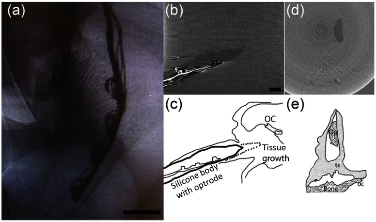
Fig. 7.
(a) An x-ray projection of an inserted optical array in situ in a cat cochlea. The thick wire is the backbone and acts as a heat sink. The thin wires connect to the anodes of the optical sources. The scale bar represents . (b) and (c) The same array after the reconstruction and its sketch. The optical sources irradiate Rosenthal’s canal. A thin layer of tissue can be seen around the electrode, which has been slightly retracted (dash line in the sketch). The organ of Corti (OC) is also marked. The scale bar represents and is used for the following panels. (d) and (e) The cross section of the same array after the reconstruction and its sketch. The optrode (Op), tissue, bone, and OC are marked in the sketch.

