Abstract
Introduction: Bronchogenic cysts may rupture or become infected, and malignant degeneration may occur. Although various types of malignant degeneration have been described, only a few reports of mucoepidermoid carcinoma arising from a bronchogenic cyst have been published. We report such a case.
Case: A 77-year-old female was referred to our institution for evaluation of left chest pain. A computed tomography scan showed an enhancing 65 × 70 mm mass of the left diaphragm. Based on the intraoperative findings of an intradiaphragmatic tumor involving the lower lobe of the left lung, the resection of the tumor with the wedge resection of left lower lobe and partial resection of the left diaphragm was performed. Histopathologic examination revealed a mucoepidermoid carcinoma arising from a bronchogenic cyst of the diaphragm with the presence of fibrous adhesion to the lower lobe.
Conclusion: We believe that complete resection of any bronchogenic cyst is justified.
Keywords: mucoepidermoid carcinoma, bronchogenic cyst, diaphragm
Introduction
A bronchogenic cyst is a congenital cyst that occurs because of an abnormal differentiation of the budding ventral foregut, which then develops into a blind fluidfilled pouch.1) While bronchogenic cysts are commonly located in the mediastinum, an intradiaphragmatic location is exceedingly rare.2) In general, bronchogenic cysts may not only be complicated by rupture or infection, but rarely they may undergo malignant degeneration.2,3) Surgical resection is a treatment option for a bronchogenic cyst.4) Although various types of malignant degeneration of bronchogenic cysts have been described previously,2,3) only a few reports of a mucoepidermoid carcinoma arising from a bronchogenic cyst have been reported.3,5) To the best of our knowledge, this is the first report describing the rare occurrence of a mucoepidermoid carcinoma arising from a bronchogenic cyst of the diaphragm.
Case Report
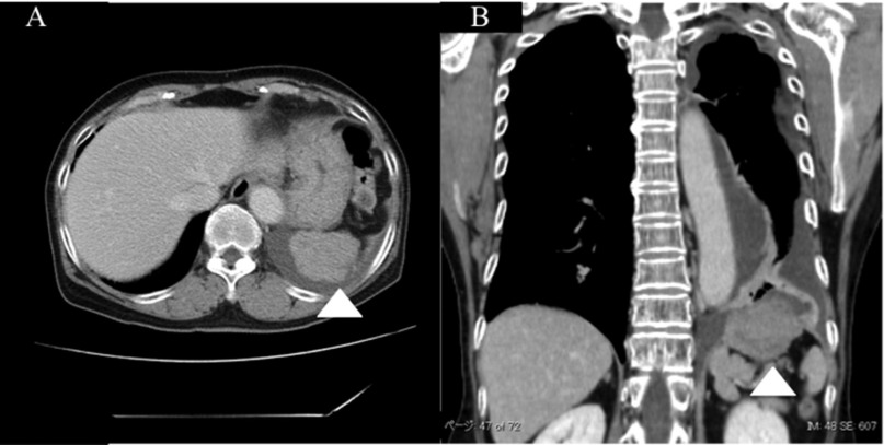
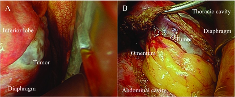
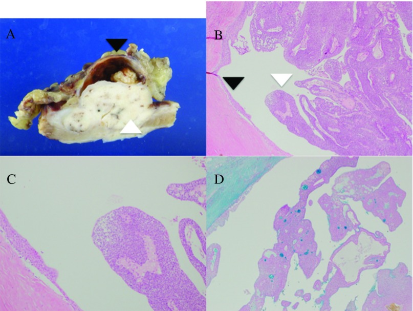
A 77-year-old female who never-smoked was referred to our institution for evaluation of left chest pain of 1-week duration, and a left pleural effusion was noted on chest X-ray. A contrast-enhanced computed tomography (CT) scan showed an enhancing 65 × 45 mm mass of the left diaphragm (Fig. 1A and 1B). Cytological examination of the pleural fluid was negative and serum tumor marker levels were within the normal range. Surgery was performed because of the possibility of malignancy. Intraoperatively, the mass was found to be an intradiaphragmatic tumor with involvement of the lower lobe of the left lung (Fig. 2). Complete excision of the tumor was accomplished with a wedge resection of the lower lobe of the left lung and a partial resection of the left diaphragm. There were no intraoperative complications, and the patient had an uneventful recovery. Histopathologic examination revealed a tumor with a maximum diameter of 7 cm, arising from a cyst lined by ciliated columnar epithelium with the presence of fibrous adhesion to the lower lobe. The tumor was composed of cells arranged in lobules, with a papillary and focal glandular pattern with mucoid and squamoid differentiation, interspersed in a fibrous vascular tissue (Fig. 3A–3D). All of the histopathologic features were diagnostic of a mucoepidermoid carcinoma arising from a bronchogenic cyst of the diaphragm (Fig. 3).
Fig. 1. Axial (A) and coronal (B) slices from a CT of the chest with contrast show a heterogeneously enhancing mass of the left diaphragm. CT: computed tomography.
Fig. 2. (A and B) Intraoperative view of the tumor.
Fig. 3. (A) Macroscopic appearance of the resected specimen showing a tumor within the cyst (white arrowhead) and the remaining cyst wall (black arrowhead). (B) Microscopic appearance of the mucoepidermoid carcinoma arising in a bronchogenic cyst (white arrowhead) and a cyst wall lined by ciliated columnar epithelium (black arrowhead) (HE stain × 40). (C) Detail of Figure 3 (B) (HE, × 100). (D) Alcian blue stain, a stain helpful for establishing the diagnosis of mucoepidermoid carcinoma, highlighting the presence of cytoplasmic mucin in the tumor cells. HE: hematoxylin and eosin.
Discussion
Although bronchogenic cysts are asymptomatic and in many cases discovered incidentally during medical checkups or workups for other diseases, intradiaphragmatic bronchogenic cysts can present with common respiratory symptoms, such as cough, or back pain from compression or irritation of adjacent structures.2) According to previous reports,2) intradiaphragmatic bronchogenic cysts have CT and magnetic resonance imaging characteristics similar to those of mediastinal bronchogenic cysts, showing hypoenhancing homogeneous soft tissue masses.2) Frequently, preoperative localization of these lesions is difficult, and based on imaging findings they may be mistakenly thought to be either within the abdominal or thoracic cavities, rather than within the diaphragm itself. The CT image findings of the present case might likely showed hyperenhancement of the diaphragmatic mass because the cyst had undergone malignant degeneration. The preoperative diagnoses of an intradiaphragmatic bronchogenic cyst or malignant degeneration of a bronchogenic cyst are often difficult.
In the present case, mucoepidermoid carcinoma arose from a bronchogenic cyst of the diaphragm. Histologically, mucoepidermoid carcinoma is similar to tumors originally described in the major salivary glands and is believed to originate from minor salivary glands lining the tracheobronchial tree.6) Bronchogenic cysts can have minor salivary gland type tissue in the wall of the cyst in addition to ciliated pseudostratified columnar epithelium, cartilage, or smooth muscle within the cyst wall although it is rare for all of these components to be present histopathologically.7) There are only a few reports of mucoepidermoid carcinoma arising from a bronchogenic cyst,3,5) and to the best of our knowledge there are no reports of mucoepidermoid carcinoma arising from a bronchogenic cyst of the diaphragm as in the present case. Some studies have suggested the potential for malignant transformation in unstable epithelial cells of the cyst wall.8) Therefore, in the present case, it is possible that mucoepidermoid carcinoma arose from salivary gland type tissue in the cyst wall.
In conclusion, we described a rare case of mucoepidermoid carcinoma arising from a bronchogenic cyst of the diaphragm.3) We believe that complete resection of any bronchogenic cyst is justified, since bronchogenic cysts may not only be complicated by rupture or infection, but also by the occurrence of malignant degeneration.
Acknowledgment
We would like to thank Junya Fukuoka, MD. PhD, Chair/professor, Department of Pathology, Nagasaki University Graduate School of Biomedical Science, for the help in establishing the diagnosis.
Disclosure Statement
Naohiro Taira and the other co-authors have no conflicts of interest and relevant financial interests to declare in this manuscript.
References
- 1).Sugarbaker David J. Overview of Benign Lung Disease: Anatomy and Pathophysiology. Adult Chest Surgery. New York: McGraw Hill Medical; 2009. [Google Scholar]
- 2).Mubang R, Brady JJ, Mao M, et al. Intradiaphragmatic bronchogenic cysts: case report and systematic review. J Cardiothorac Surg 2016; 11: 79. [DOI] [PMC free article] [PubMed] [Google Scholar]
- 3).Brassesco MS, Valera ET, Lira RC, et al. Mucoepidermoid carcinoma of the lung arising at the primary site of a bronchogenic cyst: clinical, cytogenetic, and molecular findings. Pediatr Blood Cancer 2011; 56: 311-3. [DOI] [PubMed] [Google Scholar]
- 4).St-Georges R, Deslauriers J, Duranceau A, et al. Clinical spectrum of bronchogenic cysts of the mediastinum and lung in the adult. Ann Thorac Surg 1991; 52: 6-13. [DOI] [PubMed] [Google Scholar]
- 5).Tanaka M, Shimokawa R, Matsubara O, et al. Mucoepidermoid carcinoma of the thymic region. Acta Pathol Jpn 1982; 32: 703-12. [DOI] [PubMed] [Google Scholar]
- 6).Shen C, Che G. Clinicopathological analysis of pulmonary mucoepidermoid carcinoma. World J Surg Oncol 2014; 12: 33. [DOI] [PMC free article] [PubMed] [Google Scholar]
- 7).Fraga S, Helwig EB, Rosen SH. Bronchogenic cysts in the skin and subcutaneous tissue. Am J Clin Pathol 1971; 56: 230-8. [DOI] [PubMed] [Google Scholar]
- 8).Klacsmann PG, Olson JL, Eggleston JC. Mucoepidermoid carcinoma of the bronchus: an electron microscopic study of the low grade and the high grade variants. Cancer 1979; 43: 1720-33. [DOI] [PubMed] [Google Scholar]


