Abstract
AIM
To investigate the clinical utility of alpha-fetoprotein (AFP)-producing gastric cancer (AFPGC)-specific microRNA (miRNA) for monitoring and prognostic prediction of patients.
METHODS
We performed a comprehensive miRNA array-based approach to compare miRNA expression levels between AFP-positive and AFP-negative cells in three patients with primary AFPGC. We next examined the expression levels of the selected miRNAs in five AFPGC and ten non-AFPGC tissue samples by quantitative reverse transcription-polymerase chain reaction to validate their utility. We also investigated the expression levels of the selected miRNA not only in tissue but also in plasma samples. Moreover, we investigated the relationship between plasma AFP levels and plasma selected miRNA expression levels, and also investigated the correlation of the selected miRNA expression levels and malignant potential.
RESULTS
Among the five miRNAs selected from the miRNA array results, the expression levels of miR-122-5p were significantly higher in the AFPGC patients than in the non-AFPGC patients (P < 0.05). In tissue samples, miR-122-5p expression level tended to be lower in the non-AFPGC tissue than the normal gastric mucosa. Conversely, in the AFPGC tissue, miR-122-5p expression level was significantly higher in the AFPGC tissue than both the normal gastric mucosa and the non-AFPGC tissue samples (P < 0.05). Plasma miR-122-5p expression levels were also significantly higher in the AFPGC patients than the health volunteers and the non-AFPGC patients (P < 0.05) and were strongly correlated with plasma AFP levels (r = 0.7975, P < 0.0001). Moreover, the correlation of miR-122-5p expression in tissue samples with malignant potential was stronger than that of plasma AFP level in the AFPGC patients. In contrast, no correlation was found between miR-122-5p expression levels and liver metastasis in the non-AFPGC patients.
CONCLUSION
miR-122-5p might be a useful biomarker for early detection and disease monitoring in AFPGC.
Keywords: Gastric cancer, Alpha-fetoprotein, Alpha-fetoprotein producing gastric cancer, MicroRNA, miR-122-5p
Core tip: We examined the microRNAs (miRNA) expression in alpha-fetoprotein (AFP)-producing gastric cancer (AFPGC) tissue samples using a comprehensive miRNA array-based approach, and also investigated the clinical utility of the identified AFPGC-specific miRNAs. We found the expression of miR-122-5p was significantly higher in the AFPGC tissues and plasma samples. Moreover, tissue miR-122-5p expression levels exhibited a stronger correlation with malignant potential than plasma AFP level in AFPGC patients. miR-122-5p might be a useful biomarker for early detection and disease monitoring in AFPGC.
INTRODUCTION
Gastric cancer (GC) is one of the most common solid tumors and is the third leading cause of cancer-related deaths worldwide[1,2]. Despite improvements in treatment approaches, prognosis of patients with advanced GC remains poor even after curative resection.
Among various tumor subtypes, alpha-fetoprotein (AFP)-producing GC (AFPGC) is recognized as one of the most aggressive tumors, with a high propensity for liver metastasis and subsequent poor prognosis compared with other GC subtypes[3-7]. The incidence of AFPGC is low, ranging from 1.3% to 15% of all GCs[8-12]. Therefore, recent comprehensive molecular analyses have not yet referred to this minor subtype.
MicroRNAs (miRNAs) are endogenous, small, non-coding, single-stranded RNAs of 20-25 nucleotides that regulate the expression of target genes at post-transcriptional level by binding to complementary sequences[13]. Various miRNAs were shown to play crucial roles in cancer as well as normal cells were reported to act as tumor suppressors or oncogenes in a cell type-dependent manner in various cancers[14]. In addition, certain miRNAs have been used for cancer detection, monitoring of tumor dynamics, and predicting prognosis and chemoresistance[15-20].
In the present study, we examined the miRNAs expression in AFPGC tissue samples using a comprehensive miRNA array-based approach. We also investigated the clinical utility of the identified AFPGC-specific miRNAs in monitoring and prognostic prediction of patients with AFPGC and evaluated their potential as universal biomarker for liver metastases.
MATERIALS AND METHODS
Patients and samples
A total of 492 patients underwent gastrectomy for GC at the University of Yamanashi Hospital between 2012 and 2018. Tumor specimens and resected lymph nodes obtained at the time of surgery were immediately fixed in 10% neutral-buffered formalin and embedded in paraffin after fixation. None of the patients underwent preoperative chemotherapy or radiotherapy. Tissue samples of all five patients with primary AFPGC and those from ten patients with primary non-AFPGC at various stages as controls from the same cohort were selected. The selected AFPGC samples contained all AFPGC patients who were operated in our hospital.
Pre-operative plasma samples were also obtained from four AFPGC patients and twenty non-AFPGC patients with GC who underwent surgical resection at the University of Yamanashi Hospital between 2017 and 2018. Control plasma samples were collected from 12 healthy adult volunteers. A total of 5 mL blood samples were collected into ethylenediaminetetraacetic acid-coated tubes and immediately spun at 3000 rpm at 4°C for 10 min to separate serum, which was stored at -80°C for further processing. AFPGC was defined based on a plasma AFP level above 10 ng/mL or positive AFP immunoreactivity in tissue samples. This study was approved by the Ethics Committee of Yamanashi University and performed in accordance with the ethical standards of the Declaration of Helsinki and its amendments.
RNA extraction
Formalin-fixed, paraffin-embedded tissue samples were cut into 10-µm-thick sections, and total RNA was extracted from tumor and normal gastric mucosa in each patient using RNeasy FFPE kit (Qiagen, Valencia, CA), according to the manufacturer’s protocol. In plasma samples, total RNA was extracted from 100 µL plasma using RNeasy Serum/Plasma kit (Qiagen), according to the manufacturer’s protocol.
miRNA microarray analysis
Microarray analyses of the GC tissue samples were performed using 3D-Gene miRNA oligo chips (Toray Industries, Kamakura, Japan), with 2565 genes mounted onto each DNA chip. Results were compared between the AFP-positive and AFP-negative cells among AFPGC patient samples using macro-dissection. Tissue samples from the three AFPGC patients who underwent curative surgery were mixed equally. RNAs were labeled with the 3D-Gene miRNA labeling kit (Toray Industries). Fluorescent signals were scanned using a 3D-Gene scanner 3000 (Toray Industries) and analyzed with the 3D-Gene Extraction software (Toray Industries). In the current study, expression level of each miRNA was normalized using the median signal intensity of the all genes in each chip, and median signal intensity was adjusted to 25.
Quantification of miRNA by quantitative reverse transcription-polymerase chain reaction
Levels of miRNAs were quantified by quantitative reverse transcription-polymerase chain reaction (qRT-PCR) using a Human TaqMan MicroRNA Assay kit (Applied Biosystems, Foster City, CA), according to standard procedures. Reverse transcription was conducted with a TaqMan MicroRNA Reverse Transcription kit (Applied Biosystems). Tissue miRNA levels were normalized to the endogenous control RNU6B, and plasma miRNA levels were normalized to a synthetic RNA oligonucleotide, cel-miR-39-3p (Qiagen), by spiking the samples with the oligonucleotide which does not exist in human genome. The following primers were used for the Taqman assay (Thermo Fisher Scientific, CA, United States): human hsa-miR-122-5p (cat #002245), hsa-miR-144-5p (cat #002148), hsa-miR-20a-5p (cat #000580), hsa-miR-20b-5p (cat #001014), hsa-miR-106a-5p (cat #000578), RNU6B (cat #001093), and cel-miR-39-3p (cat #000200). ΔCt values for all miRNAs relative to the control gene RNU6B and cel-miR-39-3p were determined. ΔΔCt values were calculated using mean ΔCt values in non-AFPGC tissue, normal gastric mucosa, or healthy volunteer plasma samples. Plasma miR-122-5p expression was calculated using log10(2-ΔCt).
Statistical analysis
Statistical significance was determined using GraphPad Prism® version 5 (San Diego, CA). Quantitative values were expressed as means ± SD unless noted otherwise. Statistical significance was evaluated using the Student’s t test and one-way analysis of variance for each time point, followed by Tukey’s post hoc test. Pearson’s correlation coefficient was determined to assess the correlation between plasma AFP and plasma miR-122-5p levels. P values < 0.05 were considered to indicate statistical significance.
RESULTS
Identification of miRNA candidates from a comprehensive miRNA array-based approach in AFPGC tissue
We selected miRNA candidates using a miRNA array-based approach. We compared the expression levels of each miRNA between the AFP-positive and AFP-negative cells in AFPGC patients. Of the 2565 candidates analyzed, we selected the following five miRNAs: miR-122-5p, miR-20a-5p, miR-20b-5p, miR-106a-5p, and miR-144-5p. The expression levels of these selected miRNAs were significantly different in AFP-positive cells compared with the AFP-negative cells, and the signal intensity of each miRNA was sufficient (Table 1).
Table 1.
Summary of five miRNA candidates selected by microarray analysis
|
Signal intensity |
Fold change |
||
| Gene ID | AFPGC | Non-AFPGC | AFPGC/non-AFPGC |
| hsa-miR-122-5p | 492 | 105 | 4.7 |
| hsa-miR-20a-5p | 245 | 113 | 2.2 |
| hsa-miR-20b-5p | 198 | 94 | 2.1 |
| hsa-miR-106a-5p | 304 | 195 | 1.6 |
| hsa-miR-145-5p | 712 | 1367 | 0.5 |
Expression level of each miRNA was normalized using the median signal intensity of the all genes in each chip, and median signal intensity was adjusted to 25. AFPGC: Alpha-fetoprotein-producing gastric cancer.
Validation of the expression levels of five miRNAs in AFPGC and non-AFPGC tissue samples
We examined the expression levels of the five selected miRNAs in five AFPGC and ten non-AFPGC tissue samples by qRT-PCR to validate their utility (Figure 1). Among these five miRNAs, the expression of miR-122-5p was significantly higher in the AFPGC patients than the non-AFPGC patients. Therefore, we selected miR-122-5p for further analyses in this study.
Figure 1.
Validation of five microRNAs in non-alpha-fetoprotein producing gastric cancer and alpha-fetoprotein producing gastric cancer tissue samples performed by quantitative reverse transcription-polymerase chain reaction. A: miR-122-5p; B: miR-20a-5p; C: miR-20b-5p; D: miR-106a-5p; E: miR-144-5p. The lines inside the bow plot represent the average size. AFPGC: Alpha-fetoprotein-producing gastric cancer.
miR-122-5p expression levels in tissue and plasma samples
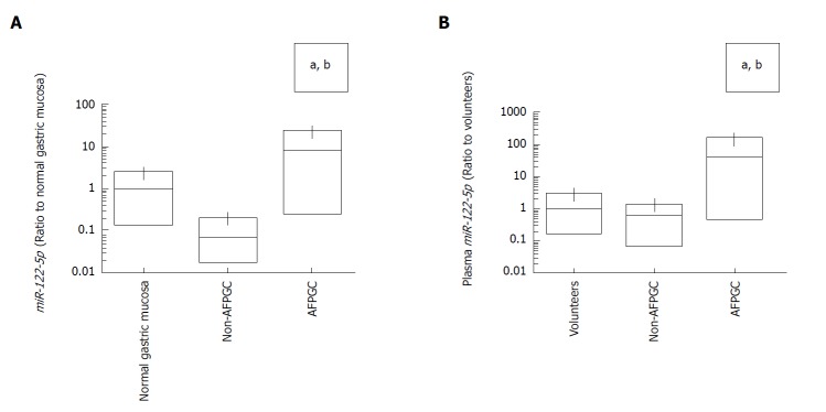
Next, we investigated the expression levels of miR-122-5p not only in tissue but also in plasma samples. In tissue samples, miR-122-5p expression levels tended to be lower in the non-AFPGC tissue samples than in the normal gastric mucosa samples. Conversely, miR-122-5p expression levels were significantly higher in the AFPGC tissue samples compared with the normal gastric mucosa and the non-AFPGC tissue samples (Figure 2A). The plasma expression levels of miR-122-5p were also significantly higher in the AFPGC patient samples than the samples from health volunteers and the non-AFPGC patients (Figure 2B).
Figure 2.
Quantification of miR-122-5p expression levels by quantitative reverse transcription-polymerase chain reaction. A: Comparison of miR-122-5p expression levels between normal gastric mucosa, non-alpha-fetoprotein-producing gastric cancer (AFPGC) and AFPGC in tissue samples. aP < 0.05, compared to normal gastric mucosa; bP < 0.05, compared to non-AFPGC; B: Comparison of miR-122-5p expression levels between health volunteers, non-AFPGC and AFPGC in plasma sample. aP < 0.05, compared to health volunteers; bP < 0.05, compared to non-AFPGC. The lines inside the bow plot represent the average size.
Plasma miR-122-5 levels are strongly correlated with plasma AFP levels in GC patients
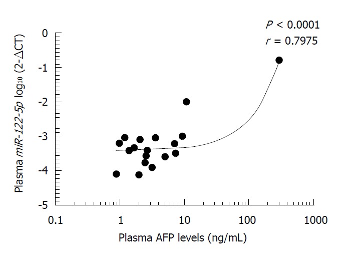
We next investigated the relationship between plasma AFP levels and plasma miR-122-5p expression levels in the AFPGC and non-AFPGC patients and found that miR-122-5p expression level in plasma was strongly correlated with plasma AFP level (r = 0.7975, P < 0.0001; Figure 3).
Figure 3.

Relationship between plasma alpha-fetoprotein levels and plasma miR-122-5p expression levels. Plasma miR-122-5p expression level was strongly correlated with plasma alpha-fetoprotein (AFP) levels in gastric cancer patients (r = 0.7975, P < 0.0001).
Prognostic utility of tissue miR-122-5p expression in AFPGC patients
Figure 4 shows the correlation between malignant potential, all biomarkers, tissue miR-122-5p expression, and plasma AFP level in the AFPGC patients. We found that the expression level of miR-122-5p in tissue exhibited a stronger correlation with malignant potential (i.e., liver metastasis) than plasma AFP level in the AFPGC patients. Two patients with malignant potential were diagnosed morphologically as poorly differentiated adenocarcinoma and mucinous adenocarcinoma, and the other current alive patients were diagnosed as poorly differentiated adenocarcinoma and hepatoid adenocarcinoma.
Figure 4.

Correlation between malignant potential and tissue miR-122-5p expression levels in alpha-fetoprotein-producing gastric cancer patients. White symbols indicate current alive and black symbols indicate current death. AFP: Alpha-fetoprotein.
DISCUSSION
AFPGC has been reported to be more likely to metastasize to liver and is therefore associated with extremely poor prognosis[8,11]. However, no genomic analyses have been conducted for AFPGC due to its rarity. Therefore, AFPGC-specific genomic and/or epigenomic alterations are not well known, which have urged us to examine the molecular characteristics specific to AFPGC. The current study investigated the molecular characteristics of AFPGC with a comprehensive analysis, with particular focus on miRNA expression.
The findings of the present study clearly demonstrated that the expression of miR-122-5p was significantly higher in the AFPGC tissues than the normal and non-AFPGC tissues. The expression levels of this miRNA were also higher in the plasma samples of patients with AFPGC compared with those of healthy volunteers and non-AFPGC patients and correlated with plasma AFP levels to a certain extent. Interestingly, the tissue expression level of miR-122-5p exhibited a stronger correlation with malignant potential than plasma AFP level in AFPGC patients, suggesting that miR-122-5p might have utility as a prognostic biomarker especially for liver metastasis in this small GC subgroup.
miR-122-5p expression has been increasingly examined in various normal and cancer tissue types. Several studies reported that miR-122-5p was specifically expressed in human liver and that hepatocyte-specific miR-122-5p regulated hepatocyte differentiation and metabolism[21-23]. Taken together, AFPGC might show characteristics of hepatocytes not only morphologically but also in its miRNA expression patterns. In fact, AFPGC was not necessarily hepatoid adenocarcinoma in this series, and two patients with aggressive development of liver metastasis were diagnosed morphologically as poorly differentiated adenocarcinoma and mucinous adenocarcinoma. Conversely, miR-122-5p was previously shown to function as a tumor suppressor and was reported to be downregulated in several cancer types such as hepatocellular carcinoma[24], non-small-cell lung cancer[25], gallbladder carcinoma[26], bladder cancer[27], and breast cancer[28]. In GC, the expression of miR-122-5p was reported to be lower in tumor tissue than the adjacent non-cancerous tissue. Furthermore, several studies reported that miR-122-5p inhibited proliferation, migration, and invasion in GC[15,29,30]. It’s not known exactly why miR-122-5p, which is known as suppressor gene, is higher in AFPGC. Some miRNA was reported that decreased in early cases and elevated again in staged-advanced cases[31]. Therefore, miR-122-5p decreased in carcinogenesis might be elevated during tumor evolution to AFPGC. However, the exact mechanism is unknown at the present time.
In the current study, we did not see a correlation between miR-122-5p in patients with non-AFPGC and development of liver metastasis, suggesting that the mechanism underlying liver metastasis might be distinct between AFPGC and non-AFPGC. We assume that AFPGC is completely different from non-AFPGC, and the mechanism of liver metastasis between AFPGC and non-AFPGC is also distinct.
Several reports demonstrated that the clinical behavior of AFPGC was distinct from that of non-AFPGC[32]. Recently, Lu et al[33] demonstrated that AFP contributed to invasion and metastasis directly. We speculate AFPGC has specific ability of liver metastasis, and correlated with miR-122-5p. Therefore, miR-122-5p might directly facilitate tumor proliferation, migration, and invasion, which raises the possibility of miR-122-5p as a potential therapeutic target in AFPGC. However, future studies are warranted to demonstrate the biological function underlying altered expression of miR-122-5p in AFPGC. The current study revealed miR-122-5p as a potentially useful biomarker for early detection, disease monitoring, and prognostic prediction in patients with AFPGC, which warrant further investigation.
ARTICLE HIGHLIGHTS
Research background
Alpha-fetoprotein (AFP)-producing gastric cancer (AFPGC) is recognized as one of the most aggressive tumors, with a high propensity for liver metastasis and subsequent poor prognosis compared with other GC subtypes. Recent comprehensive molecular analyses have not yet referred to this minor subtype because of its rareness.
Research motivation
To discover universal biomarkers for liver metastasis by researching AFPGC-specific microRNAs (miRNAs).
Research objectives
To investigate the clinical utility of AFPGC-specific miRNA for monitoring and prognostic prediction of patients.
Research methods
We performed a comprehensive miRNA array-based approach to compare miRNA expression levels between AFP-positive and AFP-negative cells, and also investigated the clinical utility of the identified AFPGC-specific miRNAs.
Research results
We found the expression of miR-122-5p was significantly higher in the AFPGC tissues than the normal and non-AFPGC tissues. The expression levels of this miRNA were also higher in the plasma samples of patients with AFPGC compared with those of healthy volunteers and non-AFPGC patients and correlated with plasma AFP levels. Moreover, the tissue expression level of miR-122-5p exhibited a stronger correlation with malignant potential than plasma AFP level in AFPGC patients.
Research conclusions
miR-122-5p as a potentially useful biomarker for early detection and disease monitoring in patients with AFPGC.
Research perspectives
We identified miR-122-5p as AFPGC-specific miRNA. miR-122-5p might be a clinical useful biomarker in AFPGC. Although studies are warranted to demonstrate the biological function underlying altered expression of miR-122-5p in AFPGC, the miR-122-5p might be a potential therapeutic target for liver metastasis in AFPGC.
ACKNOWLEGEMENTS
The Authors are grateful to Motoko Inui and Makiko Mishina for expert technical assistance.
Footnotes
Institutional review board statement: This study was approved by the Ethics Committee of Yamanashi University (approved number: 1825) and was performed in accordance with the ethical standards of the Declaration of Helsinki and its later amendments.
Conflict-of-interest statement: The authors declare that they have no conflict of interest.
Manuscript source: Unsolicited manuscript
Peer-review started: July 5, 2018
First decision: July 24, 2018
Article in press: August 31, 2018
Specialty type: Oncology
Country of origin: Japan
Peer-review report classification
Grade A (Excellent): 0
Grade B (Very good): 0
Grade C (Good): C, C
Grade D (Fair): 0
Grade E (Poor): 0
P- Reviewer: Jung Y, Takemura N S- Editor: Ji FF L- Editor: A E- Editor: Tan WW
Contributor Information
Suguru Maruyama, First Department of Surgery, Faculty of Medicine University of Yamanashi, Yamanashi 409-3898, Japan.
Shinji Furuya, First Department of Surgery, Faculty of Medicine University of Yamanashi, Yamanashi 409-3898, Japan.
Kensuke Shiraishi, First Department of Surgery, Faculty of Medicine University of Yamanashi, Yamanashi 409-3898, Japan.
Hiroki Shimizu, First Department of Surgery, Faculty of Medicine University of Yamanashi, Yamanashi 409-3898, Japan.
Hidenori Akaike, First Department of Surgery, Faculty of Medicine University of Yamanashi, Yamanashi 409-3898, Japan.
Naohiro Hosomura, First Department of Surgery, Faculty of Medicine University of Yamanashi, Yamanashi 409-3898, Japan.
Yoshihiko Kawaguchi, First Department of Surgery, Faculty of Medicine University of Yamanashi, Yamanashi 409-3898, Japan.
Hidetake Amemiya, First Department of Surgery, Faculty of Medicine University of Yamanashi, Yamanashi 409-3898, Japan.
Hiromichi Kawaida, First Department of Surgery, Faculty of Medicine University of Yamanashi, Yamanashi 409-3898, Japan.
Makoto Sudo, First Department of Surgery, Faculty of Medicine University of Yamanashi, Yamanashi 409-3898, Japan.
Shingo Inoue, First Department of Surgery, Faculty of Medicine University of Yamanashi, Yamanashi 409-3898, Japan.
Hiroshi Kono, First Department of Surgery, Faculty of Medicine University of Yamanashi, Yamanashi 409-3898, Japan.
Daisuke Ichikawa, First Department of Surgery, Faculty of Medicine University of Yamanashi, Yamanashi 409-3898, Japan. dichikawa@yamanashi.ac.jp.
References
- 1.Kamangar F, Dores GM, Anderson WF. Patterns of cancer incidence, mortality, and prevalence across five continents: defining priorities to reduce cancer disparities in different geographic regions of the world. J Clin Oncol. 2006;24:2137–2150. doi: 10.1200/JCO.2005.05.2308. [DOI] [PubMed] [Google Scholar]
- 2.Cancer Genome Atlas Research Network. Comprehensive molecular characterization of gastric adenocarcinoma. Nature. 2014;513:202–209. doi: 10.1038/nature13480. [DOI] [PMC free article] [PubMed] [Google Scholar]
- 3.Bourreille J, Metayer P, Sauger F, Matray F, Fondimare A. [Existence of alpha feto protein during gastric-origin secondary cancer of the liver] Presse Med. 1970;78:1277–1278. [PubMed] [Google Scholar]
- 4.Chang YC, Nagasue N, Abe S, Kohno H, Yamanoi A, Uchida M, Nakamura T. [The characters of AFP-producing early gastric cancer] Nihon Geka Gakkai Zasshi. 1990;91:1574–1580. [PubMed] [Google Scholar]
- 5.Motoyama T, Aizawa K, Watanabe H, Fukase M, Saito K. alpha-Fetoprotein producing gastric carcinomas: a comparative study of three different subtypes. Acta Pathol Jpn. 1993;43:654–661. doi: 10.1111/j.1440-1827.1993.tb02549.x. [DOI] [PubMed] [Google Scholar]
- 6.Chang YC, Nagasue N, Abe S, Taniura H, Kumar DD, Nakamura T. Comparison between the clinicopathologic features of AFP-positive and AFP-negative gastric cancers. Am J Gastroenterol. 1992;87:321–325. [PubMed] [Google Scholar]
- 7.Koide N, Nishio A, Igarashi J, Kajikawa S, Adachi W, Amano J. Alpha-fetoprotein-producing gastric cancer: histochemical analysis of cell proliferation, apoptosis, and angiogenesis. Am J Gastroenterol. 1999;94:1658–1663. doi: 10.1111/j.1572-0241.1999.01158.x. [DOI] [PubMed] [Google Scholar]
- 8.Kono K, Amemiya H, Sekikawa T, Iizuka H, Takahashi A, Fujii H, Matsumoto Y. Clinicopathologic features of gastric cancers producing alpha-fetoprotein. Dig Surg. 2002;19:359–365; discussion 365. doi: 10.1159/000065838. [DOI] [PubMed] [Google Scholar]
- 9.Chang YC, Nagasue N, Kohno H, Taniura H, Uchida M, Yamanoi A, Kimoto T, Nakamura T. Clinicopathologic features and long-term results of alpha-fetoprotein-producing gastric cancer. Am J Gastroenterol. 1990;85:1480–1485. [PubMed] [Google Scholar]
- 10.Chun H, Kwon SJ. Clinicopathological characteristics of alpha-fetoprotein-producing gastric cancer. J Gastric Cancer. 2011;11:23–30. doi: 10.5230/jgc.2011.11.1.23. [DOI] [PMC free article] [PubMed] [Google Scholar]
- 11.Liu X, Cheng Y, Sheng W, Lu H, Xu Y, Long Z, Zhu H, Wang Y. Clinicopathologic features and prognostic factors in alpha-fetoprotein-producing gastric cancers: analysis of 104 cases. J Surg Oncol. 2010;102:249–255. doi: 10.1002/jso.21624. [DOI] [PubMed] [Google Scholar]
- 12.McIntire KR, Waldmann TA, Moertel CG, Go VL. Serum alpha-fetoprotein in patients with neoplasms of the gastrointestinal tract. Cancer Res. 1975;35:991–996. [PubMed] [Google Scholar]
- 13.Ha M, Kim VN. Regulation of microRNA biogenesis. Nat Rev Mol Cell Biol. 2014;15:509–524. doi: 10.1038/nrm3838. [DOI] [PubMed] [Google Scholar]
- 14.Esquela-Kerscher A, Slack FJ. Oncomirs - microRNAs with a role in cancer. Nat Rev Cancer. 2006;6:259–269. doi: 10.1038/nrc1840. [DOI] [PubMed] [Google Scholar]
- 15.Chen Q, Ge X, Zhang Y, Xia H, Yuan D, Tang Q, Chen L, Pang X, Leng W, Bi F. Plasma miR-122 and miR-192 as potential novel biomarkers for the early detection of distant metastasis of gastric cancer. Oncol Rep. 2014;31:1863–1870. doi: 10.3892/or.2014.3004. [DOI] [PubMed] [Google Scholar]
- 16.Hiramoto H, Muramatsu T, Ichikawa D, Tanimoto K, Yasukawa S, Otsuji E, Inazawa J. miR-509-5p and miR-1243 increase the sensitivity to gemcitabine by inhibiting epithelial-mesenchymal transition in pancreatic cancer. Sci Rep. 2017;7:4002. doi: 10.1038/s41598-017-04191-w. [DOI] [PMC free article] [PubMed] [Google Scholar]
- 17.Hiyoshi Y, Akiyoshi T, Inoue R, Murofushi K, Yamamoto N, Fukunaga Y, Ueno M, Baba H, Mori S, Yamaguchi T. Serum miR-143 levels predict the pathological response to neoadjuvant chemoradiotherapy in patients with locally advanced rectal cancer. Oncotarget. 2017;8:79201–79211. doi: 10.18632/oncotarget.16760. [DOI] [PMC free article] [PubMed] [Google Scholar]
- 18.Imamura T, Komatsu S, Ichikawa D, Miyamae M, Okajima W, Ohashi T, Kiuchi J, Nishibeppu K, Konishi H, Shiozaki A, et al. Depleted tumor suppressor miR-107 in plasma relates to tumor progression and is a novel therapeutic target in pancreatic cancer. Sci Rep. 2017;7:5708. doi: 10.1038/s41598-017-06137-8. [DOI] [PMC free article] [PubMed] [Google Scholar]
- 19.Matsushita R, Seki N, Chiyomaru T, Inoguchi S, Ishihara T, Goto Y, Nishikawa R, Mataki H, Tatarano S, Itesako T, et al. Tumour-suppressive microRNA-144-5p directly targets CCNE1/2 as potential prognostic markers in bladder cancer. Br J Cancer. 2015;113:282–289. doi: 10.1038/bjc.2015.195. [DOI] [PMC free article] [PubMed] [Google Scholar]
- 20.Yang R, Fu Y, Zeng Y, Xiang M, Yin Y, Li L, Xu H, Zhong J, Zeng X. Serum miR-20a is a promising biomarker for gastric cancer. Biomed Rep. 2017;6:429–434. doi: 10.3892/br.2017.862. [DOI] [PMC free article] [PubMed] [Google Scholar]
- 21.Roderburg C, Benz F, Vargas Cardenas D, Koch A, Janssen J, Vucur M, Gautheron J, Schneider AT, Koppe C, Kreggenwinkel K, et al. Elevated miR-122 serum levels are an independent marker of liver injury in inflammatory diseases. Liver Int. 2015;35:1172–1184. doi: 10.1111/liv.12627. [DOI] [PubMed] [Google Scholar]
- 22.Koyama S, Kuragaichi T, Sato Y, Kuwabara Y, Usami S, Horie T, Baba O, Hakuno D, Nakashima Y, Nishino T, et al. Dynamic changes of serum microRNA-122-5p through therapeutic courses indicates amelioration of acute liver injury accompanied by acute cardiac decompensation. ESC Heart Fail. 2017;4:112–121. doi: 10.1002/ehf2.12123. [DOI] [PMC free article] [PubMed] [Google Scholar]
- 23.Satishchandran A, Ambade A, Rao S, Hsueh YC, Iracheta-Vellve A, Tornai D, Lowe P, Gyongyosi B, Li J, Catalano D, et al. MicroRNA 122, Regulated by GRLH2, Protects Livers of Mice and Patients From Ethanol-Induced Liver Disease. Gastroenterology. 2018;154:238–252.e7. doi: 10.1053/j.gastro.2017.09.022. [DOI] [PMC free article] [PubMed] [Google Scholar]
- 24.Wang N, Wang Q, Shen D, Sun X, Cao X, Wu D. Downregulation of microRNA-122 promotes proliferation, migration, and invasion of human hepatocellular carcinoma cells by activating epithelial-mesenchymal transition. Onco Targets Ther. 2016;9:2035–2047. doi: 10.2147/OTT.S92378. [DOI] [PMC free article] [PubMed] [Google Scholar]
- 25.Qin H, Sha J, Jiang C, Gao X, Qu L, Yan H, Xu T, Jiang Q, Gao H. miR-122 inhibits metastasis and epithelial-mesenchymal transition of non-small-cell lung cancer cells. Onco Targets Ther. 2015;8:3175–3184. doi: 10.2147/OTT.S91696. [DOI] [PMC free article] [PubMed] [Google Scholar]
- 26.Lu W, Zhang Y, Zhou L, Wang X, Mu J, Jiang L, Hu Y, Dong P, Liu Y. miR-122 inhibits cancer cell malignancy by targeting PKM2 in gallbladder carcinoma. Tumour Biol. 2015 doi: 10.1007/s13277-015-4308-z. [DOI] [PubMed] [Google Scholar]
- 27.Wang Y, Xing QF, Liu XQ, Guo ZJ, Li CY, Sun G. MiR-122 targets VEGFC in bladder cancer to inhibit tumor growth and angiogenesis. Am J Transl Res. 2016;8:3056–3066. [PMC free article] [PubMed] [Google Scholar]
- 28.Ergün S, Ulasli M, Igci YZ, Igci M, Kırkbes S, Borazan E, Balik A, Yumrutaş Ö, Camci C, Cakmak EA, Arslan A, Oztuzcu S. The association of the expression of miR-122-5p and its target ADAM10 with human breast cancer. Mol Biol Rep. 2015;42:497–505. doi: 10.1007/s11033-014-3793-2. [DOI] [PubMed] [Google Scholar]
- 29.Rao M, Zhu Y, Zhou Y, Cong X, Feng L. MicroRNA-122 inhibits proliferation and invasion in gastric cancer by targeting CREB1. Am J Cancer Res. 2017;7:323–333. [PMC free article] [PubMed] [Google Scholar] [Retracted]
- 30.Xu X, Gao F, Wang J, Tao L, Ye J, Ding L, Ji W, Chen X. MiR-122-5p inhibits cell migration and invasion in gastric cancer by down-regulating DUSP4. Cancer Biol Ther. 2018;19:427–435. doi: 10.1080/15384047.2018.1423925. [DOI] [PMC free article] [PubMed] [Google Scholar]
- 31.Cheng H, Zhang L, Cogdell DE, Zheng H, Schetter AJ, Nykter M, Harris CC, Chen K, Hamilton SR, Zhang W. Circulating plasma MiR-141 is a novel biomarker for metastatic colon cancer and predicts poor prognosis. PLoS One. 2011;6:e17745. doi: 10.1371/journal.pone.0017745. [DOI] [PMC free article] [PubMed] [Google Scholar]
- 32.Hirajima S, Komatsu S, Ichikawa D, Kubota T, Okamoto K, Shiozaki A, Fujiwara H, Konishi H, Ikoma H, Otsuji E. Liver metastasis is the only independent prognostic factor in AFP-producing gastric cancer. World J Gastroenterol. 2013;19:6055–6061. doi: 10.3748/wjg.v19.i36.6055. [DOI] [PMC free article] [PubMed] [Google Scholar]
- 33.Lu S, Ma Y, Sun T, Ren R, Zhang X, Ma W. Expression of α-fetoprotein in gastric cancer AGS cells contributes to invasion and metastasis by influencing anoikis sensitivity. Oncol Rep. 2016;35:2984–2990. doi: 10.3892/or.2016.4678. [DOI] [PubMed] [Google Scholar]


