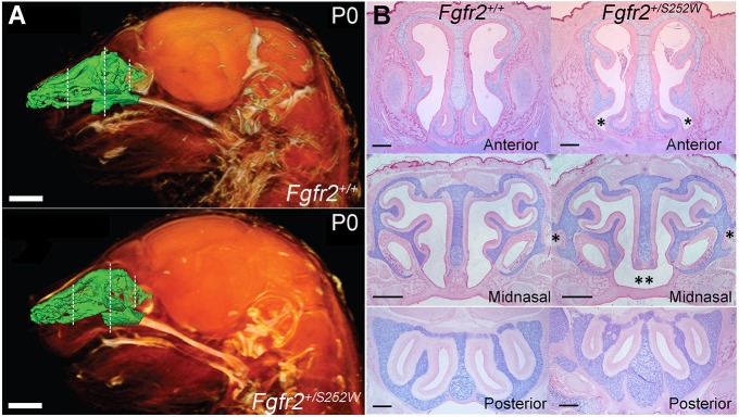
Fig. 1.
Nasal airspace and three regions of investigation in representative Fgfr2 mutant mice. (A) Lateral view of 3D reconstructions of MRM images of P0 Fgfr2+/+ and Apert Fgfr2+/S252W heads showing soft tissues (orange) and an endocast of the segmented nasal passages (green). Dashed lines indicate the three locations shown in (B) and in Movies 1 and 2. Scale bars: 1 mm. (B) Alcian Blue/Eosin staining of sections in the pars anterior (anterior), pars intermedia (midnasal) and pars posterior (posterior) regions of P0 Fgfr2+/+ and Apert Fgfr2+/S252W mice. These locations were used for quantitative analysis. Asterisks in the anterior mutant panel indicate flaring of the paraseptal region and pronounced narrowing of the adjacent nasal space. Single asterisks in the midnasal mutant panel indicate ectopic cartilage projecting from the paries nasi; double asterisks indicate lack of fusion between septum and anterior secondary palate. Scale bars: 500 µm.

