Fig. 3.
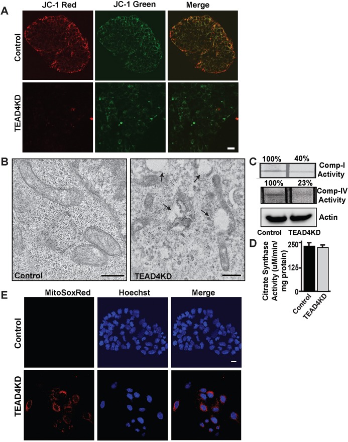
Loss of TEAD4 results in impaired mitochondrial function in mouse TSCs. (A) Control and TEAD4KD TSCs were stained with a mitochondrial membrane potential probe, JC-1, and excited simultaneously for observing hypo-polarized membrane potential (monomeric form excited by 488 nm laser, green) and hyper-polarized membrane potential (J-aggregate form excited using the 568 nm argon-krypton laser, red). Scale bar: 20 µm. (B) TEM pictures showing mitochondrial ultrastructural differences in control and TEAD4KD TSCs. Micrographs show the presence of an increased number of vacuoles and mitochondrial structural abnormalities in TEAD4KD TSCs. Scale bars: 500 nm. (C,D) Activities of mitochondrial electron-transport chain (ETC) complex I, ETC complex IV, and actin and citrate synthase were measured in control and TEAD4KD TSCs. Data show reduced ETC complex I and IV activities in TEAD4KD TSCs without any significant change in citrate synthase activity. Data are mean±s.e.m. (E) Control and TEAD4KD TSCs were stained with the mitochondrial ROS indicator MitoSox Red (red) and Hoechst (blue) to monitor mitochondrial distribution of reactive oxygen species accumulation. Scale bar: 20 µm.

