Abstract
New chemical inhibitors of protein–protein interactions are needed to propel advances in molecular pharmacology. Peptoids are peptidomimetic oligomers with the capability to inhibit protein-protein interactions by mimicking protein secondary structure motifs. Here we report the in silico design of a macrocycle primarily composed of peptoid subunits that targets the β-catenin:TCF interaction. The β-catenin:TCF interaction plays a critical role in the Wnt signaling pathway which is over-activated in multiple cancers, including prostate cancer. Using the Rosetta suite of protein design algorithms, we evaluate how different macrocycle structures can bind a pocket on β-catenin that associates with TCF. The in silico designed macrocycles are screened in vitro using luciferase reporters to identify promising compounds. The most active macrocycle inhibits both Wnt and AR-signaling in prostate cancer cell lines, and markedly diminishes their proliferation. In vivo potential is demonstrated through a zebrafish model, in which Wnt signaling is potently inhibited.
Small molecules and peptide inhibitors have their benefits and faults when it comes to inhibiting protein-protein interactions. Here, the authors designed a peptoid-peptide hybrid that inhibited β-catenin/TCF interactions, leading to inhibition of Wnt signalling in models of prostate cancer.
Introduction
Protein–protein interactions (PPIs) provide the structural and functional basis for many critical biological processes. Extensive efforts have been made to identify small molecules or peptides capable of inhibiting particular PPIs that play a role in the pathogenesis of disease1–3. Despite these important advances, additional design strategies are urgently needed. Small molecules may lack sufficient surface area to abrogate protein–protein binding effectively, as these interfaces are typically broad and flat2. In addition, peptides often lack desirable pharmacological characteristics and are readily susceptible to degradation in vivo. These shortcomings can potentially be addressed by development of peptidomimetic oligomers that exhibit steric and chemical complementarity with protein surfaces4–6. Such molecules may populate an attractive middle ground between small molecules and biomacromolecular therapeutics. Crafting peptidomimetics suitable for targeting PPIs is a formidable and intriguing challenge. However, specific binding will require conformationally ordered oligomers that present diverse chemical groups in a predictable orientation.
Foldamers are sequence-specific oligomers that can mimic a range of biomolecular secondary structure motifs7. One particularly promising class of foldamers is peptoids, which are composed of sequences of N-substituted glycine monomer units. Although peptoids are oligo-amides, like native peptides, the side chains are presented from the backbone amide nitrogens instead of the α-carbons. This backbone modification confers peptoids with a broad resistance to proteolytic cleavage8,9. Peptoid synthesis efficiently employs the use of myriad primary amines as submonomer reagents, leading to rapid solid-phase generation of oligomers that exhibit extraordinary chemical diversity and tunable physicochemical properties10,11. These synthetic and structural features endow peptoids with the capability to satisfy the physicochemical requirements for a variety of biomedical applications.
Initially, attempts to establish conformational order for peptoids proved challenging, due to the lack of backbone hydrogen bonding, along with facile isomerization about the peptide backbone amide ω dihedral angle12,13. Strategies have now been identified to direct the organization of linear peptoid secondary structures, such as inclusion of bulky branched residue types14–16. Head-to-tail macro-cyclization can further rigidify peptoids in a β-hairpin conformation and can enhance biological activity17–19. Additional structural motifs can be attained in hybrid oligomer systems upon incorporation of α-amino acid residues20. This study evaluates the potential for peptoid-containing macrocycles to address outstanding challenges in molecular pharmacology, including targets that are currently considered undruggable.
The ability to employ computational design algorithms that can reliably identify particular foldamer sequences capable of binding a specific protein surface would provide a significant advance in targeting PPIs relevant to a wide array of disease states. Computational and experimental studies have demonstrated that there are discrete low energy conformations for cyclic peptides which are highly amenable to computational prediction21–23. Recent advances have elaborated protein design tools, such as the Rosetta molecular design suite to allow the modeling, docking, and sequence optimization of both peptide and peptidomimetic macrocyclic systems19,23.
Among men, prostate cancer is the most prevalent form of cancer and the third most common cause of cancer-related death24. While patients with localized disease have a positive prognosis, those with metastatic disease have a 5 year survival rate of 30%. The main therapeutic target in prostate cancer is the androgen receptor (AR). Unfortunately, most patients treated with anti-androgens will develop resistance and continued disease progression, a state known as metastatic castration-resistant prostate cancer (mCRPC). Second generation anti-androgens, including Enzalutamide and Abiraterone, have been developed but extend life expectancy on average less than 6 months, as resistance can continue to develop25,26. There are multiple mechanisms of resistance, such as gene amplification or alternative splicing of the AR27. Additionally, the activation of auxiliary pathways can be oncogenic and/or upregulate AR signaling28. For instance, the AR gene is under transcriptional control of the Wnt signaling pathway29,30. The central Wnt regulator, β-catenin, can also interact with the AR and act as a co-activator30,31.
Recent genome-wide sequencing studies found the Wnt signaling pathway to be mutated in upwards of 20% of tumors from patients with mCRPC, making it one of the most mutated pathways after AR, P53, and PTEN32,33. The most common Wnt mutations found were in APC or the phosphorylation domain of β-catenin, both of which cause the constitutive stabilization of β-catenin34,35. Following its stabilization, β-catenin translocates to the nucleus where it binds the T-cell factor (TCF) family of transcription factors. This causes a conformational change in TCF that activates transcription of a set of genes involved in cell proliferation and differentiation. β-catenin interacts with most of its binding partners including TCF, APC, Axin, E-cadherin, and the AR through its alpha-helical armadillo domain31,36. While the protein contacts partially overlap, there are distinct differences that can potentially enable specific inhibition of one PPI without affecting others. For example, a co-crystal structure of β-catenin and TCF shows there is a unique β-hairpin loop motif in TCF that binds to a cleft in the β-catenin armadillo domain37.
Previously efforts have targeted the β-catenin:TCF interaction38,39. High-throughput screening has identified multiple classes of small-molecule β-catenin:TCF inhibitors40–43. These screens use either cell based luciferase assays or assays that monitor β-catenin:TCF interactions. Small-molecule virtual screening has also been used to identify inhibitors44,45. Some rational design techniques have been implemented that focused on modifying large peptide fragments associated with TCF’s β-catenin binding domain in order to enhance their stability46–48. Nevertheless, there is still a need to establish a robust rational design strategy for discovery of β-catenin:TCF interaction inhibitors with promising pharmacological properties that can be advanced beyond pre-clinical testing.
In this study we apply computational protein design protocols to design macrocyclic peptoid–peptide hybrids to target the N-terminal TCF β-hairpin binding pocket of β-catenin and demonstrate its potential for use as a therapeutic in prostate cancer models. Using the Rosetta suite of computational tools, we first generate a small library of peptoid–peptide macrocycles designed in silico that are predicted to bind β-catenin. We use cell culture based luciferase assays to test the oligomers and select a promising compound. We show that the compound potently inhibits binding between β-catenin and TCF proteins. It also inhibits the proliferation of prostate cancer cells with nano-molar IC50 values in both 2D and 3D cell culture models. In addition, we demonstrate that the peptoid macrocycle inhibits Wnt signaling in vivo through a zebrafish model.
Results
Design of oligomer macrocycles to bind β-catenin
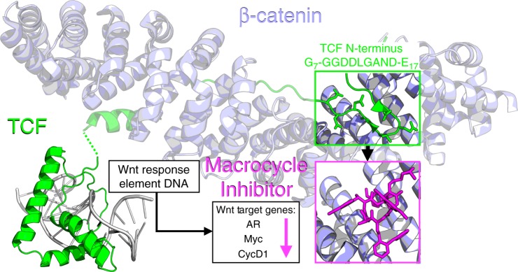
We identified a cleft on the surface of β-catenin created by armadillo repeats 8, 9, and 10 (Fig. 1) which was suitable for targeting with a cyclic oligomer. This region includes the binding site between β-catenin and the N-terminal sequence of Xenopus laevis TCF3, which forms a β-hairpin structure at the site of interaction in the X-ray structure (PDBid: 1G3J)37. The β-catenin binding domain of xTCF3 aligns very closely with the sequence of human TCF4 (Supplementary Fig. 1). This cleft is also adjacent to the G13ANDE17 region of the hTCF4 β-catenin binding sequence which had been mimicked in previous attempts to inhibit the β-catenin:TCF4 interaction48,49.
Fig. 1.
Proposed mechanism for targeting β-Catenin. The designed macrocycles inhibit the interaction of the N-terminal region of TCF and β-Catenin resulting in a downregulation of AR (Androgen Receptor), Myc, and CycD1 (Cyclin-D1)37,72 Crystal structures depicted are PDB 1G3J and 2LEF
We applied rational design techniques to target the xTCF3 β-hairpin binding pocket of β-catenin. In order to identify suitable oligomer scaffolds, we evaluated high-resolution structures experimentally determined for a set of peptoid and peptoid-α-amino acid hybrid macrocyclic structures17,50. One cyclic hexamer peptoid-α-amino acid hybrid (18-atom macrocycle Compound 1, Supplementary Fig. 2, Supplementary Table 1) was observed to provide a favorable steric complement to the targeted β-catenin cleft after modeling with the Rosetta molecular design suite51. The in silico ΔGbinding values were evaluated after several iterations of guided (The PyMOL Molecular Graphics System, Schrödinger, LLC) and automated docking protocols utilizing Rosetta’s InterfaceAnalyzer52,53.
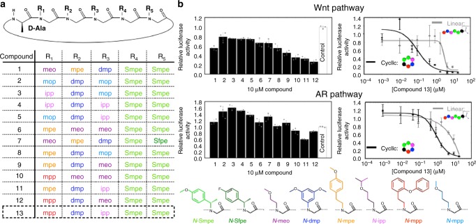
Inspection of the X-ray structure of 1 suggested that the D-Alanine and “N-Smpe” residues played a critical role in directing the overall fold of the oligomer. We hypothesize these bulky α-chiral peptoid side chains and a D-amino acid confer a unique structure to the oligomer backbone (Fig. 2a, positions colored green and black, respectively). In contrast, positions R1, R2, and R3 appeared relatively amenable for substitution to optimize complementarity to the protein surface. After thorough analysis of 1 docked into the β-catenin X-ray structure (Supplementary Fig. 3), we selected a set of side chain types at each of the designable positions based on a combination of visual inspection, modeling, aqueous solubility, and synthetic feasibility, as well as properties considered favorable for cell-permeability. A table of the shape complementarity and in silico ΔGbinding for each of the designs is provided in Supplementary Table 2.
Fig. 2.
Design of oligomer macrocylces to bind β-catenin. a Peptoid–peptide hybrid macrocycle backbone scaffold. Middle table details compound number list and chemical compositions. Bottom section details the chemical compositions of peptoid side chains. b Luciferase reporter assay of compounds relative to a “vehicle” control. Data are presented as mean and SD (n = 3). Notes: “N-meo”: N-(methoxy-ethyl)-glycine, “N-mpe”: N-(4-methoxy-phenyl-ethyl)-glycine, “N-dmp”: N-(3,5-dimethoxy-phenyl)-glycine, “N-Smpe”: N-((S)-4-methoxy-phenyl-ethyl)-glycine, “N-Sfpe”: N-((S)-4-fluoro-phenyl-ethyl)-glycine, “N-mpp”: N-(2-phenoxy-phenyl-ethyl)-glycine, “N-ipp”: N-(isopropoxy-propyl)-glycine, “N-mop”: N-(methoxy-propyl)-glycine
Compound testing and identification of a promising compound
Using a TOP-Flash Wnt luciferase reporter, we tested the 12 oligomers identified from in silico modeling for inhibitory activity (Fig. 2b). At 10 µM concentration, all compounds had some level of Wnt inhibitory activity, with varying degrees of efficacy. We also employed a 3 × -Androgen Response Element (ARE) luciferase reporter to measure the effect on AR activity. Only a few of the compounds were effective at inhibiting AR signaling, the best of which were also the most effective at inhibiting Wnt signaling. Based on the inhibitory data from other compounds containing similar side chain types, we reasoned that if we generated a macrocycle with the “N-ipp” side chain in position R3 and the “N-mpp” side chain in position R1 (Fig. 2a), we could create a more potent compound. An optimal sequence composed of these monomer types resulted in compound 13. This compound was in fact more effective at inhibiting both the Wnt and AR luciferase reporters. A comparison of the luciferase activity inhibition and the calculated ΔGbinding values for each compound yielded a correlation R2 of 0.68 (Supplementary Fig. 4). As additional controls, we obtained previously developed, commercially available Wnt inhibitors and tested them in the Wnt luciferase assay (Supplementary Fig. 5). Inhibitors upstream of β-catenin (porcupine and tankyrase) had no effect, while those targeting β-catenin/TCF (iCRT3, LF3) or β-catenin/CBP (PRI-724) did inhibit the luciferase reporter although with a weaker potency than compound 13.
Macrocycle 13 inhibits Wnt and AR signaling
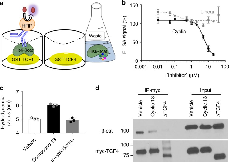
We next characterized the dose response curves for compound 13 in the Wnt and AR luciferase reporters. The linear version of 13 (linear 13: NH2-|N-mpp, N-dmp, N-ipp, N-Smpe, N-Smpe, D-Ala|-OH) was used as a control compound. It incorporates the identical chemical moieties as its cyclic counterpart, but is likely not constrained in an optimal binding conformation and was therefore anticipated to demonstrate weaker affinity. Indeed, as observed in luciferase assays (Fig. 2c), the IC50 for Wnt luciferase inhibition was 0.105 µM ± 0.040 (±standard deviation of three replicates) for 13 compared to 3.27 µM ± 0.99 for the linear oligomer. Compound 13 also showed better inhibition of the AR luciferase reporter (IC50 of 1.02 µM ± 0.20) compared to linear 13 (IC50 of 7.63 µM ± 1.22). While these values may suggest that the compounds inhibit Wnt signaling with increased potency relative to the AR pathway, the experimental conditions used with each reporter, including siRNA knockdown of APC to activate the Wnt reporter and DHT treatment to activate AR signaling, confound direct comparison of the values. To confirm that 13 does inhibit the β-catenin:TCF protein interaction, we developed a sandwich ELISA that detected recombinant β-catenin binding to a GST fusion protein containing the TCF4 N-terminal β-catenin binding domain (Fig. 3a). In this assay, 13 inhibited binding with an IC50 of 5.44 µM±0.82 (Fig. 3b). To test direct binding, we pre-formed dynamic light scattering (DLS) with β-catenin in the presence of vehicle, compound 13, and α-cyclodextrin. A significant shift in the measured hydrodynamic radius was detected with the addition of compound 13, indicative of a conformational change associated with a binding event (Fig. 3c). Addition of a similarly sized macrocyclic compound, α-cyclodextrin, was evaluated as a negative control and did not cause the same shift (additional DLS data presented in Supplemental Table 3). Inhibition of the β-catenin:TCF4 protein interaction was also confirmed in cell culture. HEK293 cells were transiently transfected with a plasmid expressing full length, myc-tagged TCF4. After 48 h treatment with either vehicle (DMSO) or 10 µM 13, cells were lysed and myc-TCF4 protein was immunoprecipitated. Western blotting was performed, revealing that endogenous β-catenin was co-immunoprecipitated. The β-catenin:TCF association was decreased in cells treated with 13 compared to vehicle treated cells (Fig. 3d).
Fig. 3.
Macrocycle 13 inhibits the β-catenin:TCF4 interaction. a Schematic for ELISA based detection of recombinant His6-β-catenin binding to a GST-TCF4 N-terminal Catenin Binding Domain fusion protein. b Dose response curves for the cyclic oligomer (black solid line) and linear oligomer (grey dashed line) in the ELISA assay. c Dynamic light scattering measurements of the hydrodynamic radius of β-catenin in the presence of vehicle, compound 13, or α-cyclodextrin. d Western blot depicting a co-immunoprecipitation (IP) for myc-TCF4 and β-catenin treated with compound. ∆TCF4: A myc-TCF4 construct that lacks the β-catenin binding domain. Data are presented as mean and SD (n = 3) of three independent experiments (c) or representative of three independent experiments (b, d)
Macrocycle 13 inhibits prostate cancer cell growth
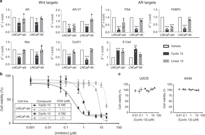
To test the effect of 13 in a model of prostate cancer, we used the LNCaP-abl and LNCaP-95 cell lines. These cell lines were derived from androgen-sensitive LNCaP cells, and passaged in androgen-deprived media to mimic the development of castration resistance in patient tumors54,55. We first isolated mRNA from LNCaP-abl and LNCaP-95 cells treated with vehicle, 10 µM 13 and linear 13 for 24 h to check the expression of endogenous Wnt and AR target genes (Fig. 4a). The mRNA of Wnt target genes cMYC, Cyclin D1, and the AR were all decreased. Transcription of E-cadherin, which has been found to be repressed by Wnt signaling, increased following treatment by 13. AR target genes PSA and FKBP5 mRNA were also decreased. In addition, the transcription of an AR splice variant, AR-V7, which lacks the ligand binding domain and as a consequence is constitutively active, was also decreased. AR-V7 is particularly challenging to target as most AR antagonists target the ligand binding domain. Similar to experiments discussed above, 13 again showed increased potency in transcriptional modulation compared to its linear analog. To check if the changes in Wnt and AR target gene expression translated into effects on growth, the viability of LNCaP-abl and LNCaP-95 cells was determined after 5 days of treatment (Fig. 4b). Compound 13 inhibited proliferation with an IC50 of 195 nM ± 52 (LNCaP-abl) and 782 nM ± 34 (LNCaP-95). We also tested compound 13 in non-prostate cell lines that have been shown to be Wnt independent (Fig. 4c)46,47. Compound 13 had no effect on the proliferation of U2OS (osteosarcoma) and A549 (lung adenocarcinoma) cells.
Fig. 4.
Macrocycle 13 affects prostate cancer cell lines. a RT-qPCR of mRNA collected from LNCaP-abl or LNCaP-95 cells treated with 10 µM 13 (black) or 10 µM linear 13 (grey) oligomer for 24 h. b Cell viability measurements using CellTiter-Fluor reagent with 5 day oligomer treatment in prostate cancer cell lines. c Cell viability measurements with cyclic 13 in two non-prostate, Wnt-independent cell lines. Data are presented as mean and SD (n = 3) of three independent experiments (a) or representative of three independent experiments (b, c). Statistical differences for RT-qPCR were calculated by unpaired t-tests, *p < 0.05, **p < 0.01, ***p < 0.001
Growth of prostate cancer spheroids slowed by macrocycle 13
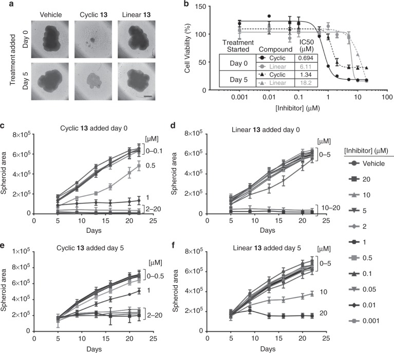
To evaluate the impact of the compounds under 3D cell culture conditions thought to be more representative of tumors, we grew LNCaP-abl cells in low attachment plates. The effects on spheroid formation and growth were evaluated following treatment with vehicle, 13, or linear 13 administered at the time of seeding at low cell density in a 384-well plate. In a separate experiment, to test the impact on pre-formed spheroids, cells were treated 5 days after initial seeding. Spheroids from both experiments were grown for 22 days, with compound refreshed on days 5, 9, 13, 16, and 20. Figure 5a shows representative images of cells treated with vehicle, 2 µM 13, or 2 µM linear 13 at 20 days. PrestoBlue was used to measure cell viability on day 22 (Fig. 5b). Compound 13 reduced spheroid proliferation with an IC50 of 0.694 µM ± 0.037 when added in the initial seeding (Day 0). It also reduced spheroid proliferation when added on day 5, with somewhat lower potency and efficacy and an IC50 of 1.34 µM ± 0.11. Figure 5c–f depicts spheroid area over time based on size measurements from the images taken throughout the experiment. Higher concentrations of 13 (20 µM, 10 µM, 5 µM, 2 µM, and 1 µM) prevented spheroid formation when added on day 0 with seeding and 0.5 µM 13 also slowed growth (Fig. 5c). Concentrations of 13 between 0.001 µM and 0.1 µM had little effect. In the case of compound addition to previously formed spheroids (treatment started at day 5), concentrations of 13 at or above 2 µM stalled the growth of pre-formed spheroids but caused less of a decrease in spheroid area (Fig. 5e) than addition at day 0. Linear 13 had similar effects with significantly reduced potency (Fig. 5d, f).
Fig. 5.
Macrocycle 13 slows prostate cancer spheroid cell growth. a Representative images of tumor spheroids imaged from above on day 20 and treated with vehicle or 2 µM oligomer. Scale bar represents 400 μm. b End point viability measurements taken on day 22 using PrestoBlue. c–f Time course of spheroid areas calculated from images taken throughout the experiment. Data are presented as mean and SD (n = 5 (c–f), n = 2 (b))
Rescue of eye development in Wnt-activated zebrafish embryos
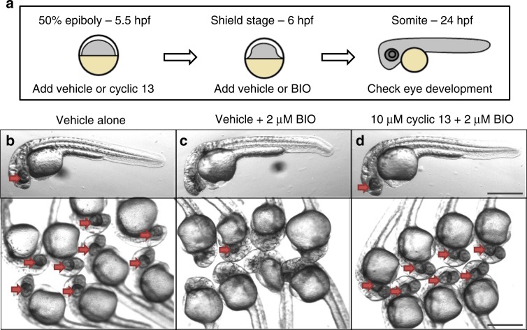
To evaluate the efficacy of 13 in vivo, we utilized a Danio rerio (zebrafish) model. The relevance of the model for our studies is that zebrafish embryos with Wnt activating mutations in Axin1, a component of the β-catenin destruction complex, fail to develop eyes and a forebrain56. The phenotype can be recapitulated by treatment with an inhibitor (termed BIO) for another β-catenin destruction complex member, GSK3, in embryos at 6 h post-fertilization (hpf)57. It would then be anticipated that 13 could inhibit the chemically overactivated Wnt signal, and potentially rescue eye and forebrain development. To test this, BIO was added to zebrafish embryos at 6 hpf with and without 13 as described in Fig. 6a. Indeed, 13 inhibited Wnt signaling as anticipated, as only one of the eight fish treated with GSK3 inhibitor BIO alone developed eyes, while all the fish treated with both BIO and 13 developed eyes (Fig. 6b–d). This result was reproduced in 2 additional independent experiments each showing that 0/8 BIO treated fish developed eyes while 8/8 BIO plus 13 treated fish developed eyes. In addition, linear 13 was tested and in each replicate 6/8 fish developed eyes although most of the eyes appeared smaller and less developed (Supplementary Fig. 6a). This result is consistent with cell culture experiments showing cyclic 13 is more potent than linear 13 (Figs. 4, 5). Images of replicate experiments are shown in Supplementary Fig. 6b.
Fig. 6.
Zebrafish phenotypic assay of Wnt signaling. a Schematic for experimental design. b–d Images of 24 h post-fertilization of zebrafish embryos treated with vehicle alone (b), vehicle plus BIO to activate Wnt signaling (c), or macrocycle 13 in combination with BIO (d). Red arrows point to developing eye structures. Scale bar represents 500 μm
Discussion
The Wnt pathway remains a tantalizing but elusive target for drug discovery38. Chemical inhibition of Wnt signaling has been achieved through multiple strategies. For example, Wnt secretion and function necessitates the post-translational addition of a palmitoyl group, catalyzed by the acyltransferase porcupine. Enzymatic inhibitors of porcupine have been developed and shown to inhibit the secretion of Wnts58,59. In addition, antibodies against frizzled receptors have also been developed to inhibit Wnt signaling at the cell surface60. Both of these methods have therapeutic potential for tumors bearing mutations in R-spondin or the ubiquitin ligases ZNRF3 and RNF43, which cause amplification of Wnt signals. Clinical trials for such Wnt inhibitory agents have been initiated. Nevertheless, the majority of activating Wnt mutations occur in the APC gene or the phosphorylation domain of β-catenin, downstream of both porcupine and frizzled. Thus, it is desirable to identify inhibitors downstream of these mutations. Multiple interactions have been described between β-catenin and components of the Wnt enhanceosome on promotors of Wnt target genes61. The central interaction between β-catenin and TCF is critical to its Wnt activation and represents an attractive target for the general inhibition of canonical Wnt signaling.
In this study, we demonstrate how computational tools can facilitate the design of oligomers that target β-catenin and disrupt its interaction with TCF. We evaluate peptoid–peptide hybrid macrocycles designed to fold into structures complementary to a region on the β-catenin surface that interacts with TCF. We demonstrate that an oligomeric macrocycle exhibits potent antiproliferative effects on prostate cancer cell lines in both 2D and 3D models. In vivo efficacy of this compound (13) is established in a zebrafish model of Wnt signaling. The hypothesis that the conformational constraint imposed on the macrocyclic oligomer enforces steric and chemical complementarity for the β-catenin surface is strongly supported by comparisons to a linear analog, which exhibits markedly diminished activities in vitro and in vivo.
In our proposed mechanism of action, the oligomer macrocycle inhibits canonical Wnt target gene expression via inhibiting the interaction between β-catenin and TCF. We also observe decreased AR pathway target gene expression, which could result either from direct inhibition of the β-catenin:AR interaction or indirectly through the decrease in AR gene transcript levels upon Wnt signal inhibition. It remains to be shown which pathway when inhibited, Wnt or AR, plays a critical role in the antiproliferative phenotype. There may be a synergistic effect of inhibiting both pathways concurrently.
The β-catenin:TCF PPI has been targeted previously38,46,47. However, there does not appear to be rapid clinical implementation of this pharmacological approach38. An example of possible difficulties is exemplified by iCRT3 whereby the compound was not systemically stable, requiring direct tumor injection in order to attain growth inhibition in mouse xenograft experiments62. In contrast, peptoids have been shown to be stable to proteolytic cleavage8,9. Additionally, peptoids have been shown to exhibit generally improved membrane permeability compared to the analogous peptide sequences63,64. Favorable permeability profiles were shown to be consistent and tolerant to variations in oligomer side chain type65. Recently, another hexameric peptoid-peptide macrocycle, designed as a CXCR7 modulator, was demonstrated to exhibit both membrane permeability and oral bioavailability in rats66. In addition to pharmacokinetic issues, there is the potential that targeting β-catenin will lead to on-target toxicity67. It remains to be seen whether it is possible to achieve a therapeutic window where the inhibitor lowers overactivated Wnt signaling, such as in β-catenin and APC mutated tumors, but does not negatively affect basal Wnt signaling levels in systems such as the intestine and bone that require it for general maintenance. The potential for achieving this balance is supported by our zebrafish experiment, in which compound 13 successfully inhibits the artificially overactivated Wnt signaling pathway but does not otherwise harm embryo development. Additional β-catenin nuclear interactions have also been targeted, such as with BCL9 and CBP68,69. The β-catenin/CBP inhibitor PRI-724 has reached the clinic, undergoing trials for colon cancer, AML, and pancreatic adenocarcinoma70. Because PRI-724 binds to CBP (and not β-catenin), it may have a different therapeutic profile than a drug targeting β-catenin:TCF and there may be opportunities for combination therapy with compound 13.
Future testing with 13 in mouse xenograft models of prostate cancer will be important to determine its therapeutic potential, as demonstrated for other bioactive peptoid oligomers71. The ability to introduce a broad range of alternative oligomer side chains should facilitate further improvements of activity along with enhanced pharmacokinetic and pharmacodynamic properties towards the identification of an optimized lead compound. In addition to promising activity versus prostate cancer, macrocycles targeting β-catenin:TCF could also be of therapeutic benefit in other forms of cancer for which Wnt signaling is similarly pro-tumorigenic, such as colon and breast cancers. More generally, this study portends further computer-assisted discovery of increasingly complex folded oligomers to address the vast number of different PPI relevant to human disease.
Methods
Synthesis of compounds 1–13
2-chlorotrityl resin (Anaspec) was incubated with Fmoc-D-Alanine (Sigma) for 1.5 h followed by washing 5× with DMF. Deprotection was carried out with 20% piperidine in DMF. Peptoid residues were then added with iterative steps of 1.2 M bromoacetic acid + DIC for 20 min and 1 M amine for 1 h. Compound 13 used the following amines: S-(-)-4-methoxy-α-methylbenzylamine (Sigma), 3-isopropoxypropylamine (Sigma), 3,5-dimethoxyaniline (Sigma, overnight incubation), and 2-phenoxyphenethylamine (Sigma). The resin was cleaved with 8:1:1 DCM:Acetic acid:HFIP for 1 h. The DCM was removed via rotary evaporation and the remaining solid was dissolved in 50:50 water:ACN and lyophilized overnight. Reverse phase HPLC was used to purify the linear product. Cyclization was carried out using 25 mg of linear compound with 5X PyBOP and 10X DIEA in dry DCM with less than 1 mg/ml compound overnight under argon. The DCM was removed via rotary evaporation and the remaining solid was dissolved in 50:50 water:ACN and lyophilized overnight. The final product was purified to > 95% by reverse phase HPLC and verified by liquid chromatography-mass spectrometry (Agilent LCMSD Trap XCT). Mass spectrometry peak identified for compound 13 (m/z): [M + Na]+ calcd. for C59H72N6O12Na 1079.5; found, 1078.346. A key for chemical abbreviations is supplied in Supplementary Table 4.
Cell culture
LNCaP-abl and LNCaP-95 cells were cultured in indicator free RPMI 1640 media (Life) supplemented with 10% charcoal stripped FBS, 1% pennicillin/streptomycin, and 1% L-Glutamine. HEK-293 Cells were cultured in DMEM media (Life) supplemented with 10%FBS and 1% pennicillin/streptomycin. A549 and U2OS cells were cultured in RPMI 1640 media (Life) and McCoy’s 5 A modified media (Life), respectively, both supplemented with 10%FBS and 1% pennicillin/streptomycin. LNCaP-abl, and LNCaP-95 cell lines were generous gifts from Z. Culig (Innsbruck Medical University, Austria) and J. Isaacs (Johns Hopkins University, Baltimore, MD), respectively. Cell lines were authenticated by STR profiling (Genetica Cell Line Testing; Burlington, NC). A549 and U2OS were purchased from ATCC (product numbers CCL-185 and HTB-96). Cell lines were routinely screened for mycoplasma. Luciferase assays were conducted using stably transfected TOP-flash and 3xARE LNCaP-abl cell lines. For the Wnt pathway, TOP-Flash LNCaP-abl cells were treated with 1 nM siAPC (a 1:1:1 mix of Ambion Silencer Silect siAPC RNA, ID#s s1433, s1434, s1435) for 24 h and seeded into 384-well plates at 5000 cells/well, followed by 24 h of treatment with compound. For the AR-pathway, 3xARE LNCaP-abl cells were seeded into 384-well plates at 5000 cells per well, treated for 48 h with compound, with the addition of R1881 for the final 8 h. Both were measured using the ONE-Glo detection kit (Promega) and data is standardized to CellTiter-Fluor (Promega) viability readings to account for potential variations in well to well cell number. Control Wnt inhibitors were purchased from Sigma with the exception of PRI-724 which was purchased from Selleckchem. Co-immunoprecipitation was carried out using HEK293 cells transfected with either myc-TCF (addgene #32738) or myc-TCF dominant negative (addgene #32739) followed by 48 h of treatment with compound. Cells were lysed using Triton lysis buffer and 1 mg total protein was incubated overnight at 4 C with 40 µl anti-cMyc magnetic beads (Thermo). Total protein concentration in cell lysates were determined using Pierce Rapid Gold BCA Protein Assay Kit (Thermo). The beads were washed 3× with lysis buffer then boiled in 2X sample loading dye. SDS-PAGE and western blotting were performed using anti-Myc (CST 2276) and anti-β-catenin (CST 8480). Both antibodies were diluted in blocking buffer 1:1000 and incubated overnight at 4 C with the membrane. Full blots from IP (Fig. 3d) are depicted in Supplementary Figure 7. RT-qPCR was performed using RNA isolated with the RNEasy kit (Qiagen) from LNCaP-abl and LNCaP-95 cells after 24 h treatment with 10 µM compound. The Verso cDNA synthesis kit (Thermo) with 1 µg total RNA was used to generate cDNA. RNA concentration was determined using a Thermo Scientific NanoDrop 2000 spectrophotometer. RT-qPCR data was collected using Fast SYBR Green Master Mix on a QuantiStudio 6 Flex (Life Technologies). RT-qPCR data are presented as averages relative to RPL19 Ct values (Δ#1) and DMSO control samples(Δ#2) as relative quantitation (2-ΔΔCt) in triplicate of biological replicates. RT-qPCR primers are given in Supplementary Table 5. 2D proliferation assays were conducted using 384-well plates seeded with 1000 cells/well for LNCaP-abl or LNCaP-95 cells and 1000 cells/well for A549 or U2OS cells. After 5 days of treatment the cell viability was measured with the CellTiter-Fluor Cell Viability Assay (Promega) and data is standardized to vehicle control.
Recombinant proteins and ELISA
The ELISA was conducted using recombinant GST-tagged catenin binding domain (CBD) of hTCF4, AA8-54, and His6-β-catenin armadillo domain, AA134-668. Both proteins were expressed in BL21 DL3 (NEB) bacteria grown to OD600 0.6 and induced with 0.5 mM IPTG (Sigma) for β-catenin and 0.1 mM IPTG for TCF. The cells were then shaken for 8 h at 30 C. The bacteria were then pelleted, resuspended in PBS, and lysed with a bacterial FRENCH pressure cell press (Thermo). The lysates were incubated overnight with Ni-NTA or glutathione affixed resin (Thermo), washed with TBS, and eluted using 250 mM imidazole or 20 mM glutathione in 20 mM Tris-HCl pH 8.0 and 200 mM NaCl. The imidazole or glutathione were removed by dialysis into a buffer containing 20 mM Tris-HCL pH 8.0, 200 mM NaCl, and 10% glycerol. Lysates were then aliquoted and frozen at −80 °C. The ELISA was initiated with adsorption of 5 µg/ml GST-TCF CBD into a 96-well Nunc Maxisorp plate (Fisher) overnight at 4 °C. BSA was adsorbed to control wells. The wells were washed, blocked with 5% milk for 1 h, washed, incubated with 5 nM His6-β-catenin with varying concentrations of compound in washing buffer for 1 h, washed, incubated with anti-His6 (Thermo MA1-135) diluted 1:10000 in blocking buffer for 1 h, washed, incubated with anti-mouse-HRP (Santa Cruz) diluted 1:15000 in blocked buffer for 1 h, washed, and then developed using 1-Step Ultra TMB-ELISA (Thermo). His6-β-catenin was pre-incubated with compound for 30 min prior to its addition to the plate.
Dynamic light scattering
Dynamic light scattering was measured with a Wyatt DynaPro NanoStar. Aliquots of recombinant frozen His6-β-catenin were thawed fresh for each experiment and centrifuged at high speed for 10 min. Compound 13 or α-cyclodextrin (Sigma) were added to a concentration of 10 μM and compared with the addition of 1% DMSO (vehicle). Ten readings were measured per experiment, each with an integration time of 5 s. Three experiments were performed per condition. Data was collected and analyzed using DYNAMICS software (Wyatt).
Spheroid 3D cell culture
LNCaP-abl cells were seeded into 384-well ultra-low attachment plates (Corning), 1000 cells/well. Varying concentrations of compound were added either on the same day as seeding or on day 5. Media was changed every 3–4 days using a Biotek automated media exchanger. Images were taken using an ArraySca VTI (Cellomics) on days 5, 9, 13, 16, 20, and 22. Images were analyzed for spheroid area with the HCS Studio Cellomics Scan Version 6.6.0 (Thermo) software. A final viability measurement was taken on day 22 using PrestoBlue reagent (Thermo).
Zebrafish experiments
The zebrafish phenotypic assay was conducted using wild-type zebrafish embryos seeded into a 96-well round bottom plate at 4 hpf in 50 µl of Fish Water (60 mg/l sea salt, Instant Ocean, Blacksburg, VA, USA) supplemented with 1 mg per L Methylene Blue (Sigma-Aldrich). The embryos were not dechorionated. At 50% epiboly, roughly 5.5 hpf, 50 µl of 30 µM 13 or Linear 13 was added. About 30 min later at shield stage, 50 µl of 6 nM (3×) BIO was added. The embryos were then visualized for the presence of eyes and imaged at 30 hpf. Zebrafish embryos were randomly assigned to non-blinded treatment groups, none were excluded. Sample size was determined with a power calculation to achieve statistical significance (alpha = 0.05, beta = 0.2) if only 60% of the fish had developed eyes. Zebrafish experiments were approved by the NYULMC Animal Care & Use Program under IACUC Protocol Number: 170105-02. All ethical regulations were complied with.
Electronic supplementary material
Acknowledgements
We thank Sarah Blosser and Dr. Paramjit Arora for the His6-β-catenin bacterial expression plasmid. We also thank Tenzin Gocha and Dr. Ram Dasgupta for the GST-TCF4 catenin binding domain bacterial expression plasmid. This research was supported by NIH CA112226 (SKL), NIH T32GM007308 (JAS), NIH 5T32CA009161 (JAS), NIH NS069839 (HK), and NSF CHE-1507964 (KK). V.S. and E.N are supported by the NIH R01GM126891, Blavatnik Family Foundation, and by the Howard Hughes Medical Institute. The High Throughput Biology Laboratory is supported by NIH/NCI P30CA16087 and NYSTEM Contract C026719. T.W.C. gratefully acknowledges the New York University Biology Department for the Fleur Strand Fellowship as well as the Graduate School of Arts and Sciences for the Horizon Fellowship in the Natural and Physical Sciences. We thank Dr. Chunhua (Tony) Hu at the NYU Department of Chemistry for assistance with X-ray structure determination.
Author contributions
J.A.S., T.W.C., C.Y., V.S., E.N., H.K., R.B., M.J.G., K.K., and S.K.L. designed research; T.W.C., K.K., and R.B. performed modeling and compound design; T.W.C., J.A.S., and A.K. synthesized compounds; M.H. crystalized compound 1; E.M.B. and J.A.S. performed luciferase screening; V.S. performed the DLS assay; J.A.S expressed and purified proteins, and performed cell culture, ELISA, IP, and zebrafish experiments; J.A.S., T.W.C., M.J.G., K.K., and S.K.L. analyzed data; and J.A.S, T.W.C, K.K., and S.K.L. wrote the paper.
Data availability
All relevant data is available from the authors upon request. The X-ray crystallographic coordinates of compound 1 have been deposited at the Cambridge Crystallographic Data Centre (CCDC), under deposition number 1866347. The structure can be obtained free of charge from The Cambridge Crystallographic Data Centre via www.ccdc.cam.ac.uk/data_request/cif.
Competing interests
The authors declare no competing interests.
Footnotes
Publisher's note: Springer Nature remains neutral with regard to jurisdictional claims in published maps and institutional affiliations.
These authors contributed equally: Jeffrey A. Schneider, Timothy W. Craven.
Contributor Information
Kent Kirshenbaum, Email: kk54@nyu.edu.
Susan K. Logan, Email: Susan.Logan@nyulangone.org
Electronic supplementary material
Supplementary Information accompanies this paper at 10.1038/s41467-018-06845-3.
References
- 1.Wojcik P, Berlicki L. Peptide-based inhibitors of protein–protein interactions. Bioorg. Med. Chem. Lett. 2016;26:707–713. doi: 10.1016/j.bmcl.2015.12.084. [DOI] [PubMed] [Google Scholar]
- 2.Arkin MR, Tang Y, Wells JA. Small-molecule inhibitors of protein–protein interactions: progressing toward the reality. Chem. Biol. 2014;21:1102–1114. doi: 10.1016/j.chembiol.2014.09.001. [DOI] [PMC free article] [PubMed] [Google Scholar]
- 3.Ran X, Gestwicki JE. Inhibitors of protein–protein interactions (PPIs): an analysis of scaffold choices and buried surface area. Curr. Opin. Chem. Biol. 2018;44:75–86. doi: 10.1016/j.cbpa.2018.06.004. [DOI] [PMC free article] [PubMed] [Google Scholar]
- 4.Rubin S, Qvit N. Cyclic peptides for protein–protein interaction targets: applications to human disease. Crit. Rev. Eukaryot. Gene Expr. 2016;26:199–221. doi: 10.1615/CritRevEukaryotGeneExpr.2016016525. [DOI] [PubMed] [Google Scholar]
- 5.Matsson P, Kihlberg J. How big is too big for cell permeability? J. Med. Chem. 2017;60:1662–1664. doi: 10.1021/acs.jmedchem.7b00237. [DOI] [PubMed] [Google Scholar]
- 6.Akram ON, et al. Tailoring peptidomimetics for targeting protein–protein interactions. Mol. Cancer Res. 2014;12:967–978. doi: 10.1158/1541-7786.MCR-13-0611. [DOI] [PubMed] [Google Scholar]
- 7.Goodman CM, Choi S, Shandler S, DeGrado WF. Foldamers as versatile frameworks for the design and evolution of function. Nat. Chem. Biol. 2007;3:252–262. doi: 10.1038/nchembio876. [DOI] [PMC free article] [PubMed] [Google Scholar]
- 8.Miller SM, et al. Comparison of the proteolytic susceptibilities of homologous L-Amino-Acid, D-Amino-Acid, and N-substituted glycine peptide and peptoid oligomers. Drug Dev. Res. 1995;35:20–32. doi: 10.1002/ddr.430350105. [DOI] [Google Scholar]
- 9.Wang Y, et al. Absorption and disposition of a tripeptoid and a tetrapeptide in the rat. Biopharm. Drug. Dispos. 1999;20:69–75. doi: 10.1002/(SICI)1099-081X(199903)20:2<69::AID-BDD153>3.0.CO;2-B. [DOI] [PubMed] [Google Scholar]
- 10.Culf AS, Ouellette RJ. Solid-phase synthesis of N-substituted glycine oligomers (alpha-peptoids) and derivatives. Molecules. 2010;15:5282–5335. doi: 10.3390/molecules15085282. [DOI] [PMC free article] [PubMed] [Google Scholar]
- 11.Zuckermann RN, Kerr JM, Kent SBH, Moos WH. Efficient method for the preparation of peptoids [Oligo(N-Substituted Glycines)] by submonomer solid-phase synthesis. J. Am. Chem. Soc. 1992;114:10646–10647. doi: 10.1021/ja00052a076. [DOI] [Google Scholar]
- 12.Butterfoss GL, Renfrew PD, Kuhlman B, Kirshenbaum K, Bonneau R. A preliminary survey of the peptoid folding landscape. J. Am. Chem. Soc. 2009;131:16798–16807. doi: 10.1021/ja905267k. [DOI] [PubMed] [Google Scholar]
- 13.Shah NH, et al. Oligo(N-aryl glycines): a new twist on structured peptoids. J. Am. Chem. Soc. 2008;130:16622–16632. doi: 10.1021/ja804580n. [DOI] [PubMed] [Google Scholar]
- 14.Stringer JR, Crapster JA, Guzei IA, Blackwell HE. Extraordinarily robust polyproline type I peptoid helices generated via the incorporation of alpha-chiral aromatic N-1-naphthylethyl side chains. J. Am. Chem. Soc. 2011;133:15559–15567. doi: 10.1021/ja204755p. [DOI] [PMC free article] [PubMed] [Google Scholar]
- 15.Wu CW, Sanborn TJ, Huang K, Zuckermann RN, Barron AE. Peptoid oligomers with alpha-chiral, aromatic side chains: sequence requirements for the formation of stable peptoid helices. J. Am. Chem. Soc. 2001;123:6778–6784. doi: 10.1021/ja003154n. [DOI] [PubMed] [Google Scholar]
- 16.Roy O, et al. Homogeneous and robust polyproline type I helices from peptoids with nonaromatic alpha-chiral side chains. J. Am. Chem. Soc. 2017;139:13533–13540. doi: 10.1021/jacs.7b07475. [DOI] [PubMed] [Google Scholar]
- 17.Shin SB, Yoo B, Todaro LJ, Kirshenbaum K. Cyclic peptoids. J. Am. Chem. Soc. 2007;129:3218–3225. doi: 10.1021/ja066960o. [DOI] [PubMed] [Google Scholar]
- 18.Huang ML, Shin SB, Benson MA, Torres VJ, Kirshenbaum K. A comparison of linear and cyclic peptoid oligomers as potent antimicrobial agents. ChemMedChem. 2012;7:114–122. doi: 10.1002/cmdc.201100358. [DOI] [PubMed] [Google Scholar]
- 19.Renfrew PD, Craven TW, Butterfoss GL, Kirshenbaum K, Bonneau R. A rotamer library to enable modeling and design of peptoid foldamers. J. Am. Chem. Soc. 2014;136:8772–8782. doi: 10.1021/ja503776z. [DOI] [PMC free article] [PubMed] [Google Scholar]
- 20.D’Amato A, et al. Cyclic peptoids as topological templates: synthesis via central to conformational chirality induction. Org. Lett. 2018;20:640–643. doi: 10.1021/acs.orglett.7b03786. [DOI] [PubMed] [Google Scholar]
- 21.Hosseinzadeh P, et al. Comprehensive computational design of ordered peptide macrocycles. Science. 2017;358:1461–1466. doi: 10.1126/science.aap7577. [DOI] [PMC free article] [PubMed] [Google Scholar]
- 22.Butterfoss GL, et al. De novo structure prediction and experimental characterization of folded peptoid oligomers. Proc. Natl Acad. Sci. USA. 2012;109:14320–14325. doi: 10.1073/pnas.1209945109. [DOI] [PMC free article] [PubMed] [Google Scholar]
- 23.Drew K, et al. Adding diverse noncanonical backbones to rosetta: enabling peptidomimetic design. PLoS ONE. 2013;8:e67051. doi: 10.1371/journal.pone.0067051. [DOI] [PMC free article] [PubMed] [Google Scholar]
- 24.Cancer Facts & Figures 2017. (American Cancer Society, Georgia, 2017).
- 25.Scher HI, et al. Increased survival with enzalutamide in prostate cancer after chemotherapy. N. Engl. J. Med. 2012;367:1187–1197. doi: 10.1056/NEJMoa1207506. [DOI] [PubMed] [Google Scholar]
- 26.de Bono JS, et al. Abiraterone and increased survival in metastatic prostate cancer. N. Engl. J. Med. 2011;364:1995–2005. doi: 10.1056/NEJMoa1014618. [DOI] [PMC free article] [PubMed] [Google Scholar]
- 27.Chandrasekar T, Yang JC, Gao AC, Evans CP. Mechanisms of resistance in castration-resistant prostate cancer (CRPC) Transl. Androl. Urol. 2015;4:365–380. doi: 10.3978/j.issn.2223-4683.2015.05.02. [DOI] [PMC free article] [PubMed] [Google Scholar]
- 28.Saraon P, Jarvi K, Diamandis EP. Molecular alterations during progression of prostate cancer to androgen independence. Clin. Chem. 2011;57:1366–1375. doi: 10.1373/clinchem.2011.165977. [DOI] [PubMed] [Google Scholar]
- 29.Li Y, et al. LEF1 in androgen-independent prostate cancer: regulation of androgen receptor expression, prostate cancer growth, and invasion. Cancer Res. 2009;69:3332–3338. doi: 10.1158/0008-5472.CAN-08-3380. [DOI] [PMC free article] [PubMed] [Google Scholar]
- 30.Yang X, et al. Complex regulation of human androgen receptor expression by Wnt signaling in prostate cancer cells. Oncogene. 2006;25:3436–3444. doi: 10.1038/sj.onc.1209366. [DOI] [PMC free article] [PubMed] [Google Scholar]
- 31.Song LN, et al. Beta-catenin binds to the activation function 2 region of the androgen receptor and modulates the effects of the N-terminal domain and TIF2 on ligand-dependent transcription. Mol. Cell. Biol. 2003;23:1674–1687. doi: 10.1128/MCB.23.5.1674-1687.2003. [DOI] [PMC free article] [PubMed] [Google Scholar]
- 32.Robinson D, et al. Integrative clinical genomics of advanced prostate cancer. Cell. 2015;161:1215–1228. doi: 10.1016/j.cell.2015.05.001. [DOI] [PMC free article] [PubMed] [Google Scholar]
- 33.Grasso CS, et al. The mutational landscape of lethal castration-resistant prostate cancer. Nature. 2012;487:239–243. doi: 10.1038/nature11125. [DOI] [PMC free article] [PubMed] [Google Scholar]
- 34.Clevers H, Nusse R. Wnt/beta-catenin signaling and disease. Cell. 2012;149:1192–1205. doi: 10.1016/j.cell.2012.05.012. [DOI] [PubMed] [Google Scholar]
- 35.Nusse R, Clevers H. Wnt/beta-catenin signaling, disease, and emerging therapeutic modalities. Cell. 2017;169:985–999. doi: 10.1016/j.cell.2017.05.016. [DOI] [PubMed] [Google Scholar]
- 36.Shapiro L. beta-catenin and its multiple partners: promiscuity explained. Nat. Struct. Biol. 2001;8:484–487. doi: 10.1038/88532. [DOI] [PubMed] [Google Scholar]
- 37.Graham TA, Weaver C, Mao F, Kimelman D, Xu W. Crystal structure of a β-Catenin/Tcf complex. Cell. 2000;103:885–896. doi: 10.1016/S0092-8674(00)00192-6. [DOI] [PubMed] [Google Scholar]
- 38.Lyou Y, Habowski AN, Chen GT, Waterman ML. Inhibition of nuclear Wnt signalling: challenges of an elusive target for cancer therapy. Br. J. Pharmacol. 2017;174:4589–4599. doi: 10.1111/bph.13963. [DOI] [PMC free article] [PubMed] [Google Scholar]
- 39.Hahne G, Grossmann TN. Direct targeting of beta-catenin: Inhibition of protein–protein interactions for the inactivation of Wnt signaling. Bioorg. Med. Chem. 2013;21:4020–4026. doi: 10.1016/j.bmc.2013.02.050. [DOI] [PubMed] [Google Scholar]
- 40.Gonsalves FC, et al. An RNAi-based chemical genetic screen identifies three small-molecule inhibitors of the Wnt/wingless signaling pathway. Proc. Natl Acad. Sci. USA. 2011;108:5954–5963. doi: 10.1073/pnas.1017496108. [DOI] [PMC free article] [PubMed] [Google Scholar]
- 41.Fang L, et al. A small-molecule antagonist of the beta-Catenin/TCF4 interaction blocks the self-renewal of cancer stem cells and suppresses tumorigenesis. Cancer Res. 2016;76:891–901. doi: 10.1158/0008-5472.CAN-15-1519. [DOI] [PubMed] [Google Scholar]
- 42.Lepourcelet M, et al. Small-molecule antagonists of the oncogenic Tcf/beta-catenin protein complex. Cancer Cell. 2004;5:91–102. doi: 10.1016/S1535-6108(03)00334-9. [DOI] [PubMed] [Google Scholar]
- 43.Zhang M, Catrow JL, Ji H. High-throughput selectivity assays for small-molecule inhibitors of beta-Catenin/T-cell factor protein–protein interactions. ACS Med. Chem. Lett. 2013;4:306–311. doi: 10.1021/ml300367f. [DOI] [PMC free article] [PubMed] [Google Scholar]
- 44.Trosset JY, et al. Inhibition of protein–protein interactions: the discovery of druglike beta-catenin inhibitors by combining virtual and biophysical screening. Proteins. 2006;64:60–67. doi: 10.1002/prot.20955. [DOI] [PubMed] [Google Scholar]
- 45.Tian W, et al. Structure-based discovery of a novel inhibitor targeting the beta-catenin/Tcf4 interaction. Biochemistry. 2012;51:724–731. doi: 10.1021/bi201428h. [DOI] [PubMed] [Google Scholar]
- 46.Grossmann TN, et al. Inhibition of oncogenic Wnt signaling through direct targeting of beta-catenin. Proc. Natl Acad. Sci. USA. 2012;109:17942–17947. doi: 10.1073/pnas.1208396109. [DOI] [PMC free article] [PubMed] [Google Scholar]
- 47.Dietrich L, et al. Cell permeable stapled peptide inhibitor of Wnt signaling that targets beta-Catenin protein–protein interactions. Cell Chem. Biol. 2017;24:958–968. doi: 10.1016/j.chembiol.2017.06.013. [DOI] [PubMed] [Google Scholar]
- 48.Huang Z, Zhang M, Burton SD, Katsakhyan LN, Ji H. Targeting the Tcf4 G13ANDE17 binding site to selectively disrupt beta-catenin/T-cell factor protein–protein interactions. ACS Chem. Biol. 2014;9:193–201. doi: 10.1021/cb400795x. [DOI] [PubMed] [Google Scholar]
- 49.Catrow JL, Zhang Y, Zhang M, Ji H. Discovery of selective small-molecule inhibitors for the beta-Catenin/T-cell factor protein–protein interaction through the optimization of the Acyl Hydrazone moiety. J. Med. Chem. 2015;58:4678–4692. doi: 10.1021/acs.jmedchem.5b00223. [DOI] [PubMed] [Google Scholar]
- 50.Vollrath SBL, Brase S, Kirshenbaum K. Twice tied tight: enforcing conformational order in bicyclic peptoid oligomers. Chem. Sci. 2012;3:2726–2731. doi: 10.1039/c2sc20473h. [DOI] [Google Scholar]
- 51.Leaver-Fay A, et al. ROSETTA3: an object-oriented software suite for the simulation and design of macromolecules. Methods Enzymol. 2011;487:545–574. doi: 10.1016/B978-0-12-381270-4.00019-6. [DOI] [PMC free article] [PubMed] [Google Scholar]
- 52.Kaufmann KW, Lemmon GH, Deluca SL, Sheehan JH, Meiler J. Practically useful: what the Rosetta protein modeling suite can do for you. Biochemistry. 2010;49:2987–2998. doi: 10.1021/bi902153g. [DOI] [PMC free article] [PubMed] [Google Scholar]
- 53.Stranges PB, Kuhlman B. A comparison of successful and failed protein interface designs highlights the challenges of designing buried hydrogen bonds. Protein Sci. 2013;22:74–82. doi: 10.1002/pro.2187. [DOI] [PMC free article] [PubMed] [Google Scholar]
- 54.Culig Z, et al. Switch from antagonist to agonist of the androgen receptor blocker bicalutamide is associated with prostate tumour progression in a new model system. Brit J. Cancer. 1999;81:242–251. doi: 10.1038/sj.bjc.6690684. [DOI] [PMC free article] [PubMed] [Google Scholar]
- 55.Pflug BR, Reiter RE, Nelson JB. Caveolin expression is decreased following androgen deprivation in human prostate cancer cell lines. Prostate. 1999;40:269–273. doi: 10.1002/(SICI)1097-0045(19990901)40:4<269::AID-PROS9>3.0.CO;2-6. [DOI] [PubMed] [Google Scholar]
- 56.Heisenberg CP, et al. A mutation in the Gsk3-binding domain of zebrafish Masterblind/Axin1 leads to a fate transformation of telencephalon and eyes to diencephalon. Genes Dev. 2001;15:1427–1434. doi: 10.1101/gad.194301. [DOI] [PMC free article] [PubMed] [Google Scholar]
- 57.Nishiya N, et al. A zebrafish chemical suppressor screening identifies small molecule inhibitors of the Wnt/beta-catenin pathway. Chem. Biol. 2014;21:530–540. doi: 10.1016/j.chembiol.2014.02.015. [DOI] [PubMed] [Google Scholar]
- 58.Liu J, et al. Targeting Wnt-driven cancer through the inhibition of Porcupine by LGK974. Proc. Natl Acad. Sci. USA. 2013;110:20224–20229. doi: 10.1073/pnas.1314239110. [DOI] [PMC free article] [PubMed] [Google Scholar]
- 59.Wang X, et al. The development of highly potent inhibitors for porcupine. J. Med. Chem. 2013;56:2700–2704. doi: 10.1021/jm400159c. [DOI] [PMC free article] [PubMed] [Google Scholar]
- 60.Gurney A, et al. Wnt pathway inhibition via the targeting of Frizzled receptors results in decreased growth and tumorigenicity of human tumors. Proc. Natl Acad. Sci. USA. 2012;109:11717–11722. doi: 10.1073/pnas.1120068109. [DOI] [PMC free article] [PubMed] [Google Scholar]
- 61.van Tienen, L. M., Mieszczanek, J., Fiedler, M., Rutherford, T. J. & Bienz, M.Constitutive scaffolding of multiple Wnt enhanceosome components byQ7 Legless/BCL9. Elife6, 10.7554/eLife.20882.001 (2017). [DOI] [PMC free article] [PubMed]
- 62.Lee E, et al. Inhibition of androgen receptor and beta-catenin activity in prostate cancer. Proc. Natl Acad. Sci. USA. 2013;110:15710–15715. doi: 10.1073/pnas.1218168110. [DOI] [PMC free article] [PubMed] [Google Scholar]
- 63.Kwon YU, Kodadek T. Quantitative evaluation of the relative cell permeability of peptoids and peptides. J. Am. Chem. Soc. 2007;129:1508–1509. doi: 10.1021/ja0668623. [DOI] [PMC free article] [PubMed] [Google Scholar]
- 64.Tan NC, Yu P, Kwon YU, Kodadek T. High-throughput evaluation of relative cell permeability between peptoids and peptides. Bioorg. Med. Chem. 2008;16:5853–5861. doi: 10.1016/j.bmc.2008.04.074. [DOI] [PMC free article] [PubMed] [Google Scholar]
- 65.Furukawa A, et al. Passive membrane permeability in cyclic peptomer scaffolds is robust to extensive variation in side chain functionality and backbone geometry. J. Med. Chem. 2016;59:9503–9512. doi: 10.1021/acs.jmedchem.6b01246. [DOI] [PubMed] [Google Scholar]
- 66.Boehm M, et al. Discovery of potent and orally bioavailable macrocyclic peptide-peptoid hybrid CXCR7 modulators. J. Med. Chem. 2017;60:9653–9663. doi: 10.1021/acs.jmedchem.7b01028. [DOI] [PubMed] [Google Scholar]
- 67.Kahn M. Can we safely target the WNT pathway? Nat. Rev. Drug. Discov. 2014;13:513–532. doi: 10.1038/nrd4233. [DOI] [PMC free article] [PubMed] [Google Scholar]
- 68.Takada K, et al. Targeted disruption of the BCL9/beta-catenin complex inhibits oncogenic Wnt signaling. Sci. Transl. Med. 2012;4:148ra117. doi: 10.1126/scitranslmed.3003808. [DOI] [PMC free article] [PubMed] [Google Scholar]
- 69.Emami KH, et al. A small molecule inhibitor of beta-catenin/CREB-binding protein transcription [corrected] Proc. Natl. Acad. Sci. USA. 2004;101:12682–12687. doi: 10.1073/pnas.0404875101. [DOI] [PMC free article] [PubMed] [Google Scholar]
- 70.Duchartre Y, Kim YM, Kahn M. The Wnt signaling pathway in cancer. Crit. Rev. Oncol. Hematol. 2016;99:141–149. doi: 10.1016/j.critrevonc.2015.12.005. [DOI] [PMC free article] [PubMed] [Google Scholar]
- 71.Wang Y, et al. Multivalent peptoid conjugates which overcome enzalutamide resistance in prostate cancer cells. Cancer Res. 2016;76:5124–5132. doi: 10.1158/0008-5472.CAN-16-0385. [DOI] [PMC free article] [PubMed] [Google Scholar]
- 72.Love JJ, et al. Structural basis for DNA bending by the architectural transcription factor LEF-1. Nature. 1995;376:791–795. doi: 10.1038/376791a0. [DOI] [PubMed] [Google Scholar]
Associated Data
This section collects any data citations, data availability statements, or supplementary materials included in this article.
Supplementary Materials
Data Availability Statement
All relevant data is available from the authors upon request. The X-ray crystallographic coordinates of compound 1 have been deposited at the Cambridge Crystallographic Data Centre (CCDC), under deposition number 1866347. The structure can be obtained free of charge from The Cambridge Crystallographic Data Centre via www.ccdc.cam.ac.uk/data_request/cif.


