



Abstract
Purpose of review
To review the salient features of the management of severe skin and soft tissue infections (SSTIs), including toxic shock syndrome, myonecrosis/gas gangrene, and necrotizing fasciitis.
Recent findings
For severe SSTIs, intensive care, source control, and broad-spectrum antimicrobials are required for the initial phase of illness. There is an increasing focus on the utility of rapid diagnostic tests to help in selection and de-escalation of antimicrobials for SSTIs. In addition, clinical prediction scores have shown promise in helping predict patients who do not require antimicrobials directed against methicillin-resistant Staphylococcus aureus. Immune status has been shown to be important in clinical outcomes of some, but not all types of SSTIs. The debate for benefits of intravenous immunoglobulin continues to be waged in the recent literature.
Summary
Severe SSTIs are common and their management complex due to regional variation in predominant pathogens and antimicrobial resistance patterns, as well variations in host immune responses. Unique aspects of care for severe SSTIs are discussed including the role of surgical consultation and source control. The unique features of SSTIs in immunocompromised hosts are also described.
Keywords: gas gangrene, necrotizing fasciitis, severe skin and soft tissue infections
INTRODUCTION
Skin and soft tissue infections (SSTIs) are a common reason for patients seeking inpatient and outpatient medical care with more than 14 million out-patient visits a year [1], and almost 900000 inpatient admissions in the United States [2]. Pathogen isolation in SSTIs is limited by currently available diagnostics and is influenced by host and geographic factors, making empiric antimicrobial therapy selection complicated [3■■,4,5]. Despite difficulties in empiric therapy selection, it is well recognized that patients with severe SSTIs require source control via surgical debridement. In this review, we summarize the salient features of the treatment of severe SSTIs.
DEFINING SEVERITY IN SOFT TISSUE INFECTIONS
Severity of illness due to SSTI loosely correlates with depth of skin structure involvement, though there is no universally agreed upon severity scoring system. For the purposes of this review, we will consider patients with toxic shock syndrome (TSS), necrotizing fasciitis, or gas gangrene/myonecrosis as having a severe SSTI. In addition, patients having any SSTI meeting criteria for severe sepsis or septic shock or having a quick Sequential Organ Failure Assessment score at least 2 will be considered to have a severe SSTI. Table 1 lists some of the common pathogens in severe SSTI, their features, and recommended antimicrobials.
Table 1.
Features of and treatment for particular organisms in severe soft tissue infections
| Organism | Features | Antibiotic therapy |
|---|---|---|
| MRSA | Can be associated with TSS and purulent infections. More common with IVDU, previous MRSA colonization, low socioeconomic status |
Vancomycin. Use linezolid or add clindamycin if suspicion for TSS. In patients with renal dysfunction, ceftaroline and daptomycin may be preferable |
|
Streptococcus pyogenes |
Predominant agent of cellulitis, type II necrotizing fasciitis |
Penicillinþclindamycin, though not for empiric therapy. IVIG may be considered in refractory shock |
| Clostridium spp. | Gas gangrene, myonecrosis. Risk factors include trauma, ‘skin popping’, neutropenia, childbirth, ’home’ abortions |
Penicillinþclindamycin, though not for empiric therapy |
| Gram-negatives | More common in lower extremity, abdominal/ perineal SSTI. More common in immunocompromised, diabetics, care facility residents, patients with recent antibiotic exposure |
Antipseudomonal carbapenem, cefepime, or piperacillin- tazobactam |
| Anaerobes | More common in head and neck, perineal/ abdominal, and lower extremity SSTI, including diabetics |
Carbapenem, piperacillin-tazobactam, or metronidazole |
IVDU, intravenous drug use; IVIG, intravenous immunoglobulin; MRSA, Methicillin-resistant Staphylococcus aureus; SSTI, Skin and soft tissue infection; TSS, toxic shock syndrome.
TYPES OF SEVERE SOFT TISSUE INFECTIONS
For all SSTIs, immune status, exposure history (animals, water, trauma), and travel history (particularly to regions with high rates of multidrug-resistant organisms) are important to inform empiric antimicrobial decisions [4,6]. Patients with severe forms of purulent SSTIs, cellulitis, or surgical site infection should receive broad-spectrum antibiotic therapy [including a Methicillin-resistant Staphylococcus aureus (MRSA) agent when high risk] and source control, when applicable.
Toxic shock syndrome
TSS is a fulminant infection typically due to Staphylococcus aureus or Streptococcus pyogenes, though similar syndromes can occur with groups B, C, and G streptococci, and Clostridium species. The annual incidence of staphylococcal TSS (SaTSS) is ~0.5/ 100 000 and ~0.4/100 000 for streptococcal TSS (SeTSS), though local rates may vary [7]. Mortality rates are less than 5% for menstrual SaTSS, 5–22% for nonmenstrual SaTSS, and 30–70% for SeTSS [7]. Clostridial toxic shock is rare and its incidence is uncertain [8,9].
When TSS is suspected, empiric therapy must cover for drug-resistant infections. Expert opinion based on retrospective studies and in-vitro data highlight vancomycin and clindamycin or linezolid alone as possible treatment regimens [10–13]. Nafcillin or oxacillin are good choices for methicillin-sensitive SaTSS, but must be used in combination with clindamycin as nafcillin alone can increase toxin production [12]. Clindamycin or linezolid are essential in treatment as they reduce superantigen production in both SaTSS and SeTSS [11–13]. When susceptibilities are available, antibiotics should be de-escalated while still including an agent that suppresses toxin production until clinical stability is achieved. For clostridial TSS, clindamycin and penicillin should be used, though there is limited data on this syndrome to guide treatment.
Intravenous immunoglobulin (IVIG) nonspecifically binds and inactivates superantigens, limiting cytokine storm in TSS, though the clinical benefits are controversial. Recruitment for randomized controlled trials (RCTs) of IVIG has been difficult due to the rarity of TSS [14]. One study found significantly improved mortality in patients that received IVIG or clindamycin for SeTSS [15]. IVIG is less studied in SaTSS, though in one study five confirmed cases received IVIG and none expired [16].
In a cohort of patients with mixed bacterial causes of necrotizing SSTI, IVIG showed no benefit in mortality or functional outcomes [17■■], though only roughly one-thirds had S. pyogenes or S. aureus. Adding further to the debate, in a recent propensity score-matched analysis of patients with necrotizing fasciitis and shock, IVIG use was rare, but not associated with improved outcomes, regardless of pathogen type [18■■]. Given the ongoing mixed evidence, IVIG can be considered in patients with TSS, but benefit is unclear and specific dosing regimens are not well studied (Fig. 1).
FIGURE 1.
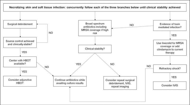
Proposed management algorithm for necrotizing soft tissue infections.
Necrotizing soft tissue infections: gas gangrene/myonecrosis and necrotizing fasciitis
Necrotizing SSTIs are difficult to treat and require aggressive surgical debridement, broad-spectrum antimicrobials, and intensive care. Table 2 and Fig. 1 demonstrate factors associated with increased likelihood of necrotizing infection and a proposed management tree [19]. Source control of infection is paramount and serial surgical debridements are generally required. The frequency and number of required debridements varies, but generally debridement should occur every 24–48 h until there is no evidence of necrosis. Daily wound dressing changes should be done to look for ongoing infection (e.g., bullae, devitalized tissue, spreading erythema) that would require repeat debridement. Increased requirements for intensive care support or laboratory parameters suggestive of worsening infection (e.g., progressive renal failure, increasing leukocytosis, increasing lactate) should prompt discussion of repeat debridement. Surgical control of infection is particularly important because diffusion of antimicrobials into affected tissues is limited due to significant tissue edema, necrosis, inflammation, and penetrating vessel thromboses [20].
Table 2.
Characteristics associated with increased likelihood of necrotizing infection
| Clinical parameters | Laboratory parameters |
|---|---|
| Pain out proportion to examination |
Serum sodium <135 mmmol/l |
| Bullae | White blood cell count >15 400 cell/ml |
| Tenderness beyond area of erythema |
Renal failure |
| Crepitus | Progressive lactic acidosis |
| Cutaneous anesthesia | |
| Cellulitis refractory to antibiotic therapy | |
| Rapid progression of cellulitis | |
| Dusky appearance of skin | |
| Systemic toxicity |
Adapted from [19].
Gas gangrene/myonecrosis
Gas gangrene or myonecrosis is caused by Clostridium species and should be managed surgically with adjunctive broad-spectrum antibiotics while awaiting culture results (Table 1). Though rare, Clostridium sordellii infections are notable as they can be associated with a toxic-shock like syndrome, particularly in patients with recent parturition or abortion [8,9,21]. TSS from clostridial infection is pathophysiologically dissimilar to SeTSS or SaTSS, making IVIG of dubitable benefit [8,9,21].
Necrotizing fasciitis
Necrotizing fasciitis (Fig. 2) is a rare SSTI that involves the deep fascia [19]. Rates of necrotizing fasciitis vary widely based on region (0.18–15.5 per 100 000) and are increasing over time [22,23]. Despite patients with necrotizing fasciitis having a higher severity of illness than patients with cellulitis, a recent study found that patients with cellulitis and necrotizing fasciitis had similar in-hospital and 90-day mortality, presumably due to higher comorbidity burden in patients with cellulitis [24■]. However, the study had a small number of patients and may not have been powered to detect a difference in mortality between the groups.
FIGURE 2.
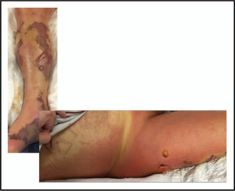
Necrotizing fasciitis of the lower extremity. Retiform purpura with bullae formation (a) or rapidly spreading erythema with bullae formation (b) should prompt urgent surgical consultation. Adapted from [19].
Type I necrotizing fasciitis is polymicrobial, including aerobic and anaerobic organisms. Type II necrotizing fasciitis is classically caused by S. pyogenes, though S. aureus also falls into this category. There are a variety of less frequently encountered agents causing necrotizing fasciitis, which makes it important for practitioners to realize the importance of surgical debridement with attendant bacterial cultures in combination with broad-spectrum antimicrobials as the first lines of therapy [25,26].
Though the classic teaching for necrotizing fasciitis is pain out proportion to physical examination findings, it is important to remember that superficial nerves can undergo necrosis, resulting in anesthesia of affected areas. A high degree of suspicion for necrotizing SSTI is required due to variability in physical examination findings and low sensitivity of imaging modalities. Imaging findings cannot rule out necrotizing fasciitis and may delay surgical intervention, which is associated with poor outcomes [27]. However, in clinically stable patients, MRI may be helpful in distinguishing necrotizing from nonnecrotizing infection [28].
Necrotizing fasciitis predominates on the lower extremity and predisposing conditions such as diabetes and peripheral vascular disease reflect this localization. Due to the relative rarity and heterogeneity of microbiologic causes, no clinical trials are available to guide duration of therapy. Based on expert opinion, recent guidelines suggest antimicrobial therapy directed against cultured organisms for at least 48–72 h after patients are clinically stable and require no further operative interventions [4].
Surgical considerations
For all patients with severe SSTIs, general resuscitative measures should be followed in accordance with institutional protocols. Source control is paramount, which may include surgical debridement, removal of invasive devices, or vaginal examination in the case of menstrual TSS. Prolonged time from presentation to first surgical intervention is associated with increased mortality [27,29]. In a mixed cohort of severe sepsis/septic shock patients that included patients with SSTIs, source control was associated with reduced mortality despite patients requiring source control having greater severity of illness [30■■].
In conjunction with serial debridements, vacuum-assisted closure of wounds may contribute to healing [31]. For cases of necrotizing infection involving the perineum or other sites with potential for stool contamination, temporary colostomy may be required to assist in wound healing. Rates of amputation in lower extremity necrotizing fasciitis vary from 15 to 72% based on comorbidities, with diabetes being a strong risk factor for amputation [32]. Although potentially life-saving, it is important to recognize that amputations, among other factors, may be associated with significant functional limitations after discharge [33].
Hyperbaric oxygen therapy
The use of hyperbaric oxygen therapy (HBOT) for necrotizing SSTI remains controversial due to mixed evidence of benefit, a lack of RCTs, and variable access to hyperbaric oxygen chambers [34–38]. In the absence of RCTs or well done propensity score analyses, we cannot recommend for or against the use of adjunctive HBOT for the management of necrotizing SSTI. For centers with HBOT readily available, its use can be considered, but should not be a substitute for or result in delays in surgical or antimicrobial therapy (Fig. 1).
Antimicrobial considerations
As a general rule, all severe SSTI should be treated empirically with broad-spectrum antibiotics directed against typical pathogens, specifically MRSA, resistant Gram-negatives, and anaerobes (Table 1 and Table 3). Notably, patients with complicated SSTI have more rapid achievement of clinical stability if empiric antimicrobials are appropriate for isolated pathogens [39■■]. All practitioners should consider local antibiograms when choosing empiric antimicrobials, as antibiograms can vary significantly. In regions such as Northern Europe with low rates of MRSA [40], it may be prudent to exclude MRSA coverage from empiric therapy in patients at low risk of MRSA infections. Preliminary work with MRSA risk prediction tools in SSTIs show promise, but more data are needed before implementing these tools and foregoing empiric MRSA coverage [41■].
Table 3.
Empiric antimicrobial dosing and duration guide for severe skin and soft tissue infections
| Organism type | First-line antimicrobials | Second-line antimicrobials | Duration of therapy |
|---|---|---|---|
| Gram-positive | Vancomycin 15 mg/kga and clindamycin 900 mg IV q8H |
Linezolid 600 mg IV q12H | At least 48–72 h after clinical stability and no further surgical debridements If bacteremic, refer to pathogen-specific guidelines, generally 14 days or more |
| Gram-negative and anaerobe |
Cefepime 1 g IV q8Hb (2 g IV q8H if BMI > 40) |
Meropenem 1 g IV q8Hb |
These recommendations are for patients in shock with risk factors for methicillin-resistant Staphylococcus aureus and multidrug-resistant Gram-negative bacterial infections. There is increasing evidence of nephrotoxicity from the combination of vancomycin and piperacillin-tazobactam, making carbapenems a more favorable second-line agent for Gram-negatives and anaerobes. IV, intravenous.
Dosing interval dependent on creatinine clearance.
Provided dose assumes a normal creatinine clearance.
De-escalation of antibiotic therapy should be based on clinical improvement, cultured pathogens, and results of rapid diagnostic tests where available. Rapid diagnostic testing for SSTIs is a relatively new area, but there is some promising data to show that their use results in increased appropriateness of therapy as well as increased rates of de-escalation [42■].
Considerations for selected antimicrobials
Dalbavancin and oritavancin are long-acting semi-synthetic lipoglycopeptides that are approved for a wide range of Gram-positive organisms. However, further studies are needed before their use can be recommended for severe SSTI. Daptomycin use may be contraindicated in patients with necrotizing fasciitis and elevated creatine kinase levels. As MRSA is one of the most common causes of SSTIs and severe illness is associated with higher rates of bacteremia, caution is advised when using linezolid, as its use in MRSA bacteremia may be associated with worse outcomes in patients with acute physiology and chronic health evaluation II scores at least 14 [43]. Tedizolid has been shown to be noninferior to line-zolid across a range of SSTI severity [44■], but there is no reason to believe it would be more efficacious in MRSA bacteremia than linezolid, so concerns about its empiric use remain. Telavancin is associated with higher rates of toxicity than other available agents for SSTI, and we therefore do not recommend its use when other agents can be employed. Though approved for SSTIs, tigecycline has been linked with worse outcomes in patients with severe illness. Tigecycline may also be a risk factor for treatment failure in patients with drug-resistant infections. As such, we recommend avoiding tigecycline therapy when other options are available.
Future therapies
There are some exciting new drugs in the pipeline for SSTI treatment, including delafloxacin and omadacycline, but discussion of their use will be covered by other articles in this issue. Nontraditional therapies for SSTIs, such as an antistaphylococcal alpha toxin antibody, have recently shown some promise in animal models, but are not available for human use [45■].
SPECIAL CONSIDERATIONS
Unusual causes of SSTI are outside the scope of this review, as most are rare and not typically associated with severe illness. For additional information, see recent reviews on this subject [19].
Immunocompromised hosts
Immunodeficiency changes the physical examination findings of SSTI, the putative pathogens, and the diagnostic and treatment plans. The differential diagnosis for dermatologic findings in the immuno-compromised host includes noninfectious causes and a broader range of infections, including invasive fungal, mycobacterial, and parasitic infections [4,19]. With a broader differential diagnosis and greater potential for decompensation, early dermatologic consultation for immunocompromised patients may be beneficial [4,46■]. Dermatology consultation can improve the diagnosis of dermato-logic findings in critically ill patients and reduce antimicrobial use [46■,47]. Many dermatologic conditions mimic infection, for which dermatologist expertise can be helpful in distinguishing [19,48].
All immunocompromised patients that are critically ill should undergo thorough cutaneous examination as immunosuppression tends to reduce physical exam findings of SSTIs. Immunosuppressed patients are more likely to have cutaneous dissemination of pathogens. A recent study showed that immunocompromised patients with S. pyogenes were more likely to have necrotizing fasciitis, septic shock, and die than immunocompetent patients [49■]. Conversely, in a cohort of patients with S. aureus infections, some of which had SSTIs, immunocompromise was not a risk factor for mortality [50■].
When possible, reduction of immunosuppression should be considered for severe infections. For patients with febrile neutropenia, Multinational Association of Supportive Care of Cancer score is important for predicting complication rates [51]. In neutropenic patients, factors to consider when contemplating surgery are probable duration of neutropenia and severity of infection. Patients with shorter durations of neutropenia have a higher likelihood of recovering from surgical interventions and are likely better candidates for surgery. Management of necrotizing SSTIs in neutropenic patients is poorly studied, and treatment strategies should be individualized.
CONCLUSION
SSTIs have a variety of presentations and can be severe enough to require intensive care. Practitioners should be familiar with the spectrum of clinical presentations for SSTI that require urgent surgical debridement to avoid delays in surgery as this can lead to worsened outcomes. Aggressive source control and broad spectrum antimicrobials are essential for all severe SSTI, with empiric therapy guided by knowledge of patient risk factors, the local antibiogram, and where available, rapid diagnostic testing.
KEY POINTS.
Severe skin and SSTIs initially require intensive care, source control, and broad-spectrum antimicrobials.
Intravenous immunoglobulin use in toxic shock syndrome remains controversial, but can be considered for severe cases.
For necrotizing skin and SSTIs, surgical consultation is paramount.
Imaging studies cannot rule out necrotizing infection and should not delay surgical interventions.
Pathogen-directed therapy and antimicrobial de-escalation should be the goal of severe skin and SSTI treatment when clinical stability is achieved.
Acknowledgements
We would like to thank Rebecca Light for her assistance in preparation of this article.
Financial support and sponsorship
M.H.K. was supported by the Barnes-Jewish Hospital Foundation.
J.P.B. reports that ‘This work was supported by the Washington University Institute of Clinical and Translational Sciences grant UL1TR000448 from the National Center for Advancing Translational Sciences (NCATS) of the National Institutes of Health (NIH). The content is solely the responsibility of the authors and does not necessarily represent the official view of the NIH.’
Footnotes
Conflicts of interest
The work was performed at Barnes-Jewish Hospital, St. Louis, Missouri.
There are no conflicts of interest.
Papers of particular interest, published within the annual period of review, have been highlighted as:
■ of special interest
■■ of outstanding interest
REFERENCES AND RECOMMENDED READING
- 1.Hersh AL, Chambers HF, Maselli JH, Gonzales R. National trends in ambulatory visits and antibiotic prescribing for skin and soft-tissue infections. Arch Intern Med 2008; 168:1585–1591. [DOI] [PubMed] [Google Scholar]
- 2.Edelsberg J, Taneja C, Zervos M, et al. Trends in US hospital admissions for skin and soft tissue infections. Emerg Infect Dis 2009; 15: 1516–1518. [DOI] [PMC free article] [PubMed] [Google Scholar]
- 3.Crisp JG, Takhar SS, Moran GJ, et al. Inability of polymerase chain reaction, pyrosequencing, and culture of infected and uninfected site skin biopsy specimens to identify the cause of cellulitis. Clin Infect Dis 2015; 61:1679–1687.■■ The reference is of interest because it describes limitations in advanced diagnostics to help determine the cause of skin and soft tissue infections (SSTIs).
- 4.Stevens DL, Bisno AL, Chambers HF, et al. Practice guidelines for the diagnosis and management of skin and soft tissue infections: 2014 update by the Infectious Diseases Society of America. Clin Infect Dis 2014; 59:e10–e52. [DOI] [PubMed] [Google Scholar]
- 5.European Centre for Disease Prevention and Control (ECDC). Annual epi-demiological report 2012: reporting on 2010 surveillance data and 2011 epidemic intelligence data Stockholm: ECDC; 2013. [Google Scholar]
- 6.Stevens DL. Reply to Gonzalez del Castillo et al. and Rashid and Kravitz. Clin Infect Dis 2015; 60:172–174. [DOI] [PubMed] [Google Scholar]
- 7.Burnham JP, Kollef MH. Understanding toxic shock syndrome. Intensive Care Med 2015; 41:1707–1710. [DOI] [PubMed] [Google Scholar]
- 8.Cohen AL, Bhatnagar J, Reagan S, et al. Toxic shock associated with Clostridium sordellii and Clostridium perfringens after medical and spontaneous abortion. Obstet Gynecol 2007; 110:1027–1033. [DOI] [PubMed] [Google Scholar]
- 9.Fischer M, Bhatnagar J, Guarner J, et al. Fatal toxic shock syndrome associated with Clostridium sordellii after medical abortion. N Engl J Med 2005; 353:2352–2360. [DOI] [PubMed] [Google Scholar]
- 10.Stevens DL, Wallace RJ, Hamilton SM, Bryant AE. Successful treatment of staphylococcal toxic shock syndrome with linezolid: a case report and in vitro evaluation of the production of toxic shock syndrome toxin type 1 in the presence of antibiotics. Clin Infect Dis 2006; 42:729–730. [DOI] [PubMed] [Google Scholar]
- 11.Carapetis JR, Jacoby P, Carville K, et al. Effectiveness of clindamycin and intravenous immunoglobulin, and risk of disease in contacts, in invasive group a streptococcal infections. Clin Infect Dis 2014; 59:358–365. [DOI] [PubMed] [Google Scholar]
- 12.Stevens DL, Ma Y, Salmi DB, et al. Impact of antibiotics on expression of virulence-associated exotoxin genes in methicillin-sensitive and methicillin-resistant Staphylococcus aureus. J Infect Dis 2007; 195:202–211. [DOI] [PubMed] [Google Scholar]
- 13.Coyle EA, Cha R, Rybak MJ. Influences of linezolid, penicillin, and clindamycin, alone and in combination, on streptococcal pyrogenic exotoxin a release. Antimicrob Agents Chemother 2003; 47:1752–1755. [DOI] [PMC free article] [PubMed] [Google Scholar]
- 14.Darenberg J, Ihendyane N, Sjolin J, et al. Intravenous immunoglobulin G therapy in streptococcal toxic shock syndrome: a European randomized, double-blind, placebo-controlled trial. Clin Infect Dis 2003; 37:333–340. [DOI] [PubMed] [Google Scholar]
- 15.Linner A, Darenberg J, Sjolin J, et al. Clinical efficacy of polyspecific intravenous immunoglobulin therapy in patients with streptococcal toxic shock syndrome: a comparative observational study. Clin Infect Dis 2014; 59:851–857. [DOI] [PubMed] [Google Scholar]
- 16.Matsushima A, Kuroki Y, Nakajima S, et al. Low level of TSST-1 antibody in burn patients with toxic shock syndrome caused by methicillin-resistant Staphylococcus aureus. J Burn Care Res 2015; 36:e120–e124. [DOI] [PubMed] [Google Scholar]
- 17.Madsen MB, Hjortrup PB, Hansen MB, et al. Immunoglobulin G for patients with necrotising soft tissue infection (INSTINCT): a randomised, blinded, placebo-controlled trial. Intensive Care Med 2017; 43:1585–1593.■■ The reference is of interest because it propagates the debate about the merits of intravenous immunoglobulin (IVIG) for the treatment of necrotizing skin and SSTIs.
- 18.Kadri SS, Swihart BJ, Bonne SL, et al. Impact of intravenous immunoglobulin on survival in necrotizing fasciitis with vasopressor-dependent shock: a propensity score-matched analysis from 130 US hospitals. Clin Infect Dis 2017; 64:877–885.■■ The reference is of interest because it propagates the debate about the merits of IVIG for the treatment of necrotizing skin and SSTIs.
- 19.Burnham JP, Kirby JP, Kollef MH. Diagnosis and management of skin and soft tissue infections in the intensive care unit: a review. Intensive Care Med 2016; 42:1899–1911. [DOI] [PMC free article] [PubMed] [Google Scholar]
- 20.Umbert IJ, Winkelmann RK, Oliver GF, Peters MS. Necrotizing fasciitis: a clinical, microbiologic, and histopathologic study of 14 patients. J Am Acad Dermatol 1989; 20(5 Pt 1):774–781. [DOI] [PubMed] [Google Scholar]
- 21.Sinave C, Le Templier G, Blouin D, et al. Toxic shock syndrome due to Clostridium sordellii: a dramatic postpartum and postabortion disease. Clin Infect Dis 2002; 35:1441–1443. [DOI] [PubMed] [Google Scholar]
- 22.Ellis Simonsen SM, van Orman ER, Hatch BE, et al. Cellulitis incidence in a defined population. Epidemiol Infect 2006; 134:293–299. [DOI] [PMC free article] [PubMed] [Google Scholar]
- 23.Das DK, Baker MG, Venugopal K. Increasing incidence of necrotizing fasciitis in New Zealand: a nationwide study over the period 1990 to 2006. J Infect 2011; 63:429–433. [DOI] [PubMed] [Google Scholar]
- 24.Cranendonk DR, van Vught LA, Wiewel MA, et al. Clinical characteristics and outcomes of patients with cellulitis requiring intensive care. JAMA Dermatol 2017; 153:578–582.■ The study is of interest because it found that patients with cellulitis fare no worse than patients with necrotizing fasciitis in the medium term due to a higher burden of comorbidities in patients with cellulitis.
- 25.Shaked H, Samra Z, Paul M, et al. Unusual ‘flesh-eating’ strains of Escherichia coli. J Clin Microbiol 2012; 50:4008–4011. [DOI] [PMC free article] [PubMed] [Google Scholar]
- 26.Cheng NC, Yu YC, Tai HC, et al. Recent trend of necrotizing fasciitis in Taiwan: focus on monomicrobial Klebsiella pneumoniae necrotizing fasciitis. Clin Infect Dis 2012; 55:930–939. [DOI] [PubMed] [Google Scholar]
- 27.Wong CH, Chang HC, Pasupathy S, et al. Necrotizing fasciitis: clinical presentation, microbiology, and determinants of mortality. J Bone Joint Surg Am 2003; 85-a:1454–1460. [PubMed] [Google Scholar]
- 28.Kim KT, Kim YJ, Won Lee J, et al. Can necrotizing infectious fasciitis be differentiated from nonnecrotizing infectious fasciitis with MR imaging? Radiology 2011; 259:816–824. [DOI] [PubMed] [Google Scholar]
- 29.Chen SC, Chan KS, Chao WN, et al. Clinical outcomes and prognostic factors for patients with Vibrio vulnificus infections requiring intensive care: a 10-yr retrospective study. Crit Care Med 2010; 38:1984–1990. [DOI] [PubMed] [Google Scholar]
- 30.Martinez ML, Ferrer R, Torrents E, et al. Impact of source control in patients with severe sepsis and septic shock. Crit Care Med 2017; 45:11–19.■■ The reference is of interest because it affirms the importance of source control in patients with a variety of infections, including those with skin and SSTIs.
- 31.de Geus HR, van der Klooster JM. Vacuum-assisted closure in the treatment of large skin defects due to necrotizing fasciitis. Intensive Care Med 2005; 31:601. [DOI] [PubMed] [Google Scholar]
- 32.Chen IW, Yang HM, Chiu CH, et al. Clinical characteristics and risk factor analysis for lower-extremity amputations in diabetic patients with foot ulcer complicated by necrotizing fasciitis. Medicine 2015; 94:e1957. [DOI] [PMC free article] [PubMed] [Google Scholar]
- 33.Pham TN, Moore ML, Costa BA, et al. Assessment of functional limitation after necrotizing soft tissue infection. J Burn Care Res 2009; 30:301–306. [DOI] [PMC free article] [PubMed] [Google Scholar]
- 34.George ME, Rueth NM, Skarda DE, et al. Hyperbaric oxygen does not improve outcome in patients with necrotizing soft tissue infection. Surg Infect 2009; 10:21–28. [DOI] [PubMed] [Google Scholar]
- 35.Massey PR, Sakran JV, Mills AM, et al. Hyperbaric oxygen therapy in necrotizing soft tissue infections. J Surg Res 2012; 177:146–151. [DOI] [PubMed] [Google Scholar]
- 36.Shaw JJ, Psoinos C, Emhoff TA, et al. Not just full of hot air: hyperbaric oxygen therapy increases survival in cases of necrotizing soft tissue infections. Surg Infect 2014; 15:328–335. [DOI] [PMC free article] [PubMed] [Google Scholar]
- 37.Devaney B, Frawley G, Frawley L, Pilcher DV. Necrotising soft tissue infections: the effect of hyperbaric oxygen on mortality. Anaesth Intensive Care 2015; 43:685–692. [DOI] [PubMed] [Google Scholar]
- 38.Levett D, Bennett MH, Millar I. Adjunctive hyperbaric oxygen for necrotizing fasciitis. Cochrane Database Syst Rev 2015; 1:Cd007937. [DOI] [PMC free article] [PubMed] [Google Scholar]
- 39.Jaaskelainen IH, Hagberg L, Forsblom E, Jarvinen A. Factors associated with time to clinical stability in complicated skin and skin structure infections. Clin Microbiol Infect 2017; 23:674.e1–674.e5.■■ The reference is of interest because it demonstrates the need for appropriate empiric antibiotics in patients with skin and SSTI, as this results in improved time to clinical stability.
- 40.Control ECfDPa. Proportion of Methicillin Resistant Staphylococcus aureus (MRSA) Isolates in Participating Countries in 2014 – See more at: http://ecdc.europa.eu/en/healthtopics/antimicrobial_resistance/database/Pages/map_reports.aspx#sthash.KnkOdOBq.dpuf2016 2016. Available from: http://ecdc.europa.eu/en/healthtopics/antimicrobial_resistance/database/Pages/map_reports.aspx. [Updated 20 July 2016].
- 41.Trinh TZE, Claeys K, Dryden M, et al. International validation of a risk-assessment tool for methicillin-resistant Staphylococcus aureus acute bacterial skin and skin-structure infections Vienna, Austria: ECCMID; 2017.■ The reference is of interest because the authors were able to find a population of patients in which they could forego Methicillin-resistant Staphylococcus aureus coverage due to their risk score.
- 42.Santiago EOR, Chong MAS, Alvarez-Uria A, et al. Clinical impact of rapid diagnostic approach (direct GeneXpert) for the management of patients with skin and soft tissue infections Vienna, Austria: ECCMID; 2017.■ The reference is of interest because the authors were able to use rapid diagnostic testing to improve antibiotic therapy in patients with skin and SSTIs.
- 43.Burnham JP, Burnham CA, Warren DK, Kollef MH. Impact of time to appropriate therapy on mortality in patients with vancomycin intermediate Staphylococcus aureus infection. Antimicrob Agents Chemother 2016; 60: 5546–5553. [DOI] [PMC free article] [PubMed] [Google Scholar]
- 44.Sandison T, De Anda C, Fang E, et al. Clinical response of tedizolid versus linezolid in acute bacterial skin and skin structure infections by severity measure using a pooled analysis from two phase 3 double-blind trials. Antimicrob Agents Chemother 2017; 61:e02687–16.■ The reference is of interest because it was found that tedizolid was noninferior to linezolid in patients with skin and SSTIs of various severities.
- 45.Le VT, Tkaczyk C, Chau S, et al. Critical role of alpha-toxin and protective effects of its neutralization by a human antibody in acute bacterial skin and skin structure infections. Antimicrob Agents Chemother 2016; 60:5640–5648.■ The reference is of interest because the authors identified a novel target for the treatment of skin and SSTIs, though it has only been tested in animal models to date.
- 46.Strazzula L, Cotliar J, Fox LP, et al. Inpatient dermatology consultation aids diagnosis of cellulitis among hospitalized patients: a multiinstitutional analysis. J Am Acad Dermatol 2015; 73:70–75.■ The reference is of interest because it demonstrates the value of dermatologic consultation in the diagnosis of skin and SSTIs.
- 47.Arakaki RY, Strazzula L, Woo E, Kroshinsky D. The impact of dermatology consultation on diagnostic accuracy and antibiotic use among patients with suspected cellulitis seen at outpatient internal medicine offices: a randomized clinical trial. JAMA Dermatol 2014; 150:1056–1061. [DOI] [PubMed] [Google Scholar]
- 48.Falagas ME, Vergidis PI. Narrative review: diseases that masquerade as infectious cellulitis. Ann Intern Med 2005; 142:47–55. [DOI] [PubMed] [Google Scholar]
- 49.Linder KA, Alkhouli L, Ramesh M, et al. Effect of underlying immune compromise on the manifestations and outcomes of group A streptococcal bacteremia. J Infect 2017; 74:450–455.■ The reference is of interest because the authors found that immunocompromised patients had worse outcomes in group A Strep bacteremia than immunocompetent patients.
- 50.Sasson G, Bai AD, Showler A, et al. Staphylococcus aureus bacteremia in immunosuppressed patients: a multicenter, retrospective cohort study. Eur J Clin Microbiol Infect Dis 2017; 36:1231–1241.■ The reference is of interest because the authors found that outcomes of S. aureus bacteremia were not influenced by immune status.
- 51.Uys A, Rapoport BL, Anderson R. Febrile neutropenia: a prospective study to validate the Multinational Association of Supportive Care of Cancer (MASCC) risk-index score. Support Care Cancer 2004; 12:555–560. [DOI] [PubMed] [Google Scholar]